

Non-ablative laser skin rejuvenation by targeting indigenous chromophores
⁎Corresponding author. khawla.s.khashan@uotechnology.edu.iq (Khawla S. Khashan)
-
Received: ,
Accepted: ,
This article was originally published by Elsevier and was migrated to Scientific Scholar after the change of Publisher.
Abstract
Three pulsed laser systems: 980 nm semiconductor diode laser, 1064 nm Nd: YAG laser and long pulse- fractional 2940 nm pulsed Er: YAG were used to treat some non-ablative skin rejuvenation applications. These three wavelengths can target the three main indigenous chromophores (melanin, hemoglobin and water) of the skin to treat different skin disorders. The efficacy of these lasers was assessed in the treatment of age spots, enlarged veins and wrinkles. The chromophore type and location of a specific disorder was taken into consideration when working out, in advance, the required laser fluence. The absorption of laser photons and the reabsorbed photon scattering were calculated and used to work out the temperature rise in the targeted tissue. The results indicated very good outcome of age spots with 980 nm laser, excellent clearance of the varicose veins with the use of 1064 nm laser and very good smoothing of fine lines wrinkles on forehead. Advances may involve conducting wavelengths or developing customizable protocols for different skin types. Long-term studies on patient outcomes to help establish standardized protocols and improve efficacy in clinical uses.
Keywords
Age spots
Enlarged veins
Wrinkles
Laser
Photons absorption and scattering

- Nd: YAG
-
Neodymium-doped yttrium aluminum garnet
- Er: YAG
-
Erbium-doped yttrium aluminum garnet
- UV
-
Ultraviolet
- HgB
-
Hemoglobin
Abbreviations
1 Introduction
Skin aging is characterized by enlarged veins, fine and coarse wrinkles, age spots and skin laxity (Piccolo et al. 2023). As someone gets older, his skin becomes prone to wrinkling and age-related problems. Skin ages because of natural deterioration and environmental factors. Collagen and elastin reduction, cell turnover slows, and diminishes of hyaluronic acid, leading to wrinkles and loss of inflexibility. Sun exposure damages skin, causing age spots and leathery texture(Mirza, Mirza, and Khatri 2021). Lifestyle influences like smoking and stress accelerate aging. (Duplechain 2023). This makes many people more concerned about the way they look and start to make efforts to reduce the age – related signs. Aesthetic laser applications rely on the way that laser light interacts with skin tissues. Anderson and Parish in 1983 presented the selective photo thermolysis theory. It is defined as the damage restricted to a selected tissue shell by specific laser wavelength, pulse width, and the rate of repetition(Lanigan 2021). The essential rule behind the use of light curative depends on the rule of selective photo thermolysis which is explained by three points: deep penetration of optical energy to attain the treated tissue, absorption of optical energy by the biological tissue and strong optical energy is enough to initiate the thermal injury of the treated biological tissue(Fodor and Sobec 2020). In selective photo thermolysis, a pulse of eradiation with suitable wavelength and duration is delivered to a targeted location. It should be effective for a broad range of treatments. the term thermal interaction includes a number of interaction types depending on the absorption of the photons by tissue contact like vaporization, coagulation, carbonization, and melting(Brown 2020). Knowing the science of light-tissue interaction help selecting the optimal laser parameters to follow a safe and effective pathway needed for the therapy (Kim et al. 2023; Mustafa, Hamoudi, and Khashan 2023). The absorption is a function of wavelength and recognized as the photon absorption eventuality in tissue per unit pathway length. Varying tissue types have varying standards of this coefficient. Blood contains two types of hemoglobin: oxyhemoglobin (HbO2) and de-oxyhemoglobin(Hb).These two types of hemoglobin have different absorption electromagnetic spectra. The Hb peak absorption is best at 420 nm and the second absorption peak is at 580 nm(Patel et al. 2024). Its spectrum increasingly decreases with light wavelength increasesHBO2 shows its greatest peak of absorption at 410 nm, and two junior peaks at 550 nm and 600 nm. At wavelengths longer than 600 nm, HbO2 absorption declines much faster than Hb absorption. Water is approximately optical clear in the domain of visible light but starts to be absorbing at the NIR section Water is an important component due to its high concentration in biological tissue. The absorption electromagnetic spectrum of water is in the range of 250–––1000 nm(Patel et al. 2024). The absorption peak of human skin melanin pigment happens around 335 nm. The absorption is almost completely attenuated for wavelengths longer than 700 nm. Fat and protein are the main contributors to the NIR spectral region. Facelift surgery, soft tissue filler, chemical peeling and laser ablative and non-ablative treatments are nowadays routine work to treat various skin disorders (Mustafa, Hamoudi, and Khashan 2023; Alhallak et al. 2023; Khalid et al. 2024). Traditional surgical besides chemical approaches to skin rejuvenation, such as facelifts and chemical peels often involve long recovery periods, significant discomfort, and a discriminating risk of side effects like scarring and uneven pigmentation(Horovitz, Clementoni, and Artzi 2021). On the other hand, laser-based therapies offer a more precise and less invasive approach, resulting in shorter downtime, reduced pain, and a lower incidence of side effects. This translates to more natural-looking effects and improved patient satisfaction (Mirza et al., 2021). Ablative lasers can be more effective than their non-ablative counterparts, but they need longer recovery time. In ablative resurfacing, the hole epidermal layer is removed and replaced by a new perfect epidermal layer in one session, but full recovery takes around six months without exposing the skin to sun light. In non-ablative procedure, on the other hand, improving texture and laxity of the skin is only achieved by collagen regeneration using at least 3 sessions. They are also associated with post inflammatory hyperpigmentation due to the excessive thermal energy transferred to adjacent tissue (Chernigovskiy et al. 2019). Infrared 980 nm semiconductor diode laser, 1064 nm Nd: YAG (neodymium-doped yttrium aluminum garnet; Nd: Y3Al5O12 laser, and 2940 nm Er: YAG lasers (erbium-doped yttrium aluminum garnet laser, erbium YAG laser) have been used in non-ablative rejuvenation of skin with variable outcomes. Infrared lasers are used in non-ablative skin rejuvenation. These lasers penetrate the skin to target specific issues like vascular lesions, pigmentation, and wrinkles. They stimulate collagen production, improve skin texture, and reduce signs of aging while effective, their results can vary depending on factors like individual skin type, laser parameters, and treatment protocol(Pour Mohammad et al. 2023). While infrared lasers have shown promise in non-ablative skin rejuvenation, a significant challenge lies in the lack of standardized treatment parameters. Optimal settings, including wavelength, pulse duration, fluence, and spot size, can vary widely depending on the specific laser system, target tissue, and desired outcome. This lack of consensus can lead to inconsistent results and potential adverse effects(Beigvand et al. 2020). Furthermore, the underlying mechanisms of action for many infrared laser treatments remain incompletely understood. While it is generally accepted that these lasers induce thermal injury to the dermis, stimulating collagen synthesis and remodeling, the precise cellular and molecular processes involved are still being investigated. A deeper understanding of these mechanisms could help optimize treatment parameters and improve patient outcomes(Topaloglu, Özdemir, and Çevik 2021). Recently Hayley and her colleagues tested this method on small numbers of participants having different skin indications who were undergone different post procedural evaluation protocols and assessments (Leight-Dunn, Chima, and Hoss 2020). Their study expressed the need for standard clinical procedures and the use of excellent post-treatment skin care. This study aims at assessing the effectiveness and efficacy of non-ablative laser treatments using 980 nm semiconductor diode laser, 1064 nm Nd laser, and 2940 nm Er laser in skin rejuvenation.
2 Laser-tissue interaction
Different interaction pathways take place when laser light interacts with human tissues. Parameters of a specific tissue (coefficients of reflection, absorption, scattering, and transmission at a certain wavelengths, heat conduction and heat capacity), as well as the laser parameters (intensity, pulse length, fluence, wavelength and laser spot diameter) are very much related to each other when considering a treatment protocol (Alhallak et al. 2023). The laser pulse duration is a very important parameter that needs to be selected carefully to decide the type of interaction. Four main categories of interaction mechanisms are classified in this field(Hussain et al. 2024). These are thermal, photochemical, photo-ablation and photo-disruption(Tanghetti 2016). The laser energy deposition is not an exclusive job of laser features like the wavelength, pulse duration, irradiance, repetition rate, and spot size, but also depends effectively on some optical tissue parameters such as the coefficients of absorption and scattering. Moreover, the tissue properties are important to describe transfer and storage of heat; like specific heat capacity and thermal diffusivity (Fisher 2020). The epidermal layer of the skin primarily absorbs the visible light by its content of melanin), while the dermal skin layer houses blood vessels, water and collagen in addition to other components (Niemz 2007). The directly absorbed light and re-absorbed internal scattering are transformed into heat which raises the temperature in the targeted tissue to a value related to the laser intensity (Peavy 2002). At the end of this cycle, the heat is diffused away, and the tissue retains its normal temperature after elapsing a time connected to the tissue's thermal relaxation time. Heat generation is determined by laser parameters such as intensity, pulse duration, and optical tissue properties. While heat transport is governing by the thermal properties of the tissue as well as exposure time (Azhdari et al. 2024). The epidermis coefficient of absorption μe merges both the absorption of melanin and the baseline skin. It is given as:
Where: μe: epidermis coefficient of absorption (cm−1), mel %: percentage of melanosomes per unit volume, μ1: coefficient of melanin absorption (cm−1), μ2: absorption of the skin baseline (cm−1) (Niemz 2007; Peavy 2002). To work out the exact value of epidermal absorption coefficient, the melanin pigmentation per unit volume is required for different skin colors (Niemz 2007).
The skin's dermis total absorption coefficient, or μa (der), is primarily influenced by hemoglobin and water absorption, with negligible effect of baseline skin absorption. The parameters of laser wavelength (nm), the percentage of water (%) and hemoglobin (%) are used to find μa (der). The absorptions of skin baseline in dermis and epidermis are similar; and can be referred to as μ2. μa is the probability per path length where a photon is absorbed. It depends on the chromophore type and the laser wavelength (Steiner 2010). For large heterogeneous tissue volumes, μa is given in accordance with the percentage of a certain chromophore. For example; the fraction of a uniformly distributed blood in dermis (f. blood) is 0.2 (Jacques 1989). Therefore the net absorption of the dermis, μa der is given as (Svaasand et al. 1995):
For water chromophore, all blood parameters go to zero. Table 1 represents the utilization of equation (1) and equation (2) and the substitution of approved published data elsewhere.
| Wavelength (nm) |
Skin tone (color) | Epidermis absorption Coefficients (cm−1) |
Dermis absorption coefficient (cm−1) |
|---|---|---|---|
| 1064 | Light | 3.19 | 3.05 |
| 1064 | Moderate | 5.6 | 3.05 |
| 1064 | Dark | 7.915 | 3.05 |
| 980 | Light | 3.44 | 2.9 |
| 980 | Moderate | 8.225 | 2.9 |
| 980 | Dark | 11.415 | 2.9 |
| 2940 | Light | − | 2400 |
| 2940 | moderate | − | 2400 |
| 2940 | Dark | − | 2400 |
A pencil-like laser beam, upon traversing a medium, undertakes scattering, dispersing its energy in multiple directions further than the incident path. While this scattering phenomenon is a well-established physical principle, its influence on the propagation of laser light within the epidermal layer of the skin is relatively limited, mostly for wavelengths within the visible and near-infrared spectrum. The relatively thin nature of the epidermis and the specific properties of light in this spectral range have a tendency to mitigate the effects of scattering, allowing the laser beam to penetrate and interact with the target tissues with minimal deviation from its intended course. (Cox 2007). The reduced dermal layer scattering coefficient combines Mie scattering by collagen and Rayleigh scattering by collagen and some cellular components(Khalil et al. 2024; Mehmood et al. 2024). The combined effect of these two types of scattering gives a scattering (µs) having an intensity attenuation similar to absorption:
Where µs; is the coefficient of scattering. In most tissues, absorption and scattering of light occur together. These tissues are known as opaque tissues. This means that light entering these tissues does not travel in a straight line but is deflected in different directions due to the presence of many small molecules within the tissue. Some of these molecules absorb the light and convert it into heat energy, while others scatter the light and change its direction. This phenomenon greatly affects the depth of light penetration into the tissue and its spatial distribution, which in turn affects the effectiveness of laser treatments and various diagnostic techniques. (Saqib et al. 2016). Scattering coefficients of dermis and epidermis for different skin tones and the total attenuation factor (µt) of different chromophores. are listed in Table 2..
| Tissue | λ (nm) | µ (cm−1) | µt (cm−1) |
|---|---|---|---|
| Epidermis (white) | 1064 | 130 | 133 |
| Epidermis (dark) | 1064 | 122 | 130 |
| dermis | 1064 | 35 | 38 |
| White epidermis | 980 | 67 | 72 |
| Dark epidermis | 980 | 60 | 70 |
| dermis | 980 | 30 | 33 |
| epidermis | 2940 | − | − |
| dermis | 2940 | − | 2400 |
At this point it is possible to calculate the exact fluence reduction through its way to the targeted tissue inside the skin. Still, the resulting fluence is not the actual value for calculating the rise in temperature(Hamoudi et al. 2021). Backscattered laser light can provide a larger fluence underneath the skin tissue than at its surface (Nouri 2018; Mahmood et al. 2013). The required treating fluence exceeds what’s been thought enough to on the skin surface because of the losses before reaching the targeted tissue, as explained hereunder.
Where F is the subsurface laser fluence, Fo is the surface laser fluence, R is the remittance of the medium. R = 0.5 for the near infrared lasers and R and R = 0 for water chromophore when using 2940 nm laser. Once the local subsurface fluence is determined, targeted tissue temperature can be predicted by thermal relaxation time, laser fluence, pulse duration, and the specific absorption of the targeted tissue (Mustafa et al., 2021a). The temperature increase of the targeted tissue will then be worked out by substituting the coefficients of scattering and absorption, laser fluence at the skin surface, target size and the laser pulse duration as explained in equation 6.
Where c is the specific heat, r is the density, g is a constant having a value of 1 for planes, 2 for cylinders, and 3for spheres, t is the thermal relaxation time of the target tissue and τ is the laser pulse length; see Table 3.. After performing simple calculations, one can estimate the temperature rise of the targeted skin tissue.
| Structure | Size (µm) | Thermal relaxation time |
|---|---|---|
| Melanosomes cells | 0.5–1 | 1 (µs) |
| 10 | 300 (µs) | |
| Blood vessels | 50 | 1 (ms) |
| 100 | 5 (ms) | |
| 200 | 20 (ms) | |
| Epidermis | − | 4 ms-10 ms |
| dermis | − | 4 ms |
In the present work, three cosmetic laser applications have been considered, in which the three main indigenous chromophores (melanin, hemoglobin and water) were accordingly targeted by three different laser wavelengths: 980 nm, 1064 nm and 2940 nm. For water chromophore, the Er: YAG laser wavelength has a high absorption coefficient 12000 cm1. This permits limited penetration depth and, in general, causes small surrounding tissue injury in hard and soft tissues (Mustafa et al., 2021b).
3 Method and strategy
Three skin disorders were treated by three different laser wavelengths. Laser wavelengths: 980 nm, 1064 nm, and 2940 nm were used to target specified skin's chromophores (melanin, hemoglobin, and water) in accordance with the principle of selective photo thermolysis. The treatment protocols (according to the calculations); designed to pre-calculate laser parameters, were based on working out the exact temperature rise required for each application.
3.1 980 nm for pigmented lesions
Pigmented keratinocytes range from dark yellow, brown, to black and can be divided into Seborrheic keratosis, Melanesia (small melanin pigmented lesions) that can be epidermal, dermal or mixed and range in color from black (superficial) to brown spots on the skin. Despite they often do not appear at birth, they are commonly referred to as “birthmarks.” It is a common skin condition, various factors contribute to its development, including hormonal changes, sun exposure, genetics, and certain medications, laser therapy has emerged as a promising treatment option. Laser therapy offers a targeted approach to addressing Melanesia. By selectively targeting the melanin pigment responsible for the discoloration, lasers can effectively reduce the appearance of these patches. In the present study hand Melanesia was treated for 70 years old male with a skin tone (II); evaluated by skin color meter before treatment. The calculations revealed an ideal treatment laser fluence and pulse durations of 12 J/cm2 and 20 ms respectively. A 980 nm diode laser with 2 mm spot size was utilized to achieve the required laser parameters in one session. The reflected fluence (related to percentage of melanin) for light skin color is about 7 % (Niemz 2007). Equation (4) was utilized to work out the laser beam losses by epidermal attenuation factors, after accounting the kind of melanin pigmentation and the total scattering and absorption attenuation. This permitted accurate laser fluence to reach the target and therefore obtaining the correct temperature increase as shown in Table 4..
| Target | skin type |
Wavelength (nm) |
Fluence (J/cm2) |
Pulse duration (sec) |
Temperature Rise (co) |
|---|---|---|---|---|---|
| Melanin pigmented lesions on hand |
II | 980 | 12 | 0.02 | 41 |
3.2 1064 Skin resurfacing of fine lines
The effectiveness and safety of using correct parameters for skin rejuvenation are evaluated in this study using 1064 nm Nd: YAG laser to reduce wrinkles and loose skin. (Elite + TM Cynosure, Westford, Massachusetts, 01886, United States) pulsed 1064 nm neodymium laser was employed for this purpose which can can penetrate to deep dermis (≈ 11 mm). The first case study was 32 years' female with skin tone (III) and fine line in the forehead. She was treated with 20 J/cm2 and 0.4 ms laser pulse duration in 4 sessions with 2-week intervals; as shown in Table 5. The parameters were chosen according to the calculation based on the mathematical formulae described above, which gave a temperature rise of 43 °C. The second case was to treat varicose vein of IV-skin tone, 50-year-old female. The laser treatment protocol was worked out to give a temperature of 65C which is perfect for shrinking the veins.
| Target | Skin type |
Wavelength (nm) |
Fluence (J/cm2) |
Pulse Duration (ms) |
Rep. Rate (Hz) |
Temperature Rise (Co) |
|---|---|---|---|---|---|---|
| Fine lines (forehead) |
III | 1064 | 20 | 0.4 | 5 | 43 |
| Enlarged vein | V | 1064 | 120 | 20 | 1 | 65 |
3.3 2940 nm Er: YAG laser for course wrinkles
Wrinkles are primarily caused by the loss of skin elasticity due to the breakdown of collagen and elastin fibers within the dermal layer. This process is mainly influenced by aging, where the skin's ability to regenerate collagen diminishes over time. Besides; (UV) radiation exposure, oxidative stress, smoking, and environmental pollutants are additional causes. Er: YAG deeper skin alterations could be brought about by laser energy without unintentionally ablating the epithelium. The application of laser energy triggers the processes of extracellular matrix synthesis, tissue remodeling, and cell activation (Hympanova et al. 2020; Vas et al. 2019). Non-invasive ACTION II™, 2940 nm Er: YAG from (Lutronic intelligent, Billerica) was utilized to treat course wrinkle in a 66 years' male. 5 J/cm laser fluence, 1.6 mm beam diameter and 0.1 ms pulse length were used in the treatment protocol Table 6.. The wrinkle lines were treated every other week by three sessions: 3 tracks in each session. The results were determined by comparative photo documentation.
| Target | Skin tone |
Wavelength (nm) |
Fluence (J/cm2) |
Pulse duration (ms) |
Rep rate (Hz) |
Temperature Rise (Co) |
|---|---|---|---|---|---|---|
| course lines (forehead) | II | 2940 | 5 | 0.1 | 3 | 88 |
4 Results and discussions
High melanin concentration in the epidermis causes melanin pigmented lesions. Heating the epidermal melanin selectively can be achieved by using laser wavelengths between 800 and 1000 nm. While longer wavelengths tend to avoid the epidermal melanin, shorter laser wavelengths can promote skin injury by raising the surface epidermal temperatures(Pour Mohammad et al. 2023). For age spot treatment, the color had immediately disappeared after the first session and no adverse reaction was noticed. Without using anesthesia, cold air was administered to avoid burning the skin surface and the patient felt very mild pain; see Fig. 1. This application philosophy presents new parameters’ values for 980 nm diode laser that are based on pre-calculated fluence and helps achieve the result in just one session.
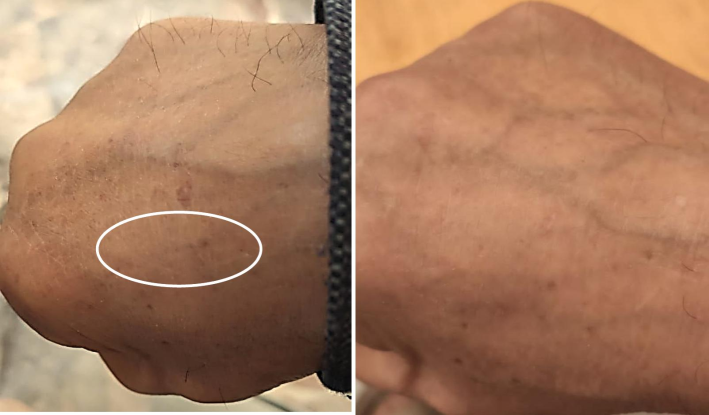
- Melanesia (a) before treatment (b) after the 980 nm laser treatment.
Fig. 2 shows the fine lines before and two months after the 1064 nm laser treatment. The result shows very good smoothing and the disappearance of the fine lines from the forehead. The patient felt little pain and redness of the treated skin, but these symptoms only lasted an hour after the end of the treatment session.
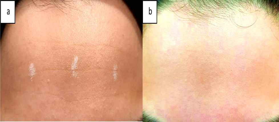
- The fine lines (a) before and (b) 2 months after the 1064 nm laser treatment.
Fig. 3 illustrates the efficacy of 1064 nm laser therapy in treating enlarged veins. The before-and-after images demonstrate a significant reduction in the visibility of the affected veins. This wavelength is particularly effective for targeting vascular lesions due to its deep penetration and selective absorption by hemoglobin. The laser energy calculates as mentioned above causing them to coagulate and eventually fade with minimum side effect.
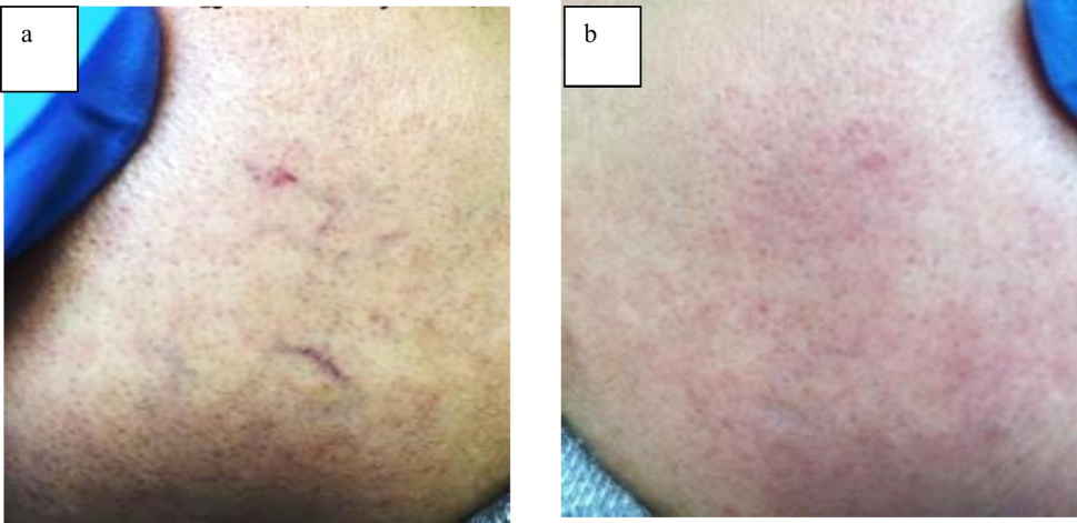
- Enlarged vein (a) before and (b) after 1064 nm laser treatment.
Fig. 4 showcases the positive impact of 2940 nm Er: YAG laser treatment on course wrinkles. The comparison between the pre-treatment and post-treatment images reveals a noticeable improvement in skin texture and a reduction in wrinkle depth. This laser's parameter selected to allow stimulating collagen production and promoting skin rejuvenation. The results indicate that multiple treatment sessions can lead to a significant aesthetic enhancement.
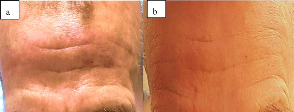
- Course wrinkle treatment with 2940 nm Er: YAG laser; (a) before and (b) after 3 sessions of laser treatment.
Indigenous absorbers of visible and infrared laser wavelengths can imitate the optical characteristics of an intermixed media. The local fluence at the targeted tissue controls the laser beam's thermal effect(Ge et al. 2024). Following their passage through the surface, the laser photons are subjected to several absorbing and scattering phenomena. The shape, size and mismatch in the particle's index of refraction with the medium have an impact on photon scattering. Absorption dominates over scattering when the particle size is smaller than 250 nm. This, however, is different for isolated absorbers like melanoma and HgB where scattering is the main way that laser light in tissue is attenuated at most wavelengths. After laser treatment, the patients experienced mild pain and redness of the treated skin, but these symptoms disappeared within an hour after the session. The patient did not experience any persistent pain or discomfort after the procedure. He did not experience any negative side effects such as swelling, redness or inflammation. On the contrary, he saw a noticeable improvement in the appearance of his skin over the next few weeks. After two months, the results showed a significant improvement in the texture and elasticity of the skin, with a clear disappearance of the fine lines that were previously present in the forehead area.
5 Conclusion
Non-ablative lasers are widely used to enhance the condition of skin. Among these non-ablative lasers, low power 980 nm diode, long pulsed Nd: YAG 1064 nm and fractional Er: YAG lasers were found effective to treat skin melanin pigmentation, fine lines and coarse wrinkles after pre-calculating the treatment laser parameters. The present study has the advantage of avoiding the “after treatment” complications such as scars and blistering that result because of the try and error techniques usually adopted by most cosmetic centers. The laser parameter used were pre calculated (for 980 nm fluence of 12 J/cm pulse duration of 0.02 sec and for 1064 nm resurfacing 20 J/cm fluence in 0.4 sec pulse duration and for 1064 vein treatment fluence of 120 pulse duration of 20 and for 2940 fluence of 5 pulse duration 0.1) based on optimum temperature raised (tissue cooking) needed for optimum laser treatment result without adverse effect. Future work will concentrate on developing new protocol to safe treat dark skins which may involve the establishment of customizable protocols.
Ethics
Clint's integrity, trust, honesty, and openness were crucial to our project. Patients received explanations about the procedure's potential risks and advantages as well as how it will impact their quality of life. Each patient gave their informed consent, ensuring they were fully informed about the operations and could make an informed decision. Every patient was informed about potential side effects, post-procedure therapies, and follow-up procedures. Establishing patient privacy was crucial to building confidence. Patients were given the freedom to discontinue treatment sessions if they felt unsafe or that the anticipated results did not meet their expectations. Every patient gave permission to have the laser operation done and promised to assist with data collection during the recovery phase.
CRediT authorship contribution statement
Muna B. Mustafa: Writing – original draft. Walid K. Hamoudi: Writing – original draft, Supervision. Khawla S. Khashan: .
Acknowledgements
The University of Technology in Baghdad, Iraq, helped, which the authors are grateful for.
Declaration of competing interest
The authors declare that they have no known competing financial interests or personal relationships that could have appeared to influence the work reported in this paper.
References
- Alhallak, Kamal, Adel Abdulhafid, Salem Tomi, Dima Omran. 2023. 'Skin, Light, and Their Interactions.' in, The Ultimate Guide for Laser and IPL in the Aesthetic Field (Springer).
- Refining thermal therapy: Temperature distribution modeling with distinct absorption in multi-layered skin tissue during infrared laser exposure. Int. Commun. Heat Mass Transfer. 2024;157:107818
- [Google Scholar]
- Assessment of laser effects on skin rejuvenation. Journal of Lasers in Medical Sciences. 2020;11:212.
- [Google Scholar]
- 'Fundamentals of Lasers and Light Devices in Dermatology', Practical Introduction to Laser. Dermatology 2020:1-52.
- [Google Scholar]
- Chernigovskiy, VV, SA Martsinukov, DK Kostrin, and VA Simon. 2019. “Calculation of the temperature effect of laser radiation on biological tissues.” In AIP Conference Proceedings, 020011. AIP Publishing LLC.
- 'Basic laser physics and interaction of laser light with soft tissue'. in, Endoscopic laser surgery handbook. (CRC Press); 2020.
- Light tissue interactions. Aesthetic Applications of Intense Pulsed Light 2020:13-23.
- [Google Scholar]
- Nonablative Laser Rejuvenation (in,). Textbook of Cosmetic Dermatology (CRC Press); 2024.
- Hamoudi, Walid K, Qusay K Alhashimi, Muna B Mustafa, and Noor R Abdulhameed. 2021. “Pre-calculated relevant Nd: YAG laser parameters for optimized varicose veins treatment.” In Journal of Physics: Conference Series, 012053. IOP Publishing.
- Nonablative laser skin resurfacing for periorbital wrinkling—a case series of 16 patients. J. Cosmet. Dermatol.. 2021;20:99-104.
- [Google Scholar]
- Hussain, Hayder S, Thuraya A Abdul Hussian, Ghufran S Jaber, Muna B Mustafa, and Ghaidaa A Khalid. 2024. “Investigation in vitro the effect of X-rays, gamma rays and beta particles on the physical and structural characteristics of human teeth.” In AIP Conference Proceedings. AIP Publishing.
- Effects of non-ablative Er: YAG laser on the skin and the vaginal wall: systematic review of the clinical and experimental literature. Int. Urogynecol. J.. 2020;31:2473-2484.
- [Google Scholar]
- Laser-tissue interactions. In: Thermal and Optical Interactions with Biological and Related Composite Materials. SPIE; 1989. p. :17-23.
- [Google Scholar]
- Unveiling peripheral symmetric acceptors coupling with tetrathienylbenzene core to promote electron transfer dynamics in organic photovoltaics. Sci. Rep.. 2024;14:21176.
- [Google Scholar]
- Exploring the potential of ZnSe nanocage as a promising tool for CO2 and SO2 sensing: A computational study. Comput. Theor. Chem.. 2024;1231:114428
- [Google Scholar]
- Laser–tissue interaction simulation considering skin-specific data to predict photothermal damage lesions during laser irradiation. J. Comput. Des. Eng.. 2023;10:947-958.
- [Google Scholar]
- 'Therapeutic Applications: Dermatology—Selective Photothermolysis'. in, Handbook of Laser Technology and Applications. (CRC Press); 2021.
- Wound Healing Treatments After Ablative Laser Skin Resurfacing: A Review. Journal of Drugs in Dermatology: JDD. 2020;19:1050-1105.
- [Google Scholar]
- Theoretical investigation for the designing of novel antioxidants. Can. J. Chem.. 2013;91:126-130.
- [Google Scholar]
- Exploring the optical properties of novel N-benzylated thiazoles based chromophores: Spectroscopic insights and computational analysis. Synth. Met.. 2024;307:117701
- [Google Scholar]
- Outcomes and adverse effects of ablative vs nonablative lasers for skin resurfacing: a systematic review of 1093 patients. Dermatol. Ther.. 2021;34:e14432.
- [Google Scholar]
- A systematic review and meta-analysis of efficacy, safety, and satisfaction rates of laser combination treatments vs laser monotherapy in skin rejuvenation resurfacing. Lasers Med. Sci.. 2023;38:228.
- [Google Scholar]
- Thermal relaxation times: an outdated concept in photothermal treatments. Lasers Med. Sci.. 2014;29:973-998.
- [Google Scholar]
- Mustafa, Muna B, Wailed K Hamoudi, Ghufran S Jaber, Mohammed Y Abbas, and Noor R Abdulhameed. 2021. 'EXACT LASER FLUENCE FOR SUCCESSFUL TREATMENT OF FACE AND LEG TELANGIECTASIA', Wiadomosci Lekarskie (Warsaw, Poland: 1960), 74: 2340-44.
- Mustafa, Muna B, Walid K Hamoudi, and Hiba H Maqdasi. 2021. “Temperature rise control for safe treatment of varicose vein by Nd: YAG laser.” In AIP Conference Proceedings. AIP Publishing.
- Optimized selection of neodymium laser parameters for successful enlarged veins treatment. Lasers Med. Sci.. 2023;38:264.
- [Google Scholar]
- Medical applications of lasers. In: Laser-Tissue Interactions: Fundamentals and Applications. 2019. p. :153-249.
- [Google Scholar]
- Niemz, Markolf H. 2007. Laser-tissue interactions (Springer).
- Nouri, Keyvan. 2018. Lasers in dermatology and medicine: dermatologic applications (Springer).
- 'Lasers and laser–tissue interaction', Veterinary Clinics: Small Animal. Practice. 2002;32:517-534.
- [Google Scholar]
- New 675 nm Laser Device: The Innovative and Effective Non-Ablative Resurfacing Technique. Medicina. 2023;59:1245.
- [Google Scholar]
- Theoretical investigation for exploring the antioxidant potential of chlorogenic acid: a density functional theory study. Int. J. Food Prop.. 2016;19:745-751.
- [Google Scholar]
- Steiner, Rudolf. 2010. 'Laser-tissue interactions.' in, Laser and IPL technology in dermatology and aesthetic medicine (Springer).
- Tissue parameters determining the visual appearance of normal skin and port-wine stains. Lasers Med. Sci.. 1995;10:55-65.
- [Google Scholar]
- The histology of skin treated with a picosecond alexandrite laser and a fractional lens array. Lasers Surg. Med.. 2016;48:646-652.
- [Google Scholar]
- Comparative analysis of the light parameters of red and near-infrared diode lasers to induce photobiomodulation on fibroblasts and keratinocytes: An in vitro study. Photodermatol. Photoimmunol. Photomed.. 2021;37:253-262.
- [Google Scholar]
- Efficacy and safety of long pulse 1064 and 2940 nm lasers in noninvasive lipolysis and skin tightening. J. Biophotonics. 2019;12:e201900083.
- [Google Scholar]


