Translate this page into:
Larvicidal property of green synthesized silver nanoparticles against vector mosquitoes (Anopheles stephensi and Aedes aegypti)
⁎Corresponding author. aarubot@gmail.com (Manickam Arumugam)
-
Received: ,
Accepted: ,
This article was originally published by Elsevier and was migrated to Scientific Scholar after the change of Publisher.
Peer review under responsibility of King Saud University.
Abstract
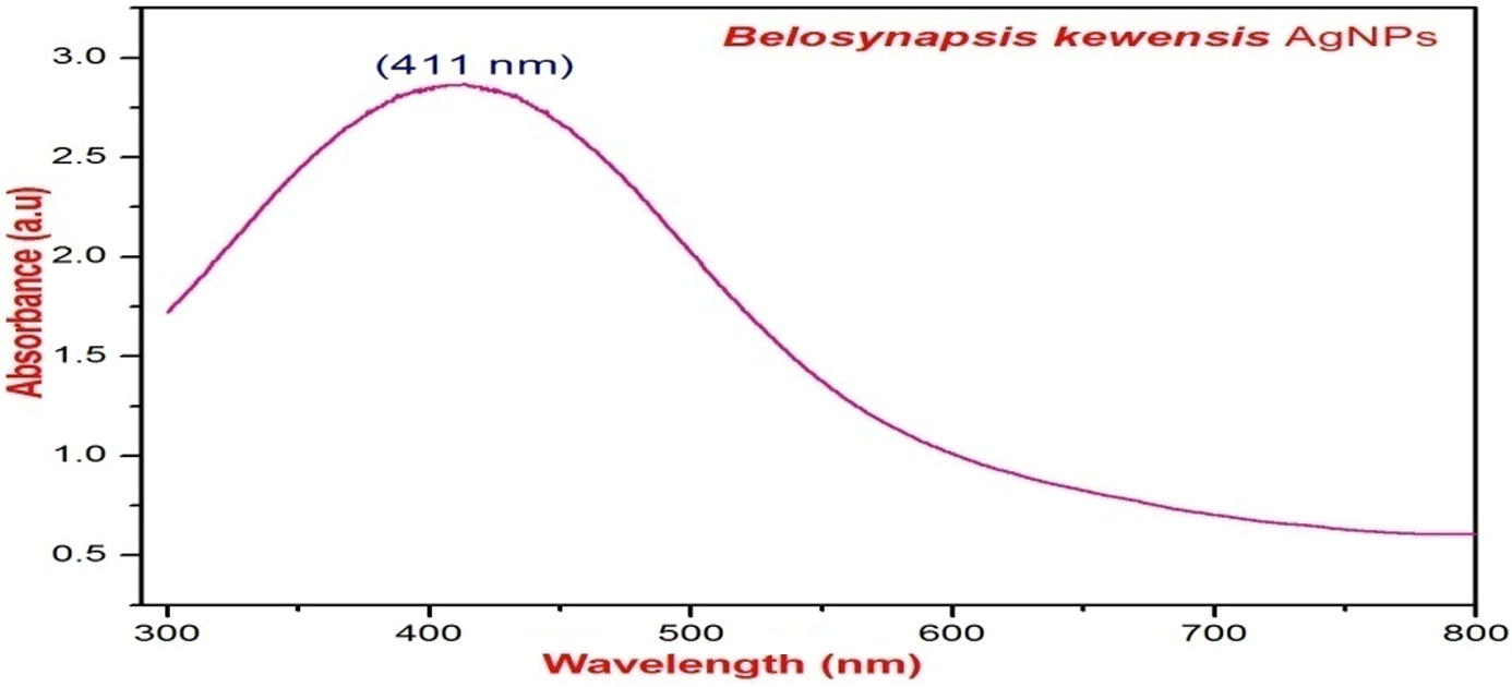
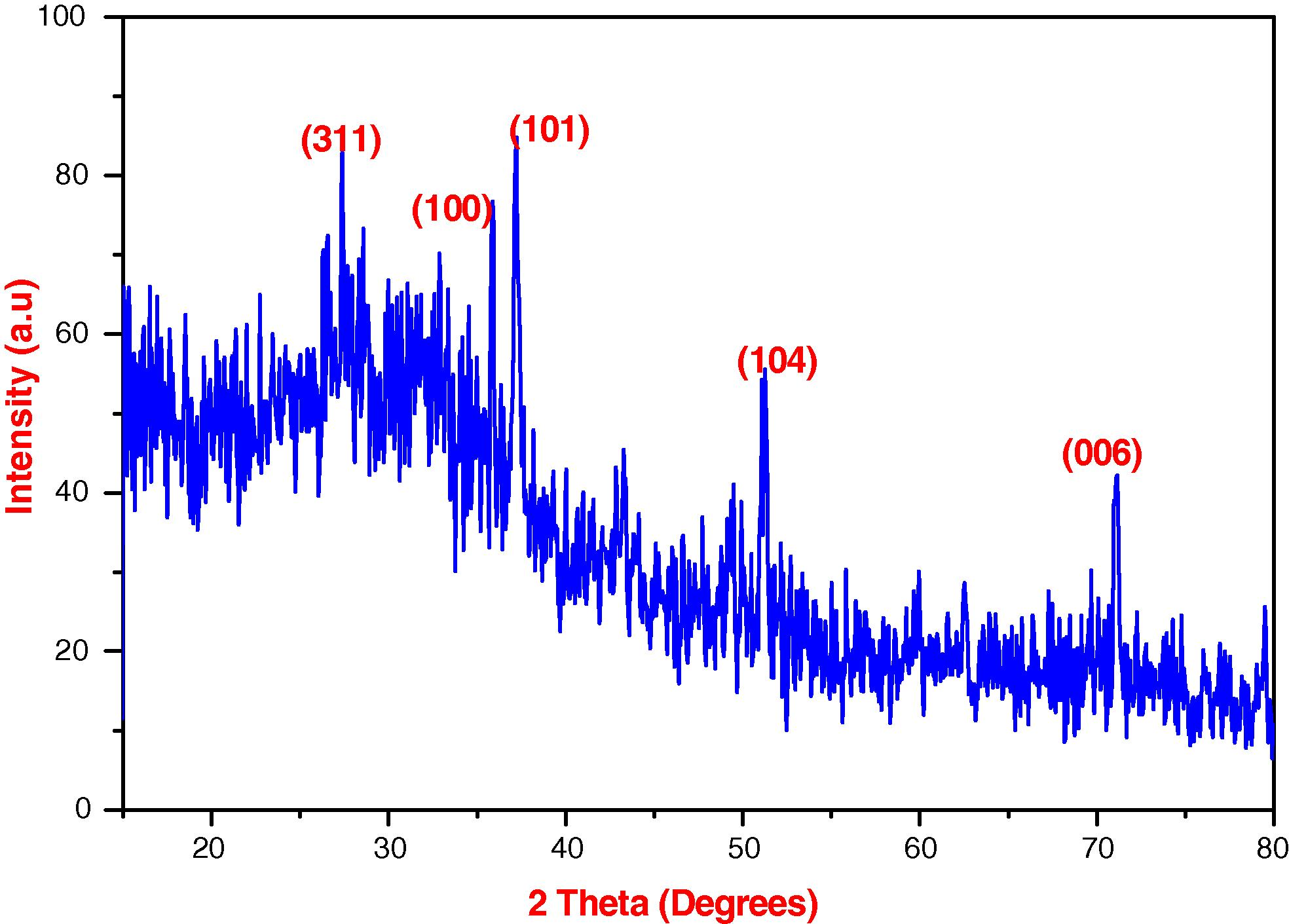
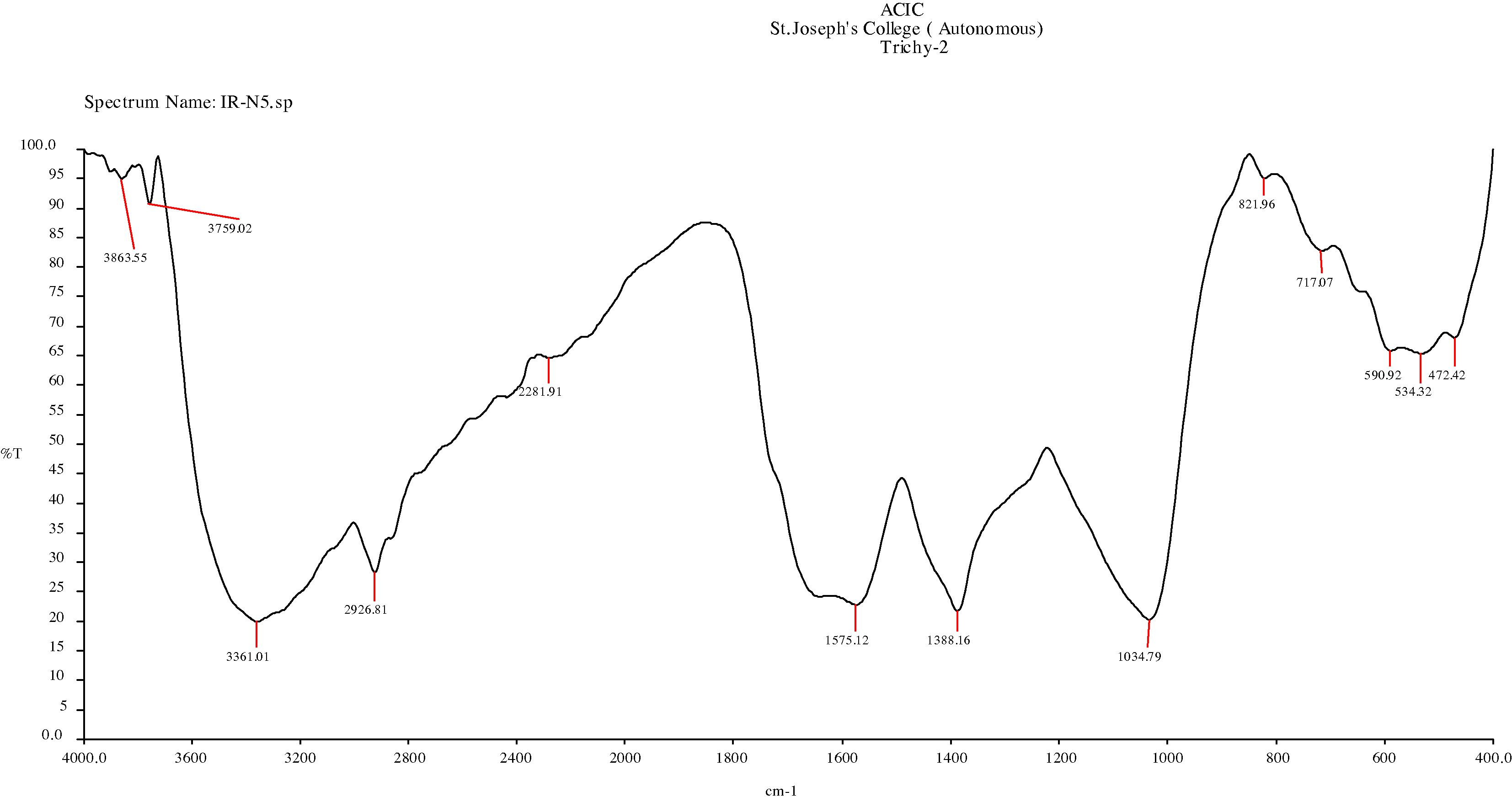
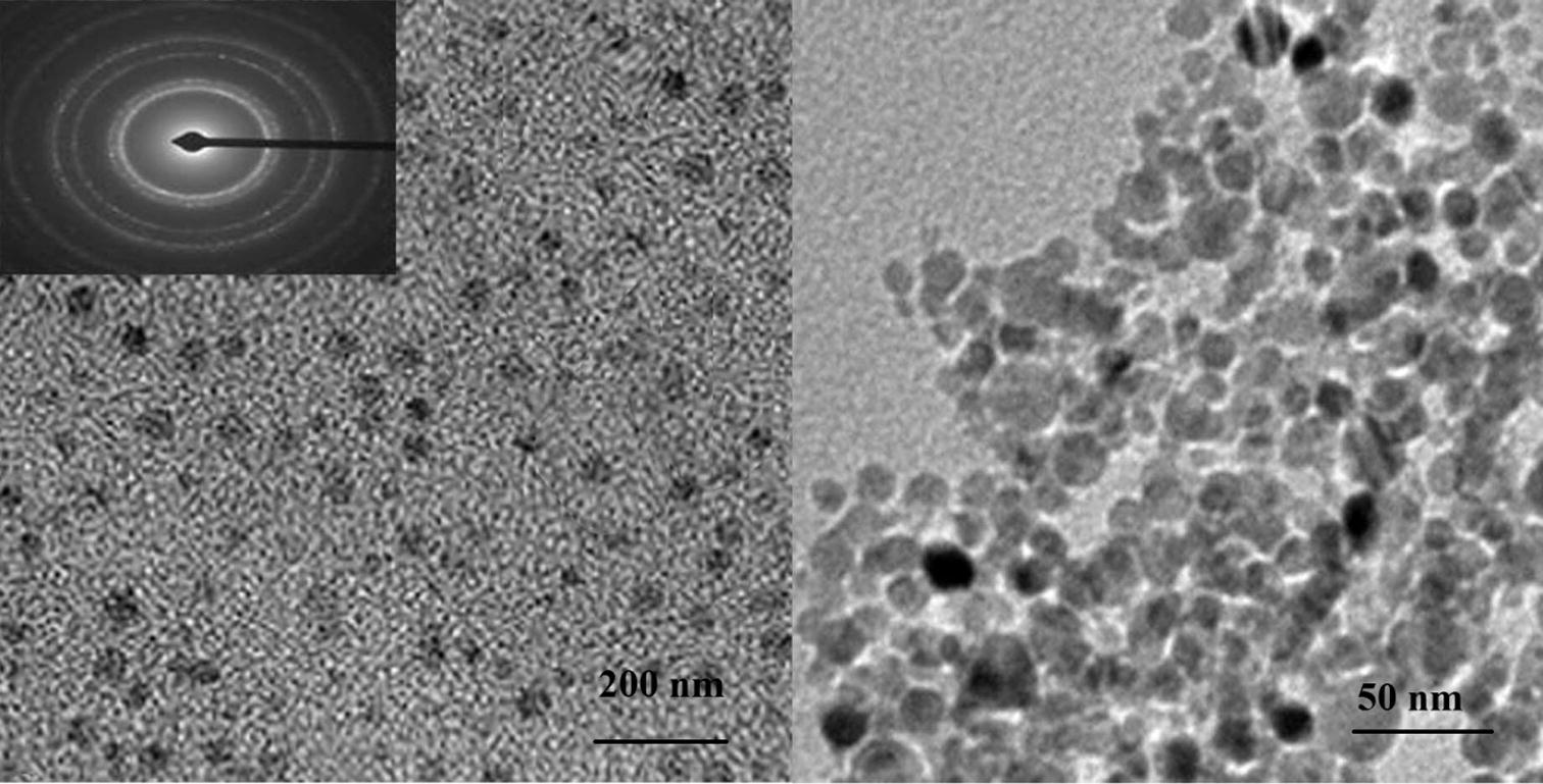
Mosquito vectors spread severe human diseases which lead to millions of deaths every year. Vector management is ultimately aimed to develop the health of every individual’s life by reducing the mosquito diversity. Control of vectors in growing counties is an important issue with various aspects. The advancement of green nanotechnology will attribute the solution for vector controlling policy. To identify the larvicidal property of silver nanoparticles (AgNPs) using Belosynapsis kewensis (B. kewensis) leaf extract against the Anopheles stephensi (A. stephensi) and Aedes aegypti (A. aegypti) in vitro study (LC50 and LC90) was analyzed. The synthesized AgNPs were characterized by UV–vis. (Ultraviolet–visible spectroscopy), FTIR (Fourier Transform Infra Red spectroscopy), TEM (Transmission Electron Microscopy), and XRD (X-ray Diffraction). Green AgNPs have a maximum absorption at 411 nm. The FTIR spectrum showed prominent peaks in (3863.55, 3759.02, 3361.01, 2926.81, 1575.12, 1388.16, 1034.79, 821.96, 717.07, 590.92, 534.32 and 472.42 cm−1) in the region of 4000–400 cm−1. The XRD peaks shown at 27.4°, 35.90°, 37.20°, 51.23° and 71.10° correspond to (3 1 1), (1 0 0), (1 0 1), (1 0 4) and (0 0 6) planes for face centered cubic (FCC). The TEM image showed the NPs with an average size of 24 ± 1.6 nm. In vitro larvicidal activity of AgNPs was used against the fourth instar of A. stephensi and A. aegypti. The LC50 and LC90 values of AgNPs showed to be effective against A. stephensi (LC50 = 78.4; LC90 = 144.7 ppm) followed by A. aegypti (LC50 = 84.2; LC90 = 117.3 ppm). These results recommend that the green synthesized AgNPs have a potential to be used as a candidate for the control of A. stephensi and A. aegypti through eco-friendly and cost effective approach.
Keywords
Belosynapsis kewensis
Green synthesis
Larvicidal activity
Silver nanoparticle
Surface plasmon resonance
1 Introduction
In tropical and subtropical regions mosquitoes contributing a major problem in public sectors especially, severe human diseases like malaria, dengue, Chikungunya, filariasis and yellow fever (Kovendan et al., 2012; Logeswari et al., 2013). All parts of the world population are being affected by vector borne diseases, but in the developing countries control of such vector is becoming a very big issue. So, green synthesized NPs may be a right opt source for mosquito control agent. The NPs consist of a variety of active compounds with many advantages like an eco-friendly, non-hazardous, greater surface, cost effect, inorganic nanomaterial and biocompatible (Albrecht et al., 2006; Harekrishna et al., 2009). Green synthesis of AgNPs is carried out by plants like Dodonaea viscosa, Elaeocarpus ganitrus, Terminalia arjuna, Pseudotsuga menziesii, Prosopis spicigera, Ficus religiosa, Ocimum sanctum and Curcuma longa (Poushpi et al., 2014) and Syzygium cumini (Mittal et al., 2014). Routine use of synthetic insecticidal products for control of mosquito has a problem with disturbing the biological system and cause to resurgences in mosquito populations (Chakkaravarthy et al., 2011; Srinivasan et al., 2014). Belosynapsis kewensis is a member of commelinaceae with high medicinal importance. Therefore, green synthesized AgNPs play a major role in the invention of larvicidal products. The present study aimed to investigate the larvicidal activity of synthesized AgNPs of B. kewensis.
2 Materials and methods
2.1 Chemicals and plant material collection
Silver nitrate (AgNO3) and dimethyl sulfoxide (DMSO) were purchased from Merck, India. The glasswares were washed thoroughly with acid and followed by rinsing with Millipore-Milli-Q water. The leaves of B. kewensis were collected from Manjolai hills, Thirunelveli district, Tamilnadu, India.
2.2 Biosynthesis of AgNPs
The collected leaves were washed thoroughly with running water and followed by de-ionized water. 10 g of cleaned leaves were boiled at 80 °C for 10 min in 100 mL with deionised water and crude extract was filtered by Whatman No.1 filter paper finally the extract was stored at 4 °C for further uses (Abou El-nour et al., 2010). The obtained extract of B. kewensis was used for synthesis of AgNPs as a reducing as well as a stabilizing agent. To facilitate the synthesis of AgNPs 10 mL of B. kewensis leaf extract was mixed with 90 mL of 1 mM AgNO3 solution. This reaction mixture was kept at room temperature. Later, the color change was observed to designate the formation of colloidal AgNPs.
2.3 Characterization of AgNPs
During the incubation period, the synthesis of AgNPs was conformed by UV–vis spectroscopy (Hitatchi-2001) in a wavelength ranges between 200 and 800 nm at 1 nm resolution. Synthesized AgNPs were diluted with deionized water and centrifuged at 10,000 rpm for 15 min. The residue was dispersed with deionized water twice to remove the biological impurities. The purified residue was dried in oven at 70 °C for overnight. The AgNPs were used for FTIR analysis to identify the functional groups on a Perkin-Elmer spectrum instrument at a resolution of 4 cm−1 in the transmission mode of 4000–400 cm−1 in KBr pellets. The particles size and nature of the green AgNPs were determined by XRD using XPERT-PRO, D-8 at 30 kV, 40 mA with Cu Kα radians at 2θ angle. XRD is a rapid analytical method to identify the crystalline structure and calculate the dimensions of unit cell using Scherrer’s formula (D = Kλ/βcos θ). For TEM study, the sample was prepared by placing a drop of the suspension onto a carbon–coated Cu grid and allowed to evaporate. Observation was done in JEM 1011, JEOL, Japan, instrument operated at 200 kV an accelerated voltage.
2.4 Larvicidal activity of AgNPs
For the larvicidal activity, the eggs of Anopheles stephensi and Aedes aegypti were collected from the field station, Center for Research in Chennai, Tamilnadu, India and they were reared under laboratory condition (25–28 °C). The larvicidal activity was studied by the standard procedure recommended by WHO (WHO, 2005). Green synthesized AgNPs were dissolved in 1 mL of methanol and prepared into different concentrations viz., 50, 100, 150, 200 and 250 ppm with double distilled water. Twenty-five reared early fourth instar stage larvae were transferred by means of the dropper to (100 mL beakers) each concentration and the experiment was performed in three replicates with the control under the laboratory conditions at 26–28°C. Mortality of the larvae was calculated at 12 and 24 h of exposure periods. During the both exposure periods, no food was supplied to the larvae and percentage of mortality was calculated. The data were presented in Mean ± Standard Deviation and all the statistical analyses were performed by SPSS version 11.5. The average of the larvae mortality LC50, LC90 and chi-square test was calculated.
3 Results and discussions
The formation of NPs was done by reduction of Ag+ ions into AgNPs with exposure of the leaf extract of B. kewensis and the formation was seen by the color change. The initial color of the suspension (AgNO3 and leaf extract) was yellow, after the incubation period, the yellow was turned into dark brown in color which indicated the surface plasmon resonance (SPR) (Vijayaraghavan et al., 2012; Karuppiah and Rajmohan, 2013; Basavegowda and Rok Lee, 2014; Mansour and Robabeh, 2014). The SPR phenomenon was very sensitive to NP’s nature, size and shape, which were formed by their inter particle distance and the surrounding media (Noginov et al., 2007; Vijayakumar et al., 2014). Fig. 1 represents the UV–vis spectrum of green synthesized AgNPs with higher peak level observed at 411 nm and SPR band also exposed at the same peak without any shifting. Muhammad et al. (2014) observed the same absorption band in seaweeds. Single peak indicated the synthesized particles were uniform in size and shape. So, the formation of AgNPs was attributed to hydrophilic and hydrophobic interaction, which, prevents the particles from aggregation by intermolecular forces (Shannahan et al., 2013). XRD analysis was employed to study the crystalline nature of the AgNPs. Fig. 2 shows the XRD pattern of AgNPs and peak values at 2θ degrees of 27.4°, 35.90°, 37.20°, 51.23° and 71.10° corresponding to (311), (1 0 0), (1 0 1), (1 0 4) and (0 0 6) planes of AgNPs. All the degrees of the peaks corresponded to a face centered cubic (FCC) crystalline structure. The intense peak 37.20° represented a high degree of crystallinity (Karuppiah and Rajmohan, 2013; Basavegowda and Rok Lee, 2014). FTIR analysis was carried out, to identify the functional groups of the synthesized AgNPs. FTIR spectrum indicated the clear peaks with (3863.55, 3759.02, 3361.01, 2926.81, 1575.12, 1388.16, 1034.79, 821.96, 717.07, 590.92, 534.32, 472.42 cm−1) different values (Fig. 3). Above the peak values they corresponded to functional groups like, amide group (N–H stretching 3361.01 cm−1), an aliphatic group (cyclic CH2 – 2926.81 cm−1), a methyl group (bend CH2–CH3 stretching 1388.16 cm−1), aliphatic amine group (C–N stretching 1034.79 cm−1), alkyl halides group (C–Cl stretching 821.96 and 717.07 cm−1) and alkyl halides (C–Br stretching 590.92 cm−1 and 534.32 cm−1). The functional groups such as alcohol, amines, amides, alkanes, methyl, aliphatic and halides confirmed their presence in AgNPs and these are the main classes in most of the functional groups. They were denoted as possible biomolecules responsible for stabilizing, capping and reducing agents of the AgNPs (Cho et al., 2005; Vijayaraghavan et al., 2012; Srinivasan et al., 2014). The terpenoid groups have a high potential to convert the aldehyde groups into carboxylic acids in the Ag+ medium. Additionally, amide groups are also responsible for the presence of the some enzymes which, may be responsible for the synthesis of metal particles. Further, polyphenols are also proving that they have the potential to reduce the silver metals (Srinivasan et al., 2014). TEM micrograph (Fig. 4) indicated the synthesized AgNPs with 50 and 200 nm scales and most of the AgNPs were spherical in structure. The particle sizes vary from 10 to 28 nm and the average size of the AgNPs was 24 nm (Aguilare et al., 2011; Thomas et al., 2014). The Larvicidal activity of synthesized AgNPs was studied, which showed the effective larvicidal activities (Table 1) against the fourth instar larvae at 12 and 24 h of exposure time. After the 12 h of exposure time the larvicidal activity of AgNPs was LC50 = 147.6; LC90 = 285.9 ppm against A. stephensi and LC50 = 178.4; LC90 = 344.7 ppm against A. aegypti. The larvicidal activity after the 24 h of exposure time was LC50 = 112.7; LC90 = 217.3 ppm against A. stephensi and LC50 = 149.9; LC90 = 288.4 ppm against A. aegypti. The plant B. kewensis mediated AgNPs were expressed as 98.3% of larvicidal activity at 250 ppm. Srinivasan et al. (2014) reported the larvicidal activity of biosynthesized AgNPs of Avicennia maria leaf extract against An. aegypti (LC50 = 4.374 and LC90 = 4.928 ppm) and A. stephensi (LC50 = 7.40 and LC90 = 9.865 ppm). The larvicidal activity of synthesized AgNPs of Ficus racemosa was noted against Culex quinquefasciatus and Culex gelidus, which were LC50 = 67.72 and LC90 = 63.70 ppm. The larvicidal activity was observed in AgNPs of Tinospora cordifolia against fourth instar larvae of Anopheles subpictus and C. quinquefasciatus LC50 = 6.43 ppm. The AgNPs showed (using Eclipta prostrata) maximum efficacy of larvicidal activity against the fourth instar larvae of C. quinquefasciatus (LC50 = 27.49 and LC90 = 70.38) and A. subpictus (LC50 = 27.85 and LC90 = 71.45 ppm) (Velayutham et al., 2013). In the present investigation, the larvicidal activity of AgNPs showed higher activity against A. stephensi than A. aegypti. Vector management is one of the major issues due to the capacity of resistance against the usual insecticides. Therefore, an urgent need has emerged to develop the new insecticides (Krishnamoorthy et al., 2015). Hence, the invention of nanometals using B. kewensis as represented here, could be provided a new product to control the mosquitoes by replacing the synthetic larvicidal products and this route would make available for larvicides to prevent several dreadful diseases. Control-nil activity, SE-standard error, LCL-lower confidence level, UCL-upper confidence level.
UV–vis spectrum of silver nanoparticles using B. kewensis leaf extract.

XRD pattern of synthesized silver nanoparticles using B. kewensis leaf extract.

FTIR spectrum of synthesized silver nanoparticles using B. kewensis leaf extract.

TEM image of 200 nm and 50 nm magnification showing the silver nanoparticles using B. kewensis leaf extract.
Name of the mosquito species
Exposure period (h)
Conc.
(ppm)% mortality ± standard error
LC50
(LCL-UCL)a
LC90
(LCL-UCL)a
χ2 (d = 4)b
Anopheles stephensi
12
Control
0 ± 0
92.4
(76.2−134.5)181.6
(160.8−192.6)8.3
50
19.1 ± 0.18
100
36.6 ± 0.37
150
48.2 ± 0.75
200
62.4 ± 0.34
250
88.5 ± 0.75
24
Control
0 ± 0
78.4
(63.2−96.5)144.7
(124.0−150.2)1.42
50
28.0 ± 0.98
100
42.1 ± 0.56
150
58.2 ± 0.13
200
88.6 ± 0.55
250
98.3 ± 0.87
Aedes aegypti
12
Control
0 ± 0
104.2
(68.5−93.7)166.7
(96.1−136.4)4.57
50
16.3 ± 0.28
100
29.4 ± 0.57
150
41.1 ± 0.73
200
54.4 ± 0.39
250
78 ± 0.81
24
Control
0 ± 0
84.2
(63.5−81.4)117.3
(80.1−140.7)4.6
50
21.0 ± 0.14
100
32.0 ± 0.45
150
46.0 ± 0.38
200
64.0 ± 0.15
250
37.0 ± 0.67
4 Conclusion
The green synthesized AgNPs using the leaf extract of B. kewensis offered an excellent larvicidal activity. Green route of AgNPs synthesis shows to be an eco-friendly, time consuming, cost-effective, and alternative to chemical methods of NPs production would be an appropriate for extending the biological route for large-scale production of the biolarvicidal product.
Acknowledgment
We are grateful to the Department of Biotechnology, Ministry of Science and Technology, New Delhi, Govt. of India for providing the major research project fund (BT/PR-10002/BCE/08/591/2011). The authors are thankful to Prof. R. Panneerselvam, Dean, Faculty of Science (Rtd.), Annamalai University, Annamalai Nagar, Tamil Nadu, India for his valuable guidance and support to complete the work.
References
- Synthesis and applications of silver nanoparticles. Arabian J. Chem.. 2010;3(3):135-140.
- [Google Scholar]
- Synthesis and characterization of silver nanoparticles: effect on phytopathogen Colletotrichum gloesporioides. Nanopart. Res.. 2011;13(6):2525-2532.
- [CrossRef] [Google Scholar]
- Green chemistry and the health implications of nanoparticles. Green Chem.. 2006;8:417-432.
- [Google Scholar]
- Synthesis of silver nanoparticles using Satsuma mandarin (Citrus unshiu) peel extract: a novel approach towards waste utilization. Mater. Lett.. 2014;109:31-33.
- [Google Scholar]
- Bioefficacy of Azadirachta indica (A. juss) and Datura metel (Linn.) leaves extracts in controlling Culex quinquefasciatus (Diptera: Culicidae) Trends Appl. Sci. Res.. 2011;8:191-197.
- [Google Scholar]
- The study of antimicrobial activity and preservative effects of nanosilver ingredient. Electrochim. Acta. 2005;51:956-960.
- [Google Scholar]
- Green synthesis of silver nanoparticles using latex of Jatropha curcas. Colloids Surf. A. 2009;339:134-139.
- [Google Scholar]
- Green synthesis of silver nanoparticles using Ixora coccinea leaves extract. Mater. Lett.. 2013;97:141-143.
- [Google Scholar]
- Studies on larvicidal and the pupicidal activity of Leucas aspera Willd. (Lamiaceae) and bacterial insecticide, Bacillus sphaericus against the malarial vector, Anopheles stephensi Liston. (Diptera: Culicidae) Parasitol Res 2012
- [Google Scholar]
- Identification of chemical constituents and larvicidal activity of essential oil from Murraya exotica L. (Rutaceae) against Aedes aegypti, Anopheles stephensi and Culex quinquefasciatus (Diptera: Culicidae) Parasitol Res. 2015
- [CrossRef] [Google Scholar]
- Ecofriendly synthesis of silver nanoparticles from commercially available plant powders and their antibacterial properties. Sci. Iranica. 2013;20:1049-1054.
- [Google Scholar]
- Plant mediated green synthesis and antibacterial activity of silver nanoparticles using Crataegus douglasii fruit extract. Ind. Eng. Chem.. 2014;20:739-744.
- [Google Scholar]
- Biosynthesis of silver nanoparticles: elucidation of prospective mechanism and therapeutic potential. J. Colloid Interface Sci.. 2014;415:39-47.
- [Google Scholar]
- Silver nanoparticles: green synthesis, characterization, and their usage in determination of mercury contamination in seafoods. Nanotechnology. 2014;5:1-5.
- [CrossRef] [Google Scholar]
- The effect of gain and absorption on surface plasmos in metal nanoparticles. App. Phys. B. 2007;86:455-460.
- [Google Scholar]
- Phytofabrication characterization and comparative analysis of Ag nanoparticles by diverse biochemicals from Elaeocarpus ganitrus Roxb., Terminalia arjuna Roxb., Pseudotsuga menzietii, Prosopis spicigera, Ficus religiosa, Ocimum sanctum and Curcuma longa. Ind. Crops Prod.. 2014;54:22-31.
- [Google Scholar]
- Silver nanoparticle protein corona composition in cell culture media. PLoS One. 2013;8(9):e74001.
- [CrossRef] [Google Scholar]
- Biosynthesis of silver nanoparticles from mangrove plant (Avicennia marina) extract and their potential mosquito larvicidal property. Parasit. Dis. 2014
- [CrossRef] [Google Scholar]
- Evaluation of antibacterial potential of silver nanoparticles (SNPS) produced using rhizome extract of Hedychium coronarium j. Koenig. Int. J. Pharm. Pharm. Sci.. 2014;6(10):92-95.
- [Google Scholar]
- Larvicidal activity of green synthesized silver nanoparticles using bark aqueous extract of Ficus racemosa against Culex quinquefasciatus and Culex gelidus. Asian Pac. J. Trop. Med.. 2013;6(2):95-101.
- [CrossRef] [Google Scholar]
- Green synthesis and characterization of AgNPs using Boerhavia diffusa plant extract and their antibacterial activity. Ind. Crops Prod.. 2014;52:562-566.
- [Google Scholar]
- Biomimetic synthesis of silver nanoparticles by aqueous extract of Syzygium aromaticum. Mater. Lett.. 2012;75:33-35.
- [Google Scholar]
- Guidelines for Laboratory and Field Testing of Mosquito Larvicides. World Health Organization document WHO; 2005. cds/WHO-pes/gcdpp/13


