

Translate this page into:
Isolation, identification and molecular characterization of Newcastle disease virus using SDS-PAGE
⁎Corresponding author at: Department of Zoology, College of Science, King Saud University, Riyadh 11451, Saudi Arabia. mushahid@ksu.edu.sa (Shahid Mahboob)
-
Received: ,
Accepted: ,
This article was originally published by Elsevier and was migrated to Scientific Scholar after the change of Publisher.
Peer review under responsibility of King Saud University.
Abstract
Field isolates of Newcastle disease virus (NDV) were subjected to polypeptide analysis using SDS-PAGE. On electrophoresis the NDV proteins were settled into several peptide components related to structural elements of the virus. The field isolates of NDV were recovered from different outbreaks in commercial chicken flocks. The virus grew well into 9th day old embryonated chicken eggs. The presence of virus in harvested allantoic fluids was assessed by Haemagglutination Test (HA). The HA titer of virus ranged from 1:512 to 1:1024. All the field isolates were purified by chloroform extraction method. Same quantities of samples were loaded on 12.5% discontinuous SDS-PAGE system. There were 6 to 13 polypeptides recorded with a molecular weight ranged from 12 KDa to 160 KDa. The results of SDS-PAGE denoted that the indigenous isolates of NDV are almost identical and comparable with vaccines. SDS-PAGE, using 12.5 percent gel is a simple, economical, and easy way for the characterization of NDV and other viruses. Furthermore, the potency of the vaccines can easily be checked through this technique and comparison can be made between field and vaccine ND viruses. It is recommended that more work should be done at the molecular level of local isolates of NDV to confirm the molecular nature of these isolates.
Keywords
Newcastle disease virus
Chicken
SDS-PAGE
Molecular weights

1 Introduction
The poultry industry is one of the major industry after textile in Pakistan (Ismail, 2017). Recently, the government has announced certain incentives to expand this industry, which has resulted in high density poultry farming. The massive contribution of the poultry industry to healthy nation and GDP is threatened by the problems, which may be viral, bacterial, parasitic and nutritional, or of unknown etiology (Siddique and Javed, 1989; Alsahami et al., 2018). Around the world, poultry has faced many socioeconomic crises due to viral poultry diseases with every passing year like Newcastle disease (ND) is one of them (Yune and Abdela, 2017).
The respiratory problems are major hazard to the development of this industry. Among these respiratory problems ND is one, which causing heavy economic losses in Pakistan poultry (Ashraf et al., 2016). Newcastle disease (ND) is a fatal disease of poultry in many parts of the world (Leslie, 2000). It causes some great economic losses, particularly in developing countries and acts as a trade barrier (King, 1996; Westbury, 2001). NDV is a single stranded RNA virus with a genome length of about 15.2 kb (Aldous et al., 2003; Ashraf et al., 2016). Losses from production inefficiencies are usually of greater concern than losses from mortality, although the later and reported to be economically significant in broilers.
Haemagglutination Test is widely used for the diagnosis of ND virus in morbid samples, but it cannot diagnose sub serotypes of the virus. For serotyping of isolated virus SDS-PAGE may be used as a useful tool for an identification of different serotypes of the same virus, depending on the molecular weight of the protein fractions (Hemmatzadeh and Kazemimanesh, 2017). In this way we can also isolate and identify new serotypes of a virus which emerges in a specific area. The objective of this research work was to characterize Newcastle disease virus by using SDS-PAGE molecular technique to improve the rapid diagnosis of the disease and continued surveillance of the commercial and non-commercial poultry.
2 Materials and methods
2.1 Collection of samples
Broilers, layers, and non-descript birds were examined for, the evidence of ND and for the isolation of the virus. The source for collecting samples included the birds that brought for treatment at the diagnostic laboratory in Samundri, Punjab, Pakistan. In addition, outbreaks of the disease reported from poultry farms, located in and around Faisalabad, were also attended and material collected for isolation of the virus. All the dead birds were autopsied and those showing lesions of the disease their internal organs (spleen, lungs and liver) were collected and stored in a deep freezer till required for processing (Jordan, 1990).
2.2 Isolation of virus
The samples, prior to their processing, were thawed at room temperature. Thereafter a 20% solution of the tissues was made in physiological saline and homogenized with the help of pestle and mortar. Antibiotics (penicillin 200 I.U and Streptomycin 200ug/ml) were added to suppress the possible bacterial life, gaining entrance through extraneous contamination or already present in the pathological material (Anonymous, 1989). The material so processed was subjected to filtration, for eliminating traces of particulate matter, and dispensed in sterilized containers (Reddy et al., 1997).
2.3 Fertilized eggs and Haemagglutination Test
Eight-day old chicken embryonated eggs were obtained from the Poultry Experimental Station, University of Agriculture, Faisalabad, Pakistan and kept them in the incubator at 37 °C. The incubated eggs were candled and a 1 ml sterile tuberculin syringe containing inoculum (0.2 ml) was inserted into the allantoic cavity of each egg through the shell (Senne, 1989). After 48 h. The allantoic fluid was aspirated with the help of sterile Pasteur pipette and was stored at −20 °C until further use (Buxton and Fraser 1977). 1% RBCs were made and Haemagglutination Test performed for NDV. Discontinuous Buffer System of Lammli (1970) was used for separation of NDV proteins.
2.4 Qualitative analysis of proteins by sodium-dodecyl-sulphate
The vertical gel electrophoresis system was used for the separation of polypeptides of different molecular weights of NDV. All the purified virus samples were by following the methodology of Reddy et al. (1997). “The standard curve was plotted by calculating the Rf values of each standard protein against the log 10 of its molecular weight (Fig. 1). The approximate molecular weights of the unknown polypeptides were determined by finding its Rf value on the standard curve and reading the log 10 molecular weight from the ordinate. The anti-log of this number was the molecular weight of the protein” (Reddy et al., 1997).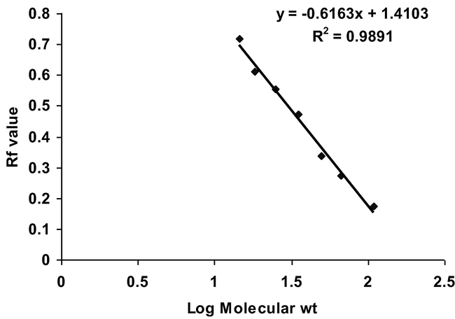
The Standard Curve plotted by the Rf values of each Standard Protein against the Log 10 of its Molecular Weight.
3 Results and discussion
All the allantoic fluids harvested from the chick embryos were initially tested for haemagglutination. The haemagglutination titre ranged from 1:64–1:1024 (Table 1).
Sr. No.
Well numbers
1
1:22
1:43
1:84
1:165
1:326
1:647
1:1288
1:2569
1:51210
1:102411
1:204812
(con.)
A
+
+
+
+
+
+
+
+
+
−
−
−
B
+
+
+
+
+
+
+
+
−
−
−
−
C
+
+
+
+
+
+
+
−
−
−
−
−
D
+
+
+
+
+
+
+
+
+
−
−
−
E
+
+
+
+
+
+
−
−
−
−
−
−
F
+
+
+
+
+
+
+
+
+
+
−
−
G
+
+
+
+
+
+
+
+
+
−
−
−
H
+
+
+
+
+
+
+
+
+
+
−
−
In lane A, the field isolates of NDV presented nine peptide bands having Mol. wt. ranged from 103 to 23.9 KDa. In lane B, the field isolates of NDV presented ten peptide bands having Mol. wt. ranged from 103 to 16.0 KDa. In lane C, the field isolates of NDV presented twelve peptide bands having Mol. wt. ranged from 158.75 to 13.8 KDa. In lane D, the field isolates of NDV presented twelve peptide bands having Mol. wt. ranged from 159.00 to 13.3 KDa. In lane E, the field isolates of NDV presented ten peptide bands having Mol. wt. ranged from 98.77 to 8.4 KDa. In lane F, the field isolates of NDV presented four peptide bands having Mol. Wt. range 98.03–13.7 KDa. In lane G, the field isolates of NDV presented three peptide bands having Mol. wt. ranged from 98.03 to 19.4 KDa. In lane H, the field isolates of NDV presented three peptide bands having Mol. wt. ranged from 98.03 to 19.2 KDa. In lane I, the field isolates of NDV presented twelve peptide bands having Mol. wt. ranged from 115.55 to 14.7 KDa. In lane A, the field isolates of NDV presented seven peptide bands having Mol. wt. ranged from 103 and 13.99 KDa. In lane B, the field isolates of NDV presented seven peptide bands having Mol. wt. range 103 and 13.99 KDa. In lane C, the field isolates of NDV presented four peptide bands having Mol. wt. ranged from 106.43 to 53.52 KDa (Tables 2A–C). M = Marker; A-E = Field isolates of NDV. M = Marker; F-I = Field isolates of NDV.
Band
M
A
B
C
D
E
MW (KDa)
MW (KDa)
MW (KDa)
MW (KDa)
MW (KDa)
MW (KDa)
1
116.0
103
103
158.75
159
98.77
2
66.2
92.4
91.3
120.4
117.7
88.3
3
45.0
67.9
70.3
102.9
100.6
67.5
4
35.0
62.4
60.8
82.3
84.4
59.9
5
25.0
51.2
52.5
74.5
74.9
48.6
6
18.4
40.4
41.2
57.7
58.1
39.4
7
14.4
35.5
35.2
44.9
44.6
32.3
8
28.4
27.3
37.4
37.1
26.5
9
23.9
20.9
27.1
28
18.2
10
16
23.6
22
10.5
11
19.2
18.7
8.4
12
15.7
15.3
13
13.8
13.3
Band
M
F
G
H
I
MW (KDa)
MW (KDa)
MW (KDa)
MW (KDa)
MW (KDa)
1
114.7
98.03
98.03
98.03
115.55
2
69.7
69.3
68
66.2
96.2
3
54.7
31.1
34
32.3
83.2
4
33.55
20.2
19.4
19.2
77
5
24.88
13.7
65
6
19.88
57
7
13.33
46.8
8
41.2
9
36.2
10
27.3
11
23.3
12
19.9
13
14.7
The isolated virus grew well into 9th day old chicken embryos. The infected chicken embryos were alive till 48 h. post-inoculation. The embryo death might be due to the virus induced agglutination of erythrocytes. The HA titers of AAF harvested from the chick embryos 48 h ranged from 1: 64–1:1024. Similar findings were also reported by Manin et al. (2002) and Hemmatzadeh and Kazemimanesh (2017). The chick embryos supported the extensive virus replication. This could be the possible reason of the wide use of chick embryos for ND Virus growth.
Among several protein fractions of field strains fragmented by SDS-PAGE, the peptide of Mol. wt. of 160 KDa was found to be present in Lanes B, C and I (Table 1) and that resembled in Mol. wt. of large protein (L) as described by Parker and Collier, (1990). The Mol. wt. of approximately 93 KDa was found in lanes A, B, D, E, F, G and H (Table 2C) which resembled the haemagglutinin-neuraminidase (HN) protein in their electrophoretic mobility, reported by Bolen et al. (1982). The polypeptides with Mol. wt. of 20, 28, 30 and 34 KDa were similar (Alsahami et al., 2018). Apart from the above mentioned proteins of Mol. wt. 120, 80, 74, 60 and 45 KDa were also found in local isolates. These findings were upheld by the results of Vijayarani et al. (1992) and Al-Hadid (2016). M = Marker; A-C = Field isolates of NDV.
Band
M
A
B
C
MW (KDa)
MW (KDa)
MW (KDa)
MW (KDa)
1
116.0
103.3
103.3
106.43
2
66.2
90.64
90.64
93.04
3
45.0
61.22
61.22
61.22
4
35.0
52.14
52.14
53.52
5
25.0
41.2
41.2
6
18.4
28.46
28.46
7
14.4
13.99
13.99
4 Conclusion
It has been concluded that the NDV can be isolated from the field outbreaks. It can be propagated in chicken eggs and identified by its stable and differential haemagglutination property. The results of SDS-PAGE denoted that the indigenous isolates of NDV are almost identical and comparable with vaccines. SDS-PAGE, using 12.5 percent gel is simple, economical, and easy way for the characterization of NDV and other viruses. Furthermore, the potency of the vaccines can easily be checked through this technique and comparison can be made between field and vaccine ND viruses. It is recommended that more work should be done at the molecular level of local isolates of NDV to confirm the molecular nature of these isolates.
Acknowledgements
The authors (SM and KAAG) express their sincere appreciation to the Deanship of Scientific Research at the King Saud University for its funding of this research through the Research Group Project No. RG-1435-012.
Declaration of Competing Interest
The authors declare that they have no known competing financial interests or personal relationships that could have appeared to influence the work reported in this paper.
References
- A molecular epidemiological study of avian paramyxovirus type 1 (Newcastle disease virus) isolates by phylogenetic analysis of a partial nucleotide sequence of the fusion protein gene. Avian Pathol.. 2003;32:239-256.
- [Google Scholar]
- Isolation, identification and molecular characterization of Newcastle disease viruses in vaccinated chickens from commercial farms in the Sultanate of Oman. Int. J. Vet. Sci. Med.. 2018;6(2):248-252.
- [Google Scholar]
- Evaluation of antiviral activity of different medicinal plants against Newcastle Disease Virus. Am. J. Agri. Biol. Sci.. 2016;11(4):157-163.
- [Google Scholar]
- Anonymous, 1989. A laboratory manual for the isolation and identification of avian pathogens. In: third ed., The American Association of Avian Pathologist (AAAP), Kendall/Hunt Pub. Co., Iowa, USA. pp. 114–120, 124–128.
- Isolation, identification and molecular characterization of highly pathogenic newcastle disease virus from field outbreaks. Br. Arch. Biol. Technol.. 2016;59(0)
- [CrossRef] [Google Scholar]
- Detection and quantitation of Newcastle disease virus proteins in infected chicken embryo cells. Appl. Environ. Microbiol.. 1982;43(1):193-199.
- [Google Scholar]
- Animal Microbiology. London: Blackwell Scientific Publication Ltd.; 1977. p. :498.
- Detection of specific antigens of Newcastle disease virus using an absorbed Western blotting method. IJV. 2017;18:92-96.
- [Google Scholar]
- Biochemical and hematological studies on the effect of neem (Azadirachta indica) leaves aqueous extract on Newcastle Disease vaccine and infection in broiler chickens. Int. J. Rec. Sci. Res.. 2017;30:15876-15884.
- [Google Scholar]
- Poultry Diseases (third ed.). Bailliers Tindall, London, UK: English Language Book Society (ELBS); 1990. p. :121-167.
- Lammli, U. K. 1970. Cleavage of structural proteins during the assembly of the head of bacteriophage T4. Nature (London), P. O. Box 100, New York. 173–185.
- Newcastle disease: outbreak losses and control policy costs. Vet. Rec.. 2000;146:603-606.
- [Google Scholar]
- Characteristics of field isolates of Newcastle disease virus isolated in the course of outbreaks in the poultry plant in the Leningrad region in 2000. Vopr. Virusol.. 2002;47(6):41-43.
- [Google Scholar]
- Parker, M.T., Collier, L.H., 1990. Toply and wison, Principles of Bacteriology, Virology and Immunity, eighth ed., Edward Arnold Malborn.
- Pattern of infectious bursal disease in commercial white leghorn chicken. Ind. Vet. J.. 1997;74:1019-1021.
- [Google Scholar]
- Virus Propagation in Embryonating Eggs in Isolation and Identification of Avian Pathogens (third ed.). Iowa, USA: Kendall Hunt Publishing Company; 1989. p. :176-181.
- Prevalence, diagnosis and control of common poultry diseases. J. Anim. Health Prod.. 1989;9:18-27.
- [Google Scholar]
- Characterization of field isolated of Newcastle disease virus. Indian Vet. J.. 1992;69:584-588.
- [Google Scholar]
- Update on epidemiology, diagnosis and control technique of Newcastle disease. J. Vet. Sci. Technol.. 2017;8:429.
- [CrossRef] [Google Scholar]


