

Translate this page into:
Impact of heavy metals on the environment and human health: Novel therapeutic insights to counter the toxicity
⁎Corresponding authors. talhabmb@bgctub.ac.bd (Talha Bin Emran), jsimal@uvigo.es (Jesus Simal-Gandara)
-
Received: ,
Accepted: ,
This article was originally published by Elsevier and was migrated to Scientific Scholar after the change of Publisher.
Peer review under responsibility of King Saud University.
Abstract
Heavy metals are well-known environmental pollutants owing to their toxicity, longevity in the atmosphere, and ability to accumulate in the human body via bioaccumulation. The pollution of terrestrial and aquatic ecosystems with toxic heavy metals is a major environmental concern that has consequences for public health. Most heavy metals occur naturally, but a few are derived from anthropogenic sources. Heavy metals are characterized by their high atomic mass and toxicity to living organisms. Most heavy metals cause environmental and atmospheric pollution, and may be lethal to humans. Heavy metals can become strongly toxic by mixing with different environmental elements, such as water, soil, and air, and humans and other living organisms can be exposed to them through the food chain. Plenty of experimental studies were performed to appraise the promising treatment options from natural products. Additionally, nanotechnology based treatment options are being constantly developed. As an emerging field, nanotechnology is making substantial advances in the analysis and removal of heavy metals from complicated matrices. Removal of heavy metal has been accomplished by the use of a variety of nanomaterials, including graphene and its derivatives, magnetic nanoparticles, metal oxide nanoparticles, and carbon nanotubes, to name a few. Using nanotechnology for heavy metal analysis and removal from food and water resources provides many benefits over traditional methods. These advantages include a broad linear range, low detection and quantification limits, a high sensitivity, and high selectivity. Therefore this review aimed to explore the environmental consequences of the heavy metals, toxicity to the human health, as well as novel therapeutics development from the natural resources. Additionally, nanotechnological and nanomedicinal applications to treat heavy metal toxicity are also highlighted in this review.
Keywords
Heavy metals
Environmental health
Toxicity
Nanotechnological approaches
Nanomedicine

1 Introduction
Periodic table consists of heavy metals to a notable portion with high density and atomic weight. Among them, the majorities are found in the biosphere, such as in water, soils, and rocks, and are also released into the surroundings from anthropogenic resources, mostly commercial and industrial. The toxic principles of heavy metals have been known for decades. However, recent experimental investigations show that some, including nickel, copper, and zinc, are vital for humans and are widespread in nature (Azeh Engwa et al., 2019). Manganese is found all over the world and makes about 0.1 percent of the earth's crust. However, heavy metals have such a number of undesirable repercussions on the environment; for example, the conversion of mercury into methylmercury in the presence of water creates sediments with high toxicity (Rice et al., 2014). Chromium is used extensively in industry and can be carcinogenic (Coetzee et al., 2020). However, some heavy metals are involved in the control of certain physiologic bodily functions. Naturally found vital heavy metals penetrate into the body via food, air, and water, where they regulate numerous biological activities (Chasapis et al., 2012; Roohani et al., 2013).
Most of the toxic heavy metals including lead, thallium, cadmium, and antimony, are common in industrial operations and are substantial polluters of the environment. Thallium has a more severe effect than other heavy metals, but is less abundant in nature (Karbowska, 2016); it is a cause of alopecia in humans. The benefits of heavy metals are generally outweighed by their hazards; for example, carcinogenicity is promoted by high exposure to antimony and chromium (Sun et al., 2015; Sundar and Chakravarty, 2010), lead poisoning causes intellectual abnormalities in children (Hou et al., 2013). Mercury toxicity causes Minamata disease, while cadmium poisoning causes itai-itai disease. Heavy metals can also cause toxicity in certain organs of the human body, such as nephrotoxicity, neurotoxicity, hepatotoxicity, skin toxicity, and cardiovascular toxicity, among other things. To avoid toxic effects, it is necessary for people to move away from industrial areas where heavy metal emission is considerable.
Many treatment procedures have been developed to counteract the toxicity of heavy metals. Natural products are being efficiently used to treat the adverse consequences (Singh et al., 2011; Tchounwou et al., 2012). Medicinal herbs and natural products for the treatment of various diseases have been around for almost the entire survival of mankind. One of the most significant advantages of traditional or plant-based medicine seems to be its perceived effectiveness, as well as its low frequency of severe adverse responses and its relatively cheap cost. Experimentally induced heavy metal toxicity in laboratory animals was significantly reduced using a variety of medicinal herbs and natural products (Bhattacharya, 2018).
Nanotechnological approaches are also seeking attention due to promising benefits on eradication of the adversative consequences of heavy metals. Nanotechnology is the use of interdisciplinary methods of creating nanoscale materials or devices that include concepts from physics, chemistry, engineering, and biology. With the rise of nanotechnology, nanomaterials for heavy metal detection and removal have been painstakingly developed and manufactured, providing countless benefits (He et al., 2019b).
This review aims to provide a clear explanation of the toxic consequences of heavy metals and the impact on the environment. Therefore, nanotechnological, nanomedicinal, and natural products based therapeutic strategies to counter the toxicity are also highlighted in this review.
2 Heavy metals in the environment
Heavy metals naturally occur in the environment and are vital for survival, but they may become hazardous when they accumulate in organisms. A few of the most frequent heavy metals that contaminate the environment include mercury, cadmium, arsenic, chromium, nickel, copper, and lead (Hazrat et al., 2019).
Cadmium is released into the atmosphere as a result of natural or manmade activities and animals and humans can be exposed to it differently. Cadmium pollution of the aquatic environment occurs through absorption, industrial waste, and surface runoff into sediments soil and sediments. People can be poisoned by cadmium via ingesting food, breathing air, or drinking water rich in the metal. Cadmium does not have any attributes that are helpful for plant growth and metabolic processes (Hayat et al., 2018).
Mercury is an extremely hazardous heavy metal that may be found in biosphere. Due to human activities, it has also become a widespread contaminant and is increasing in the atmosphere. Mercury converts to the highly toxic methylmercury when in contact with aquatic sediments (Gworek et al., 2020). Methylmercury enters the human body through the food chain via fish, seafood, and wildlife, which become contaminated after ingestion of toxic microorganisms. It penetrates the circulation after being absorbed into the human body and causes a variety of neurological problems (Rice et al., 2014).
Lead is a non-biodegradable metal that is available in nature and found in relatively low amounts. Atmospheric lead levels are increasing continuously because of the human activities including manufacturing, mining, and fossil fuel burning. Lead is toxic to the human body when exposed to amounts greater than the optimum. Children are at higher risk of lead poisoning; when they come into contact with dust laden with environmental lead, the severity of poisoning increases (Loh et al., 2016).
Manganese, the most plentiful of the toxic heavy metals, is found in various oxidation states in nature. During combustion of methylcyclopentadienyl manganese tricarbonyl (MMT), an additive in gasoline, manganese oxides are emitted into the air. Although manganese is required for a variety of physiological activities, excessive consumption results in substantial toxicity (Loranger and Zayed, 1995; O’Neal and Zheng, 2015).
Chromium is a cancerous and toxic element. In the environment, it exists in two stable oxidation states: chromium (III) and chromium (IV) (VI). Chromium (III) is a less hazardous form of chromium (VI). They can interconvert to each other during industrial operations. However, conversion of chromium (VI) to chromium (III) is less harmful to the environment because the latter is lower in toxicity. Chromium is used in many industries that pose a threat to regional climates. In comparison to natural chromium emissions from the environment, ferrochrome industry emissions are at the highest level (Fig. 1) (Coetzee et al., 2020; Kimbrough et al., 1999).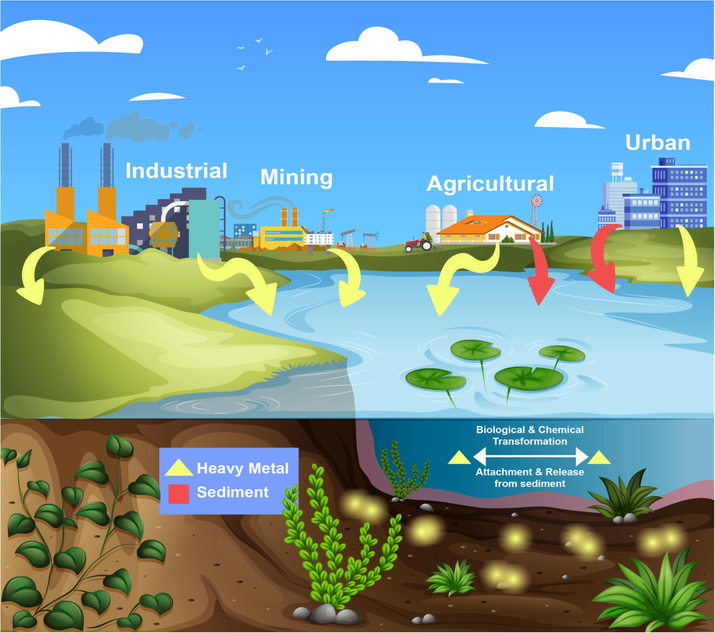
Diagrammatic explanation about heavy metals in the environment.
Cobalt is found in abundance across the environment, such as vegetation, soils, rocks, and water and is utilized to make alloys. Although its rate of discharge is low, it is highly dangerous to humans. Cobalt affects the human body in both beneficial and harmful aspects. The little amounts of cobalt usually have no negative consequences, but massive discharges into the environment can cause fatalities (Domingo, 1989).
Nickel is a naturally abundant element and has extensive industrial uses. It is emitted from both natural and anthropogenic sources into the atmosphere (Li et al., 2016a). It has many adverse effects on humans, and causes allergies, nasal and lung cancer, and kidney and cardiovascular diseases owing to the inhalation of contaminated air (Genchi et al., 2020; Lu et al., 2005).
Copper is recognized as a vital micronutrient for living organisms. It has a role in normal physiological functions of plants, such as formation of chlorophyll, photosynthesis, and carbohydrate and protein metabolism. Copper deficiency alters important metabolic processes, and elevated exposure causes toxicity (Schwartz et al., 2003).
Zinc is a fundamental and omnipresent metal. It is associated with plenty of enzymatic reactions via acting as a cofactor. Zinc toxicity depends on the manner and quantity of exposure. Smelting and mining are major the sources of zinc. A large amount of zinc emitted into the environment originates from the activities of mineral processing and affects ecosystems as well as living organisms (Zhang et al., 2012).
Antimony is a poisonous element that may be found in nanogram amounts in the air. Natural occurrences, including volcanic activity and weathering, as well as anthropogenic activities, cause emissions into the atmosphere (He et al., 2019a). Antimony toxicity develops in those working in industrial areas from inhalation (Fig. 1). Antimony poisoning causes physiological shortcomings, including pancreatitis, cardiotoxicity, and respiratory problems (pleural adhesions, chronic emphysema, chronic bronchitis, respiratory irritation, and inactive tuberculosis). It is also carcinogenic and affects reproduction (Sundar and Chakravarty, 2010).
Thallium is found in the environment in many forms and is hazardous to biological organisms. Thallium's toxicity is greater than any other heavy metal. Monovalent thallium ions arise in natural water and are emitted into the air via aqueous route (Fig. 1) (Peter and Viraraghavan, 2005). In addition, industrial emissions are a major contributor to the increase in thallium levels in the atmosphere. Exposure to thallium is extremely harmful to humans (Kazantzis, 2000).
3 Bioaccumulation of heavy metals in living system
Any essential or non-essential trace elements that are present in excess of the safe levels may result in physiological or morphological abnormalities or genetic mutations, such as slowing or stopping growth or causing mutations (Khan et al., 2010; Li et al., 2010; Luo et al., 2011). Food crops are one of the most essential components of our nutrition, and they may include a variety of both necessary and hazardous metals (Waqas et al., 2015; Yang et al., 2011), based on the properties of the growth medium used. Human exposure to heavy metals comes mostly through edible vegetables, which account for around 90% of the overall intake, while the remaining 10% comes from skin contact and breathing of polluted dust (Khan et al., 2014; Kim et al., 2009; Martorell et al., 2011). Because of the growing demand for food in recent decades, food safety has become a major public health concern in terms of human health. This scenario serves to motivate researchers and scientists to do study on the health risks linked with the ingestion of heavy metals, pesticides, and toxin-contaminated food products (Jaishankar et al., 2014).
Our food chain is constantly being replenished with essential and non-essential materials as a result of the excessive use of agrochemicals, municipal wastewater, industrial effluents, and raw sewage for irrigation (Tongesayi et al., 2013). In accordance with the Agency for Toxic Substances and Disease Registry's toxicity classification system, heavy metals and metalloids such as arsenic, lead, and cadmium present in the environment are classed as 1, 2, and 7 on a scale from 1 to 7 (ATSDR, 2007).
Mineral resources and elements such as copper, chromium, iron, manganese, and zinc, among others, are essential for both animals and humans because they are involved in a variety of metabolic functions, enzyme activities, receptor sites, hormonal function, and protein transport at specific concentrations (Antoine et al., 2012). Another group of elements, such as arsenic, cadmium, lead, and mercury, are non-essential and play no useful role in plants, animals, or people. They also serve no nutritional purpose since they are exceedingly poisonous (Khan et al., 2015). To set quality standards and identify the hazards to human health and food safety, it is required to describe the sources and amounts of heavy metals in soil (Sun et al., 2013). Environmental pollution caused by heavy metals is persistent, covert, and long-term (Ali et al., 2019). Because metals are nonbiodegradable and have a lengthy half-life, biological species are unable to decompose them, and they remain in their body parts and surroundings, posing health risks (Nabulo et al., 2011). Bioaccumulation of heavy metals in vegetables poses a health hazard due to their potential to transfer from polluted land and water into the food chain (Khan et al., 2015; Stasinos and Zabetakis, 2013).
Soil properties are crucial in food production, and heavy metal pollution of this critical resource, as well as their subsequent absorption and bioaccumulation in food crops, poses substantial environmental and health concerns, especially in poor countries. Heavy metal concentrations are influenced by soil type, plant genotype, and their interactions (Ding et al., 2013). In comparison to organic manure, mineral fertilizers contain increased concentrations of heavy metals; as a consequence, the use of mineral fertilizers leads in increased levels of heavy metal pollution in soil (Hu et al., 2013).
The health risks associated with toxic metals are dependent on the concentrations of these metals in certain media and the length of exposure. Even at low quantities of hazardous metals, long-term and chronic exposure may cause health problems (Mahalakshmi, 2012). Heavy metal toxicity is one of the principal abiotic stressors on plants, and it is based on heavy metal physiochemical features (Saxena and Shekhawat, 2013).
4 Toxicity of heavy metals
4.1 Neurotoxicity
As an essential element, manganese is involved in several physiological functions of the body. Its acute exposure exerts potential neuroprotective action by reducing apoptotic cellular death but exposure to a large quantity may induce harmful conditions alike neurological complications, such as alzheimer and parkinson disease which leads to apoptotic cell death and alteration of homeostasis (Goldhaber, 2003). Homeostasis of the cellular Mn relies on sufficient intake, storage, as well as excretion via different cell receptors and ion channels. The receptors associated with metal uptaking are down-regulated by homeostatic pathways in terms of excessive Mn exposure, whereas those engaging in its discharge from this cell become up-regulated. However, ongoing Mn accumulation leads to the increased formation of ROS, which contributes to mitochondrial dysfunction. Mitochondrial dysfunctions result in the discharge of cytochrome c, stimulating the apoptosis precursor caspase-9, by which caspase-3 cleaves. The cleaved caspase-3 fragment binds to a pro-apoptotic protein, PKCδ (protein kinase C delta). The proteolytic cleavage of PKCδ induced by Caspase-3 contributes to DNA fragmentation, as well as apoptosis (Fig. 2) (Harischandra et al., 2019).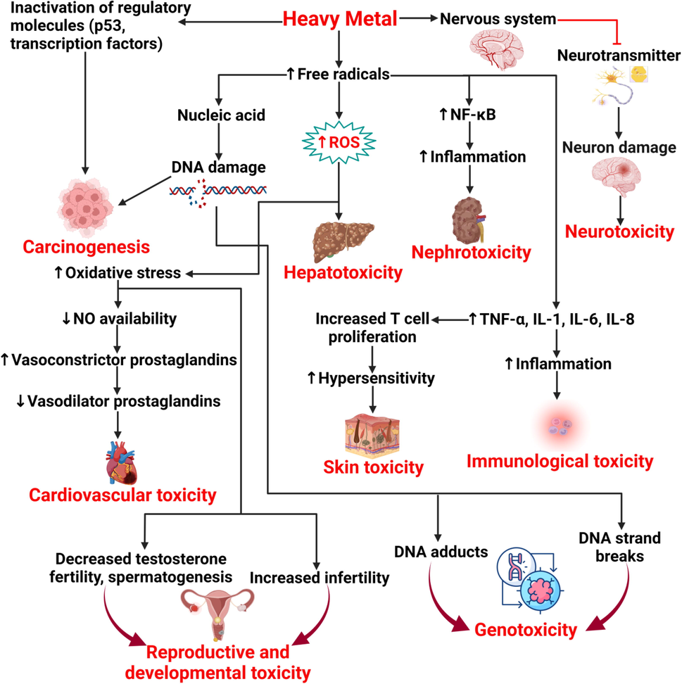
Mechanism of heavy metal toxicity in human.
The central nervous system suffers from cognitive impairment when arsenic is consumed. It's also linked to a number of neurological illnesses, including neurodevelopmental changes, and leads to excessive of neurodegenerative diseases. Arsenic poisoning also causes changes in synaptic transmission and neurotransmitter balance (Garza-Lombó et al., 2019). Additionally, the neurotoxic effects of arsenic are attributed to induce multiple apoptotic mechanisms. Firstly, arsenic and its methylated metabolites facilitates caspase- induced apoptosis in neural cells via the MAPK signaling pathways that include the ERK2, JNK, or p38; follow the intrinsic mitochondrial-apoptotic mechanisms. Besides, arsenic initiates intracellular calcium upturn that arbitrates apoptosis. On the contrary, cellular apoptosis can be mediated by activation of autophagy via the stimulation of the AMPK as well as inhibition of the mammalian target of rapamycin (mTOR). Autophagy is referred to a homeostatic manner where double-membraned autophagosome erupt cellular constituents to be eventually degraded when fused with lysosomes (Garza-Lombó et al., 2019).
Neurodegenerative defects, including amyotrophic lateral sclerosis, Parkinson's disease, Alzheimer's disease, and multiple sclerosis, result from neurotoxicity induced by cadmium (Branca et al., 2018). Numerous preclinical evidences have revealed that cadmium severely affects the functionalities of PNS (Miura et al., 2013) and CNS (Marchetti, 2014), with many clinical manifestations, such as peripheral neuropathy, olfactory dysfunctions, neurological disturbances, learning disabilities, and mental retardation, along with the impairment of motor function and behavioral changes in both adults and children. Additionally, many types of cellular activity, such as cell differentiation, proliferation, and cell death, are affected. The neurotoxicity of cadmium arise from neural cell death via apoptosis; providing plenty of apoptosis-induction factors, including impairment of neurogenesis, inhibition of neuron gene expression, offering epigenetic effect, endocrine disruption, etc. (Wang and Du, 2013).
Pathological investigations of poisoned animals and humans demonstrate that thallium toxicity causes damage to the brain and peripheral nerve. It produces edema and vascular engorgement in the cerebral hemispheres, capillary alterations in the brain, cerebellar edema with pyknotic Purkinje cells, and isolated regions of necrosis (Davis et al., 1981).
In addition to manganese, arsenic, and cadmium, a lot of heavy metals have been established for their neurotoxic consequences. As well, copper and zinc, like iron, act as impediments to neurodevelopment when an excessive amount enters the brain (Prohaska, 2000). Excess copper retention causes Wilson's disease, a hereditary condition which causes neurobehavioral abnormalities similar to schizophrenia. Zinc deficiency has an adverse influence on neurodevelopment, but the consequence of vast quantities is not clear (Cai et al., 2005). An experimental study by Ken-ichiro Tanaka and Masahiro Kawahara showed that copper augments zinc-induced neurotoxicity (Tanaka and Kawahara, 2017).
4.2 Nephrotoxicity
Nephrotoxicity induced by cadmium leads to intense clinical symptoms such as glucosuria, Fanconi-like syndrome, phosphaturia, and aminoaciduria (Hazen-Martin et al., 1993; Reyes et al., 2013). The proximal tubular epithelium is affected by direct exposure to the kidneys, resulting in a significant level of cadmium in urine, aminoaciduria, 32-microglobulinuria, and glucosuria, as well as impaired renal tubular phosphate reabsorption (Goyer, 1989). Renal tubular acidosis, renal failure, and hypercalciuria can all result from excessive exposure (Friberg et al., 2019; Jacquillet et al., 2007).
Lead has deleterious effects in all organs, but it has the greatest influence on the kidneys. Acute lead nephropathy causes proximal tubular dysfunction, resulting in Fanconi-like syndrome. Chronic lead nephropathy can be characterized by hyperplasia, interstitial fibrosis, atrophy of the tubules, renal failure, and glomerulonephritis (Fig. 2).
Acute exposure of the kidneys to mercury causes acute tubular necrosis, and has many clinical symptoms, such as acute dyspnea, altered mental status, abdominal pain, profuse salivation, tremors, vomiting, chills, and hypotension. In contrast, chronic exposure to mercury causes injury to the epithelium and necrosis in the pars recta of the proximal tubule. Tubular failure, higher urine excretion of albumin and retinol-binding protein, and a nephritic state with a characteristic of membranous nephropathy are all symptoms of mercury-induced chronic kidney injury (Lentini et al., 2017).
Thallium sulfate excretion via the kidneys is delayed and can take up to two months after consumption. Toxic injury to the kidneys is indicated by albuminuria and hematuria. However, thallium poisoning does not cause gross diminishment of renal function (Yumoto et al., 2017).
4.3 Carcinogenicity
Arsenic causes epigenetic alterations, damage to DNA, changes in the p53 protein's expression, histone modifications, DNA methylation, and reduced p21 expression (Fig. 2) (Martinez et al., 2011; Park et al., 2015). Arsenic poisoning raises the risk of cancer by attaching to DNA-binding proteins and slowing down the DNA-repair process (Garcia-Esquinas et al., 2013).
Lead is a carcinogenic substance that causes damage to the DNA repair mechanism, cellular tumor regulating genes, and chromosomal structure and sequence by releasing ROS (Fig. 2). It disrupts transcription by shifting zinc from certain regulatory proteins. (Silbergeld et al., 2000).
Mercury's peroxidative activity generates a significant quantity of reactive oxygen species (ROS), which can aid protumorigenic signaling and cancerous cells growth. ROS can contribute to carcinogenesis by damaging cellular proteins, lipids, and DNA, resulting in cell damage (Reczek and Chandel, 2017; Zefferino et al., 2017).
Nickel works as a carcinogen via controlling a variety of carcinogenic mechanisms, including gene regulation, transcription factor management, and free radical generation. It controls the expression of particular long non-coding RNAs, mRNAs, and microRNAs. It participates in the methylation of the promoter and the downregulation of gene 3 (MEG3) to increase the modulation of hypoxia-inducible factor-1, both of which contribute to carcinogenesis (Zambelli et al., 2016; Zhou et al., 2017).
4.4 Hepatotoxicity
The toxicity of lead on liver cells is well established. Exposure to it increases oxidative stress resulting in liver damage. Organic solvents, combined with lead, also cause injury to the liver because of having the same characteristics as lead (Farmand et al., 2005; Malaguarnera et al., 2012). Chronic lead exposure is potentially toxic to liver cells, resulting in glycogen depletion and cellular infiltration, which can result in chronic cirrhosis (Fig. 2) (Hegazy and Fouad, 2014).
Cadmium has two human target tissues: the renal cortex and the liver (Bernard, 2004). During acute exposure, it accumulates in the liver and is linked to a variety of hepatic dysfunctions. Cadmium changes the cellular redox balance, resulting in oxidative stress and hepatocellular damage (Zalups, 2000). Hepatotoxicity induced by cadmium, both acute and chronic, causes liver failure and therefore can increase the risk of cancer (Hyder et al., 2013).
Copper is well known to accumulate in the liver due to Wilson's disease. Increased levels of copper may cause oxidative stress; therefore, hepatic copper deposition is not only pathognomonic, but also pathogenic. Elevated hepatic copper levels are also observed in cholestatic liver diseases. However, they result from diminished biliary excretion of copper and are not a cause of hepatic infection (Deering et al., 1977; Gross et al., 1985; Yu et al., 2019).
Numerous studies have shown that Cr(VI) can harm the liver, and histopathological changes such as steatosis of hepatocytes, parenchymatous degeneration and necrosis were already identified. Elevated ROS levels, lipid peroxidation, suppression of DNA, RNA, and protein synthesis, DNA damage, decrease of antioxidant enzyme activity, mitochondrial dysfunction, such as impaired mitochondrial bioenergetics, cell growth arrest, and apoptosis are all associated with Cr(VI) hepatotoxicity (Hasanein and Emamjomeh, 2019).
4.5 Immunological toxicity
Acute and chronic lead exposure leads to several toxic effects on the immune system and causes many immune responses, such as increased allergies, infectious diseases, and autoimmunity, as well as cancer (Dietert et al., 2004; Hsiao et al., 2011). A high risk of lung, stomach, and bladder cancer in several demographic groups has been linked to lead exposure (Rousseau et al., 2007; Steenland and Boffetta, 2000). Exposure to lead triggers the production of B and T-cells as well as MHC activity (Kasten-Jolly et al., 2010). It can influence cellular and humoral responses by modifying the role of T-cell and increasing susceptibility to development of autoimmunity and hypersensitivity (Fig. 2) (Mishra, 2009).
Occupational and environmental exposure to cadmium may induce immunosuppressive effects based on varying exposure conditions. Humoral immune responses are amplified at low exposure, whereas the effects at higher exposures are not yet established. However, phagocytosis, natural killer cell activity, and host resistance in experimental infections are notably reduced in most cases.
Laboratory studies on exposure of mice and rodents to heavy metals resulted in many immunological defects, including immunosuppression and immunostimulation. Injection of mercury chloride into mercury-insensitive strains of laboratory animals’ reduced cellular function in the immune system, i.e., the animals showed immunosuppression. When mercury was applied to mercury-sensitive rodent strains, cellular activity in the immune system was enhanced, i.e., the animals showed immunostimulation. Both immunosuppression and immunostimulation lead to infections, allergies, and autoimmune diseases. However, mercury does not appear to have an impact on the human immune system, although Swedish authors have concluded that amalgam, a mercury alloy, affects the immune system.
Chromium is known to have many adverse effects on the human immune system. The influence of chromium on the immune system has been explored in numerous experimental studies. According to Faleiro et al. who used CoCrMo disc samples, lymphocyte proliferation is obstructed. High doses of hexavalent chromium reduce the phagocytic action of alveolar macrophages and the humoral immune response (Glaser et al., 1985). In addition, chromium induces two types of hypersensitivity reactions: type I (anaphylactic type); and type IV (delayed type). Development of allergic contact dermatitis due to chromium exposure has also been found in many studies (Bruynzeel et al., 1988; Leroyer et al., 1998).
4.6 Cardiovascular toxicity
Lead exposure, either acute or chronic, produces a variety of abnormalities in the human body. Chronic exposure to lead may cause arteriosclerosis and hypertension, thrombosis, atherosclerosis, and cardiac disease by increasing OS, reducing NO availability, increasing vasoconstrictor prostaglandins, altering the renin–angiotensin system, lowering vasodilator prostaglandins, disrupting vascular smooth muscle Ca2+ signaling, increasing inflammation and endothelium-dependent vasorelaxation, and adjusting the vascular response to vasoactive agonists. Exposure for a long time also increases arterial pressure (Hertz-Picciotto and Croft, 1993; Vaziri, 2008, 2002).
Cadmium is a toxicant and carcinogenic metal. In addition to its carcinogenic properties, cadmium induces kidney disease, bone disease, and cardiovascular disease (Toxicological Profile for Cadmium, 2002). Low to moderate cadmium exposure results in hypertension (Tellez-Plaza et al., 2008), diabetes (Schwartz et al., 2003), carotid atherosclerosis (Messner et al., 2009), peripheral arterial disease (Navas-Acien et al., 2004), chronic kidney disease (Hellström et al., 2001), myocardial infarction (Everett and Frithsen, 2008), stroke, and heart failure (Peters et al., 2010). In prospective studies, cadmium was linked to an increased risk of cardiovascular death in the general population of the United States (Tellez-Plaza et al., 2013, Tellez-Plaza et al., 2012).
Mercury has been shown to cause neurotoxicity, nephrotoxicity, and hepatotoxicity in humans. Cardiovascular toxicity has also been discovered in recent research. Levels of mercury in hair have been linked to oxidized LDL levels in atherosclerotic lesions, acute coronary failure and atherosclerosis (Yoshizawa et al., 2002). Paraoxonase, an extracellular antioxidative enzyme linked to HDL dysfunction, is likewise inactivated by mercury (Gonzalvo et al., 1997; Salonen et al., 1999); this is directly linked to the progression of atherosclerosis and the increased risk of a coronary heart disease, cardiovascular disease, acute myocardial infarction, coronary heart disease, and carotid artery stenosis (Kulka, 2016).
Cobalt exposure causes reversible systolic cardiac depression, which may be distinguished from other cardiomyopathy disorders. Cardiomyopathy caused by cobalt can be slow and fatal. However, the cardiac function of survivors usually recovers (Packer, 2016). Increased T cell proliferation
4.7 Skin toxicity
Chronic arsenic exposure promotes a lot of possible skin diseases, including hyperkeratosis, hyperpigmentation, and several types of skin cancer. Hyperpigmentation is the most prevalent skin change caused by prolonged arsenic exposure. Arsenic exposure can potentially cause Bowen's disease, a type of early skin cancer. Arsenic hyperkeratosis is usually widespread, affecting the soles and palms, but it can also affect the legs, toes, fingers, arms, and dorsum of the hands. Some hyperkeratotic and Bowen's disease lesions have the potential to develop into invasive malignancies (Huang et al., 2019).
The skin, the body's outermost organ, serves as a barrier against different contaminants. Contact with chromium causes a variety of acute and chronic severe dermatological consequences, including contact dermatitis, systemic contact dermatitis, and skin cancer. Contact dermatitis is a common skin disorder characterized by delayed hypersensitivity as a result of recurrent dermal contact with allergens (haptens) (Fig. 2). Systemic contact dermatitis is a kind of dermatitis induced by systemic exposure to an allergen, which causes the skin to become sensitive through direct dermal contact at first (Matthews et al., 2019; Menné et al., 1994; Uter et al., 2018; Winnicki and Shear, 2011; Yoshihisa and Shimizu, 2012).
Many skin infections are caused by mercury and mercury-containing compounds, including such acrodynia (pink disease), a common dermatological ailment in which the skin becomes pink when exposed to heavy metals, particularly mercury (Horowitz et al., 2002). People tattooed with the red pigments cadmium sulfide and mercury sulfide may have inflammation restricted to specific areas typically within six months after getting tattooed (Boyd et al., 2000). Moderate swelling, scaling, vesiculation, and irritation are symptoms of acute contact dermatitis caused by mercury-containing substances. According to several studies, mercury poisoning is the most prevalent cause of dermatological problems (Boyd et al., 2000).
4.8 Reproductive and developmental toxicity
Arsenic is a known reproductive toxin in humans, and it causes abnormalities in experimental animals, particularly neural tube anomalies (Wang et al., 2006). Inorganic arsenic impairs male reproduction by reducing the weights of the testes, the accessory sex organs, and the number of sperm in the epididymis. Aside from affecting sperm production, inorganic arsenic exposure also causes variations in testosterone and gonadotropin levels, as well as disturbances in the steroidogenesis process (Kim and Kim, 2015). In females, arsenic consumption is associated with an increased incidence of endometrial cancer (Salnikow and Zhitkovich, 2008). Endometrial angiogenesis, which is critical for embryo development, is then impaired by arsenic exposure during pregnancy. Symptoms of endometriosis, subfertility, prematurity, sterility, and spontaneous abortions are all caused by these conditions (Milton et al., 2017).
Several studies conducted by the World Health Organization (WHO) have found that more than 10% of women are at risk of infertility because of their exposure to heavy metals such as lead, cadmium, mercury, and other pollutants, which are the most common environmental contaminants that can cause reproductive disorders (Apostoli and Catalani, 2011). According to a study conducted by the WHO, the condition of infertility is mostly more prevalent in women than in men. Ovulation disturbances are a frequent cause of subfertility in women (Naz and Batool, 2017; Upadhyay et al., 2020). Ovulation disturbances are characterized by irregular or absent menstrual cycles, and they may be corrected with the use of reproductive hormones. The risk of infertility in women elevated as a result of increasing levels of toxin exposure, which resulted in hormonal disruption, delayed ovulation, and chromosomal abnormality in oocytes. Female infertility is caused mostly by hormonal imbalance, which is exacerbated by endocrine disruption caused by heavy metal poisoning, which is the most common cause of female infertility currently (Fig. 2) (Rattan et al., 2017).
4.9 Genotoxicity
Several investigations have shown significant interindividual variability in receptiveness to arsenic poisoning, with genetic factors as the fundamental source of this variability being identified. The genotoxicity of arsenic results in deoxyribonucleic acid alteration, which includes chromosomal abnormalities, mutation, micronuclei production, deletion, and sister chromatid exchange (Roy et al., 2018). Numerous investigations have been conducted to determine the mechanism of arsenic's genotoxic impact, which includes the induction of oxidative stress and the disruption of DNA repair (Pierce et al., 2012). Arsenic has been shown to have no direct effect on DNA and is regarded as a weak mutagen because, despite its low mutagenicity, it impacts the mutagenicity of other carcinogens. In human cells, for example, an increased mutagenicity of arsenic has been found when exposed to UV light (Yin et al., 2019).
The genotoxic effects of human exposure to chemical compounds cause changes in the genetic material primarily via two processes: teratogenesis and carcinogenesis. Teratogenesis is a process in which a chemical compound causes changes in the genetic material. The first of these may manifest itself in the offspring in the form of congenital abnormalities, whilst the second manifests itself in the development of malignancies in those who have been exposed directly. The development of the central nervous system is especially affected by certain mercury compounds, known as teratogenic agents, which are toxic to the developing neurological system (Young et al., 2008). However, the relationship between mercury exposure and carcinogenesis (one of the most serious outcomes of DNA-induced damage) is still debated, since some experiments appear to show that mercury has genotoxic activity, while others have not proven such DNA-damaging effects (Fig. 2) (Crespo-López et al., 2009).
In yeast and animal cells, chromium's genotoxicity and carcinogenicity are being studied extensively. Those who work in the mining and consuming sectors that use Cr have also been shown to be at risk for cancer (Li Chen et al., 2012; Thompson et al., 2012). Researchers found that Cr(VI) causes a wide range of genetic material structural changes, including DNA chromosomal protein cross-links, inter-DNA strand cross-links, and nucleotide strand breakage in living and cultured cells (Fang et al., 2014).
5 Treatment options from natural resources
5.1 Neurotoxicity treatment
Taking into account Mn-related toxicity mechanisms and pharmacokinetics, several therapeutic approaches, and neuroprotective compounds have been investigated to evaluate their efficacy in alleviating the Mn-induced neurotoxicity. Anti-inflammatory compounds, natural and synthetic antioxidants, glutamate protectors, and ATP/ADP ratio protectors have been used to decrease Mn-induced neurotoxicity. Also, the efficacy and mechanisms of several therapeutic interventions such as ethylene-diamine-tetra acetic acid (EDTA), levodopa, and para-aminosalicylic acid (PAS) have been established. An analysis with the polyphenolic extract Euphorbia suppina (PPEES) from a Korean prostrate spurge has shown that PPEES can effectively inhibit Mn-induced neurotoxicity by antioxidants by modulation of endoplasmatic reticule (ER) stress and ER stress-mediated apoptosis. The amounts of ROS and malondialdehyde (MDA), which are products of lipid peroxidation, were also significantly reduced. The antioxidant activities of GSH and SOD and catalase (CAT) were enhanced at the same time. Improvement of Mn-induced histopathological alterations in the striatum and cerebral cortex by PPEES was also seen in vivo work (Bahar et al., 2017). A research has shown that curcumin and arsenic could substantially protect arsenic-induced dopaminergic changes and oxidative stress in rat's brain (Fig. 3). In another study, Yousef et al. (2008) also found that carcinogen-induced metabolic changes in the brain and liver of rats could be protected by curcumin (Rahaman et al., 2021; Yousef et al., 2008). Aware of the fact that the behavioral and neurochemical roles of brain biogenic amines and NOs have a significant role to play, this research explores the neuro-protective impact of curcumin against modifications of the arsenic on biogenic amines, their metabolites and NO amounts in rats (Yadav et al., 2010). A study demonstrates about a bio-hazard which is known as cadmium. It is also known as a strong neurotoxin. Nuts provide vital nutrients required to sustain human brain function. Cadmium was delivered by mouth at a dosage of 50 mg/kg per week with or without almond and walnut supplementation in rats. Cadmium-induced depression, anxiety and memory decline were greatly attenuated by dietary consumption of almonds and walnuts at a dosage of 400 mg/kg/day. Following supplementation of certain nuts in rats, neurochemical aberrations have stabilized. The current research indicates that long-term almond and walnut supplementation provides vital nutrients that can overcome dietary shortages and thereby decrease heavy-metal intoxication (Batool et al., 2019). PCA has also stopped inflammation caused by Cd by minimizing cytokines, including tumor necrosis factor-α and interleukin-1β, pro-inflammatory. In comparison, PCA supplementation relieved neuronal death caused by Cd by increasing Bcl-2 and reducing cortical tissue levels of Bax and Cas-3 (Al Olayan et al., 2020). A research investigated the potential involvement in Tl(+) mediated toxicity of a glutamatergic portion in rats by dizocilpine (MK-801) receptor through Nmethyl-d-aspartate (NMDA). Early (24-hour) motor shifts, decreased Glutathione (GSH) levels, lipid peroxidation, and GSH peroxidase activity induced by Tl(+) acetate (32 mg/kg, ip) of adult rats were examined for the effects in MK-801 (1 mg/kg, intraperitoneally [ip]). In rat striatum, hippocampus or midbrain, the Tl(+)-induced hyperactivity and lipid peroxidation were diminished, and mild effects were generated at other endpoints (Osorio-Rico et al., 2015). The effects of curcumin are antioxidant, anti-inflammatory and antidepressant (Rahaman et al., 2021). The rescue function for curcumin in Copper2+ mediated toxicity of D. Melanogasters was assessed in this report. For 7 days, Cu2+ (1 mM) and/or Curcumin (0.2 to 0.5 mg/kg Feed) is exposed to adult, wilderness flies in the diet. The findings showed Cu2+- flies were less likely than the control group to survive. Around the same time as the activity of rising acetylcholinesterase, nitric oxide and Dopamine levels, the toxicity of copper was also associated with a substantial decline in overall Thiol (T-SH) as well as the catalase and glutathione S-transferase activities. In addition, curcumin has restored the rates of outbreak and the status of cellular antioxidants and alleviated nitric oxide level accumulation in the fly. Curcumin has improved oxidative damage to the flies, as shown by survival rates, durability testing and antioxidant status restoration (Fig. 3) (Abolaji et al., 2020). Apigenin is also used to treat copper induced neurotoxicity (Dourado et al., 2020). The small dipeptide of carnosine (β-alanyl-L-histidine) has many positive impacts, including preservation of the acid-base balance, anti-glycemia, chelating agents, anti-crosslinking and antioxidants. The skeletal muscle and the hippocampus have elevated amounts of carnosine and its analog anserine (1-methyl carnosine). In pathogenesis of vascular dementia (VD), the neurotoxicity of zinc (Zn), caused by carnosine, plays a key function, and inhibits neuronally-borne death by Zn. Lead (Pb) is an all-encompassing pollutant for the atmosphere and for the industry. It causes neurotoxicity and cell mortality by disturbing pro- and anti-oxide balance; however, it is still not yet completely known the mechanisms of its toxicity. The isoflavonoid genistein (GEN) derived from soy has been reported to have neuroprotective and antioxidant effects. The research explored the pathways for in vivo and in vitro Pb-induced neurotoxicity to guard against Pb-induced toxicity of GEN. Cell viability was decreased and cell apoptosis was increased with the exposure of the Pb. In vitro production of reactive oxygen species (ROS), and GEN pretreatment significantly reduced Pb-induces oxidative injury by increasing main antioxidant enzyme expression, nuclear factor 2 p45-related, antioxidant transcription factor 2 (Nrf2). The activation of the PKC-α in vitro and pretreatment PB attenuated ROS generation by PKC-α inhibition was then established following Pb exposure and pretreatment. GEN also inhibited MAPK-NF-B activation induced by Pb (Su et al., 2016). The levels of lipid permeation, protein carbonyl, ROS production were substantially increased by the exposed rats to lead, and the activity in glutathione peroxidase, superoxide dismutase, and catalase in the cerebellum and brain cortex respectively decreased compared to controls. Abnormal histopathology and a rise in blood and brain lead levels in contrast with controls were also observed. The extent of lipid peroxidation, the volume of protein carbonyl, the production of ROS and increased glutathione peroxidase, the superoxide dismutase and catalase activity have been decreased and safety were shown in histopathological tests (Kumar Singh et al., 2018).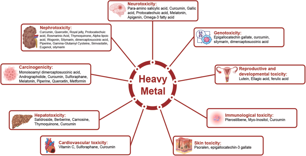
Diagrammatic explanation of heavy metal toxicity treatment by natural bioactive molecules.
5.2 Nephrotoxicity treatment
Chronic cadmium toxicity (Cd) is acquired by oral Cd administration, causes serious damage to the kidneys. The histologic modifications triggered by Cd have been improved by curcumin pretreatment. Urinary excretion substantially decreased from the Molecules of Kidney Damage (KIM-1). Osteopontin (OPN), metalloproteinase 1 tissue inhibitor (TIMP-1), lipocaline associated neutrophil gelatinase (NGAL) and netrin-1. The use of curcumin has a significant protective effect against nephrotoxicity caused by Cd (Kim et al., 2018). The levels of renal function markers, lipid peroxidation, renal injury molecules-1 (KIM-1), metallothionein, interleukin-1β, tumor necrosis factor-ш, nitric oxide, and apoptosis regulators Bax and caspases-3 also increased significantly in the renal tissues of royal-jelly treated mice, while antioxidant enzyme activity, the levels of glutathione, and apoptosis inhibitor Bcl-2 were important (Fig. 3). Vacuation and congested glomeruli in the kidney tissue of royal-jelly treated mouse is seen in histopathological tests (Almeer et al., 2019). Moreover, overall protein level was decreased in the cadmium-induced toxicity which was improved when treated with protocatechuic acid (Adefegha et al., 2015).
A study demonstrates that silymarin and dimercaptosuccinic acid reduce blood lead levels and provide protection against genotoxic effects (Alcaraz-Contreras et al., 2016). Besides, gamma-glutamyl cysteine, piperine, and Spirulina platensis are also used in this regard. To eradicate chromium induced nephrotoxicity Spirulina platensis, eugenol, extra virgin olive oil and simvastatin are usually used. A substantial decrease in MDA content and ACP activity and improved LDH and ALP activity relative to HgCl2 treated community was observed in comparison with spirulina with HgCl2. Spirulina also greatly decreases pathological changes in the kidneys before and following treatment with mercury (Sharma et al., 2007). Quercetin and L-α-Phosphatidylcholine also play vital role in this context (Elblehi et al., 2019; Kim et al., 2014). Besides these, diallyl sulfide and curcumin and zinc sulfate and silymarin can eradicate thallium induced nephrotoxicity (Abdel-Daim and Abdou, 2015; Al-jawad et al., 2017).
5.3 Carcinogenicity treatment
A recent study shows that sodium arsenite and DMA intensify chronic exposure to the urinary bladder. The expression of the metallopeptidase 9 matrix (MMP-9) and surviving are linked to carcinogenesis. These biomarkers may be used to mark the mediated carcinogenesis of the bladder. Oxidative damage that minimizes carcinogenesis is minimized as a burden. MiADMSA may be beneficial for blood carcinogenesis caused by systemic arsenic. The data revealed that the tissue-arsenic content, ROS, TBARS level, catalases, SOD activities (Islam et al., 2021) and GSH level, which can induce an eighth OHdG rise, were dramatically enhanced by sodium arsenite and DMA exposure. These improvements may have improved pro-oncogenic biomarkers such as MMP-9 and serum, bladder tissue, NBT-II and T-24 cells survived. In NBT-II and T-24 arsenic-exposed cells, a high cell migration and clonogenic ability indicate important carcinogens. In MiADMSA therapy, substantial recuperation was observed in these biomarkers (Sathua et al., 2020). Another study demonstrates that the levels of lead mediated hepatic and renal damage products and lipid peroxidation have been greatly suppressed prior to therapy using extract of Rosmarinus officinalis. Rosmarinus officinalis has also retained the composition and renal and hepatic cells in blood cells (Mohamed et al., 2016). Cd increased dose-dependent proliferation of lines of ovarian cancer cells. Cd-induced OVCAR3 and SKOV3 cell lines were blocked by melatonin. In addition, CdCl2 increased dramatically ERα expression compared with control in both the OVCAR3 and SKOV3 cell lines. Cd induction effect on ERα expression of OVCAR3 and SKOV3 cells was substantially inhibited by melatonins. In conclusion, Cd may have a significant role in the etiology of ovarian cancer by developing cells of ERα expression because of the proliferative effect on the lines of cancer in the ovary (Ataei et al., 2018). Besides, piperine, curcumin, beta-cryptoxanthin and sulforaphane can help in this regard (Liu et al., 2016; Mohajeri et al., 2017; Verma et al., 2020; Wang et al., 2018b). Quercetin protects against nickel-induced hepatic dysfunction, elevates the histology modifications in the liver of nickel, decreases the expression of inflammatory markers in nickel exposed livers of mice, decreased the overall DNMT activity and Nrf2 DNA methylation of nickel exposed livers of the rat, and reduced carcinogenicity (Fig. 3) (Liu et al., 2015). Metformin is another candidate for treating nickel-induced carcinogenicity (Kang et al., 2017).
5.4 Hepatotoxicity treatment
Salidroside (SDS) shows clear antioxidant activity and can treat lead acetate-induced liver damage by reducing oxidative stress and boosting antioxidant stress activity, therefore improving liver tissue structure. In this approach, it is possible to eliminate lead-induced hepatotoxicity (Chen et al., 2019). Furthermore, berberine was discovered to raise serum albumin, reducing lead-induced hepatotoxicity (Hasanein et al., 2017; Rauf et al., 2021). Carnosine, curcumin, and thymoquinone also significantly reduced lead-related histological and hepatological complications (Fig. 3). In a research, selenium (Se) was found to be an efficient chemo-protectant of Cd. Se therapy was shown to significantly reduce Cd-induced hepatocyte mortality and morphological changes. Simultaneously, Se decreased ROS generation, increased reduced glutathione (GSH) levels, and increased selenoenzyme (glutathione peroxidase, GPX) activity, all of which helped to attenuate Cd-induced oxidative stress. Finally, it was discovered that Se may protect against Cd-induced hepatotoxicity by inhibiting the ER stress response (Zhang et al., 2020). In another study, the impact of M. oleifera-based diets on nickel (Ni)-induced hepatotoxicity in rats was investigated. Male rats were divided into six groups and administered 20 mg/kg body weight nickel sulfate in normal saline before being fed either a standard diet or an M. oleifera-based diet for 21 days. All of the animals were anesthetized and killed 24 h following the final treatment. The activities of alanine transaminase, aspartate transaminase, and alkaline phosphatase in rat plasma were considerably increased after exposure to nickel. Ni also increased triglyceride, total cholesterol, and low-density lipoprotein cholesterol levels while lowering high-density lipoprotein cholesterol levels. Furthermore, Ni exposure increased rat plasma malondialdehyde while depleting glutathione levels. The histopathology findings indicated that Ni exposure induced inflammation and cellular damage. M. oleifera-based diets were shown to protect rats against Ni-induced hepatotoxicity by increasing liver function indicators, lipid profile, and restoring cellular architecture and integrity.
5.5 Immunological toxicity treatment
Pterostilbene (PT) is a naturally occurring chemical found mostly in blueberries. PT has been shown in studies to be an effective anti-inflammatory and anti-oxidant agent. Epidermal Cr(VI) delivery produced cutaneous inflammation in mice ear skin in an in vivo investigation, and pro-inflammatory cytokines TNF-α and IL-1 were identified in the epidermis, indicating a level of rise following Cr(VI) therapy. Meanwhile, the findings of another in vitro experiment revealed that treatment of HaCaT cells with varying doses of Cr(VI) promoted apoptosis and endoplasmic reticulum (ER) stress (human keratinocyte). Cr(VI) triggered the p38 mitogen-activated protein kinase (MAPK)/MAPK-activated protein kinase 2 (MK2) pathways, which elevated TNF-α and IL-1 mRNA expression. In the epicutaneous elicitation test, the intensity of the skin responses was dramatically reduced when the mouse was given PT. In addition, PT treatment reduced inflammation and apoptosis in HaCaT cells in vitro. Furthermore, recent studies revealed that the NLRP3 inflammasome may be implicated in Cr(VI)-mediated apoptosis and inflammation in allergic contact dermatitis (ACD) (Wang et al., 2018a). Besides, nuclear factor (erythroid-derived 2)-like 2 (Nrf2) attenuated autoimmunological problems induced by chromium (Banerjee et al., 2020). In combination with Se, myo-inositol protects mice from Cd-induced thyroid injury. When utilizing the follicular structure as a read-out for comparison, the effectiveness of this combination was larger than that of resveratrol (Benvenga et al., 2020). In another study, curcumin (diferuloylmethane), a natural substance derived from the rhizomes of Curcuma longa, was evaluated in a guinea pig model of airway hyper responsiveness for its anti-asthmatic properties. Curcumin was given to guinea pigs during sensitization (to see whether it may prevent airway obstruction) or after they had symptoms of airway obstruction (to examine its therapeutic effect). Specific airway conductance (SGaw) was measured using a non-invasive method called constant-volume body plethysmography to detect the status of airway constriction and hyper reactivity. OVA-induced airway constriction (p=0.0399) and airway hyper reactivity (p=0.0043) are dramatically reduced when curcumin (20 mg/kg body weight) is given. Curcumin is useful in correcting defective airway characteristics in OVA-sensitized guinea pigs, according to the findings (Ram et al., 2003).
5.6 Cardiovascular toxicity treatment
Mercury and cadmium are very hazardous elements that may cause serious cardiac problems in both animals and humans. In a study, the therapeutic impact of vitamin C against these metals in rabbits was investigated, and favorable findings for heart toxicity were discovered (Ali et al., 2020). In another study, the histological and biochemical alterations detected in the rat heart after exposure to K2Cr2O7 were greatly reduced when the rats were given C. aurantium peel extract (300 mg/kg). Because of its antioxidant activity, C. aurantium peel extract was able to prevent K2Cr2O7-induced myocardial damage, according to their findings (Chaabane et al., 2017). A recent study demonstrates that oxidative stress, hematological changes, structural disorder, cardiomyocyte apoptosis, and heart malfunction, caused by K2Cr2O7 were all reduced by Sulforaphane (SFN). SFN decreased the levels of p53, cleaved Bcl2-associated X protein, caspase-3, interleukin-1, and nuclear factor kappa-B, while increasing the levels of Sesn2, NAD(P)H quinone oxidoreductase-1, heme oxygenase-1, nuclear factor erythroid 2-related factor 2 (Nrf2), and phosphorylated adenosine 5′-mon. The Sesn2/AMPK/Nrf2 signaling pathway is activated by SFN, which reduces Cr(VI)-induced cardiotoxicity. SFN might be a potential therapeutic for chronic Cr(VI) exposure as well as a defender against Cr(VI)-induced cardiac impairment (Yang et al., 2020).
5.7 Skin toxicity treatment
Research was conducted to explore if Solanum melongena peel extract could help with arsenic-induced Bowen's disease. From the two arsenic-endemic sites, a total of eight individuals with arsenic-induced Bowen's illness were chosen. Each patient was given a peel extract containing ointment and advised to apply it twice daily on the lesion site for 12 weeks. Significant improvement was observed in reducing the lesion of Bowen’s disease (Sarah and Misbahuddin, 2018). In another research, PUVA therapy was used to treat two male patients who had long-term contact sensitivity to chromium. One patient with simultaneous photosensitivity had a very positive response; his skin lesions cleaned up and his light tolerance improved. On PUVA-exposed (pigmented) skin, this was accompanied by a reduction in photopatch test reactivity and the extinction of patch-test reactivity. In both cases, PUVA therapy reduced the amount of rosette-forming T cells while increasing lymphocyte stimulation. While PUVA-therapy may have some systemic immunological effects, it appears that its abating impact on contact sensitivity and photosensitivity is mostly mediated through local mechanisms in the skin (Jansén et al., 1981). Besides, Epigallocatechin-3 gallate is used to treat acne vulgaris (Yang et al., 2017).
5.8 Reproductive and developmental toxicity treatment
A recent study demonstrated the importance of grape seed proanthocyanidin extract which reduced oxidative stress damage in mouse testis, counteracting arsenic-induced reproductive toxicity by activating Nrf2 signaling (Li et al., 2015). Another study investigated the impact of lutein over arsenic-induced reproductive toxicity. The findings revealed that lutein reduces arsenic-induced reproductive toxicity in male mice via Nrf2 signaling, indicating a plausible mechanism for lutein in avoiding reproductive harm and elucidating that ingesting lutein-rich plant sources can reduce chemical-induced reproductive toxicity (Li et al., 2016b; Mitra et al., 2021b).
A considerable rise in tissue indicators of oxidative stress, severe histopathological alterations, seminal tubule destruction, poor sperm parameters, low spermatogenesis index, tubular desquamation, and decreased sperm mitochondrial function were all signs of lead poisoning in the reproductive system. In the rat reproductive system, carnosine and histidine supplementation reduced lead-induced oxidative stress and mitochondrial dysfunction. Carnosine and histidine's antioxidative and mitochondria-protective capabilities thus act as main defense mechanisms against lead-induced reproductive damage (Ommati et al., 2019). Male reproductive protection was demonstrated by reduced Pb-induced testicular injury, higher sperm count and motility, and a lower sperm abnormality rate. Anthocyanin from purple sweet potato (APSP) also helps in Pb-induced decreases in enzymatic and non-enzymatic antioxidants. Furthermore, APSP inhibited Pb-induced Bax mRNA and protein expression, decreased caspase-3 activation, increased Bcl-2 protein expression, and avoided DNA damage caused by Pb. Pb-induced testicular JNK signaling was similarly disrupted by APSP administration, which inhibited Jun N-terminal kinase (JNK) mRNA expression and phosphorylation, leading in c-Jun expression suppression. Pb was able to reverse the effects of APSP. Finally, because of its antioxidant and anti-apoptotic characteristics, as well as its regulation of the JNK signaling pathway, APSP may be a viable therapeutic treatment for avoiding Pb-induced reproductive damage (Zhou et al., 2021).
5.9 Genotoxicity treatment
Arsenic is a well-known genotoxicant that creates an overabundance of reactive oxygen species (ROS) and inhibits antioxidant enzyme systems, resulting in cell damage via the activation of oxidative sensitive signaling pathways. Green tea's primary polyphenolic catechin, epigallocatechin gallate (EGCG), has shown significant antioxidant, free radical scavenging, and genoprotective activities in vivo. By controlling arsenic-induced oxidative stress in mice, the researchers intended to examine the antioxidant and genoprotective effectiveness of EGCG. For 15 days, animals were given prophylactic and therapeutic dosages of EGCG (25 and 50 mg/kg b.wt.) orally, and then arsenic was delivered intraperitoneally at a dose of 1.5 mg/kg b.wt. (1/10th of LD50) for 10 days. Increased ROS production (114%) in lymphocytes; increased levels of LPO (2–4 fold); reduced levels of hepato-renal antioxidants (approx. 45%) and augmented genomic fragmentation in hepato-renal tissues; increased chromosomal anomalies (78%) and micronucleation (21.93%) in bone marrow cells; and comet tailing (25%) in lymphocytes of mice after arsenic intoxication (Kaushal et al., 2019). Researchers explored if curcumin nanoformulations might protect against arsenic-induced genotoxicity better than free curcumin. The emulsion process was used to make curcumin-loaded poly(lactic-co-glycolic acid) nanoparticles (CUR-NP). The CUR-NP was water soluble and released in two phases. The rats were placed into five groups, each with six rats. Chromosome abnormalities, micronuclei production, and DNA damage were all exacerbated by arsenic. Both free curcumin and CUR-NP reduced the genotoxic effects of arsenic. However, at the same dosage level, the results imply that nanoformulations has a better protective impact than free curcumin (Sankar et al., 2014).
Researchers looked examined how silymarin and dimercaptosuccinic acid (DMSA), a chelating agent, worked against lead (Pb) poisoning in rats whether given singly or in combination. Wistar rats (n = 200) were put into five groups at random. Group A was used as a control group. For 8 weeks, groups B–E were exposed to 2000 ppm of lead acetate in their drinking water. A positive control group, Group B, was used. For 8 weeks, Group C was given silymarin (100 mg kg−1 orally). For the final 5 days of therapy, Group D got DMSA (75 mg kg−1 orally) once daily. Groups C and D were given DMSA and silymarin, respectively. The impact of Pb was assessed, and therapies were estimated based on blood lead levels (BLLs), renal system, and genotoxic effects using the comet assay. Following exposure to lead acetate, the BLLs considerably increased. BLLs were reduced after silymarin and DMSA were administered. Silymarin and DMSA were found to give considerable protection against Pb's genotoxic effects (Alcaraz-Contreras et al., 2016). The experimental evidences regarding the treatment options for heavy metal induced toxicities are highlighted in Table 1.
Field of Study
Treatment for
Treatment procedure/element
Study model
Dose
Assay
Outcomes
References
Heavy Metal-induced neurotoxicity
Manganese
Polyphenolic extract of Euphorbia supina (PPEES)
Human neuroblastoma SKNMC cells and SD male rats
(50, 100 and 200 µg/mL)
Western blot and RT-PCR analyses
PPEES significantly increased the cell viability and decreased LDH activity of Mn-exposed cells, which could protect the SKNMC cell from cytotoxicity
(Bahar et al., 2017)
Para-amino salicylic acid (PAS)
Female aged 50
6 g
n.m.
Symptoms were significantly alleviated, and handwriting recovered to normal
(Jiang et al., 2006)
Arsenic
Curcumin
Rats
100 mg/kg
Reversed phase HPLC and electrochemical detector
Significant increase in the levels of DOPAC (30%), 5-HT (70%), DA (42%), and HVA (50%) in corpus striatum as compared to those treated with arsenic alone
(Yadav et al., 2010)
Gallic acid
Sprague Dawley male rats (36)
Saline + GA (50 mg/kg/mL)
Elevated plus maze, Light dark activity box, Forced swim test and Morris water maze
Increased brain LP, brain antioxidant enzymes activity, brain acetylcholinesterase activity
(Samad et al., 2019)
Saline + GA (100 mg/kg/mL)
iAS + saline
iAS + GA (50 mg/kg/mL)
iAS + GA (100 mg/kg/mL) iAS
Cadmium
Almond and walnut
Rats
400 mg/kg/day
Elevated plus maze, novel object recognition task, and Morris water maze
Attenuated depression, paranoia and impairments in memory due to cadmium.
(Batool et al., 2019)
Protocatechuic acid
Adult male Wistar rats
100 mg/kg
ELISA kits
Cd concentration decreased dramatically and activity of cortical acetylcholinesterase and neurotropic factor derived from the brain increased
(Al Olayan et al., 2020)
Melatonin
Mouse neuroblastoma cells (Neuro-2a cells)
12.5, 25 and 50 μM
CTSB activity fluorometric assay
Increased “TFEB-responsive genes”, preserved lysosomal protease activity, maintained the lysosomal pH level, enhanced autophagosome-lysosome fusion
(M. Li et al., 2016)
Thallium
MK-801
Rats
1 mg/kg
Fluorometric assay, Lipid Peroxidation.
Reduced the Brain Regional Content of Tl+, mitigated the Tl+-Induced Brain Regional lipid peroxidation
(Osorio-Rico et al., 2015)
Copper
Curcumin
Drosophila melanogaste
0.2 and 0.5 mg/kg
n.m.
Ameliorates Cu2+- induced oxidative stress
(Abolaji et al., 2020)
Apigenin
AD cell model
n.m.
MTS assay
Attenuates copper-mediated β-amyloids by antioxidation, mitochondrial protection and inactivation of the MAPK signal
(Zhao et al., 2013)
Lead
Omega-3 fatty acid
Rats
750 mg/kg body weight
n.m.
Reduced the lipid peroxidation level, the protein carbonyl content, the development of ROS and increased glutathione peroxidase, superoxide dismutase and catalase activities and demonstrated safety from lead-alone rats in the histopathology study
(Kumar Singh et al., 2018)
Heavy metal induced nephrotoxicity
Cadmium
Curcumin
Sprague-Dawley rats
50 mg/kg
TUNEL assay
Protects against cadmium-induced renal damage, lowers AKI biomarker urinary excretion, and inhibits cadmium-induced apoptosis in the kidney
(Kim et al., 2018)
Royal jelly
Male mice
85 mg/kg
Western blot analysis, Quantitative RT-PCR
The levels of renal function markers, lipid peroxidation, renal injury molecules-1 (KIM-1), metallothionein, interleukin-1β, tumor necrosis factor-ш, nitric oxide, and apoptosis regulators Bax and caspases-3 also increased significantly in the renal tissues
(Almeer et al., 2019)
Protocatechuic acid
Rats
10 and 20 mg/kg
n.m.
Overall protein level was decreased in the cadmium-induced toxicity which was improved when treated with protocatechuic acid
(Adefegha et al., 2015)
Rosmarinic Acid
Mouse proximal tubular epithelial cells
5–60 µM
Flow cytometric analysis
Excessive ROS development imparted oxidative stress to renal cells, NADPH oxidase activation, cellular redox protection mechanism impairment, and triggering NO level
(Joardar et al., 2019)
Thymoquinone
Rats
50 mg/kg body weight
TUNEL assay
Significantly decreased Cd toxicity and maintained the usual histological tissue architecture, the over expression of the nuclear factor-κB in renal tissue caused by Cd substantially decreased
(Erboga et al., 2016)
Alpha lipoic acid
Rats
50 mg/kg
Western blot analysis
Small malondialdehyde and Cd content reductions, restore endogenous enzyme activity, renew the mitochondrial function
(Chen et al., 2018)
Wogonin
Sprague Dawley rats
10 and 20 mg/kg
TUNEL assay and western blot analysis
Wogonin demonstrated antioxidative and anti-inflammatory effects in Cd- intoxicated rats by obstructing OS and activation of NF-kB via restricting the stimulation of upstream kinases inclusive of MAPKs
(Jiao et al., 2019)
Lead
Silymarin, dimercaptosuccinic acid
Rats
100 mg kg−1 and 75 mg kg−1
Comet assay
Reductions in blood lead levels and protection against genotoxic effects.
(Alcaraz-Contreras et al., 2016)
Spirulina platensis
Newborn rats
n.m.
n.m.
Alleviated lead induced hematotoxicity by restoring the biochemical markers to near normal levels
(Gargouri et al., 2018)
Piperine
Male rats
50 mg; 100 mg and 200 mg/kg
n.m.
Significantly improved decreased amounts of blood urea nitrogen, creatinine and malondialdehyde, renal histopathology and also substantially increased superoxide Dismutase and glutathione Peroxidase in the kidneys of lead acetate-treated rats
(Sudjarwo et al., 2017)
Gamma-Glutamyl cysteine
Rat
65 mg/kg
n.m.
γ-Glutamyl cysteine lowers the contents of kidney lead. Thus, promotes body weight gain, lower creatinine and urea serum levels, modulates oxidative stress markers in kidney tissue, decreases kidney tissue amount of pro-inflammatory cytokines
(Salama et al., 2016)
Chromium
Spirulina platensis
Male Sprague-Dawley rats
300 mg/kg
Avidin–biotin peroxidase method
Histopathological improvements, antioxidants and renal markers have been substantially restored
(Elshazly et al., 2015)
Extra virgin olive oil (EVOO)
Sprague Dawley rats
15 mL/kg
ANOVA method
Ameliorated DNA damage and some of the kidney diseases induced by Cr(VI) toxicity.
(Saber et al., 2015)
Simvastatin
Adult male Wister rats
20 mg/kg/day
Hematoxylin-eosin (H&E) staining technique, ANOVA method
Simvastatin clearly reversed the microscopic damage; it also shows protective effects against kidney injury induced by Cr(VI)
(Goodarzi et al., 2017)
Eugenol
Wister rats
15 mg/kg
ELISA and ANOVA method
Decrease in serum urea, creatinine and lactate dehydrogenase levels; this was accompanied with significant increase in renal glutathione, and superoxide dismutase contents, and increase in renal malondialdehyde and TNF-α
(Barhoma, 2018)
Mercury
Spirulina fusiformis
Swiss albino mice
800 mg/kg
MDA (malondialdehyde) assay
Significantly eliminates kidney pathological shifts
(Sharma et al., 2007)
Quercetin (QC)
Sprague Dawley rats
250 mg/kg
Western blot analysis and TUNEL assay
Metallothionin and Hg urinary excretion were substantially improved by QC pretreatment. Histopathology has demonstrated that QC protects from proximal tubular damage to the kidneys caused by HgCl2
(Kim et al., 2014)
L-α-Phosphatidylcholine
Rats
100 mg/kg
ELISA method
Attenuates the expression of the hematologic and serum biochemical alternations known as pro-inflammatory cytokines known as factor-α and interleukin-6 tumor necrosis, decreases OS and pro-inflammatory cytokine levels, and increases histopathological improvements in livers or kidneys of HgCl2-treated rats
(Elblehi et al., 2019)
Thallium
Diallyl sulfide and curcumin
Rats
200 mg/kg
MDA assay
Reduced hepatic toxicity by thallium effectively
(Abdel-Daim and Abdou, 2015)
Zinc sulfate and silymarin
Albino rats
20 mg/kg zinc sulfate and 25 mg/kg silymarin
n.m.
The creatinine levels have shown substantial decreased, an enhancement in renal tissue parts that have an effect on thallium toxicity
(Al-jawad et al., 2017)
Heavy Metal-Induced Carcinogenicity
Arsenic
Monoisoamyl dimercaptosuccinic acid
Male rats
50 mg/kg
MTT assay, RT-PCR assay, ELISA method
Significantly increased the tissue arsenic contents, ROS, TBARS levels, catalase, SOD activities and significantly decreased GSH level which might be responsible for an increased 8 − OHdG level
(Sathua et al., 2020)
Lead
Rosmarinus officinalis
Albino rabbit
30 mg/kg
Gas chromatography/mass spectrometry analysis
The levels of lead mediated hepatic and renal damage products and lipid peroxidation have been greatly suppressed prior to therapy. REE has retained the composition and renal and hepatic cells in blood cells
(Mohamed et al., 2016)
Mercury
Andrographolide, curcumin and melatonin
Human blood
Different concentrations
n.m.
These 3 elements ameliorated mercury induced genotoxicity and carcinogenicity due to their excellent antioxidant properties and their mixture seems efficient
(Patel and Rao, 2015)
Cadmium
Sulforaphane
Cd-transformed cells
Different concentrations
n.m.
Protects against lung carcinogenesis caused by cadmium
(Wang et al., 2018b)
Melatonin
OVCAR3 and SKOV3 cells
1–100 nM
Western blot analysis
Have a significant role in the etiology of ovarian cancer by developing cells of ERα expression because of the proliferative effect on the lines of cancer in the ovary
(Ataei et al., 2018)
β-cryptoxanthin
Rat testis
10 μg kg−1
ELISA method, TUNEL assay
Improved the resistance to Cd-mediated oxidative damages to rats along with cancer and indicated promising defense against testicular toxicity induced by Cd as a pharmacological agent
(Liu et al., 2016)
Curcumin
Rats
200 and 400 mg/kg
ELISA method
Ameliorated Cd-induced carcinogenicity
(Mohajeri et al., 2017)
Piperine
Human blood
Different concentrations
Comet assay
Ameliorated Cd-induced oxidative stress and carcinogenicity
(Verma et al., 2020)
Nickel
Quercetin
Male mice
20 mg/kg
Western blot analysis
Quercetin protects against nickel-induced hepatic dysfunction, decreases the expression of inflammatory markers in nickel exposed livers of mice, decreased the overall DNMT activity and Nrf2 DNA methylation of nickel exposed livers of the rat
(Liu et al., 2015)
Metformin
Lung cells
Different concentrations
n.m.
Chemoprevention of lung cancer
(Kang et al., 2017)
Heavy Metal-Induced Hepatotoxicity
Lead
Salidroside
Sprague-Dawley Rats
150 mg/kg and 300 mg/kg
Analysis of serum biochemical parameters
Serum levels of AST, ALT, ALP, and TB reduced significantly in a dose-dependent manner
(Chen et al., 2019)
Berberine
Rats
50 mg/kg
Analysis of serum biochemical parameters
Significantly prevented the lead-induced elevation in ALT, AST, and ALP of treated rats along with the concentration of serum albumin was significantly elevated
(Hasanein et al., 2017)
Carnosine
Male adult Wistar rats (27)
10 mg/kg/day
Hepatic biomarkers analysis
Significant reductions in the serum levels of hepatic enzymes like ALT, AST and ALP and increase in the level of albumin and total proteins
(Hasanein et al., 2016)
Curcumin
Male Wistar rats (30)
200 mg/kg
Determination of liver function markers with histological examination
Curcumin ameliorated ALT, AST and LDH level and markedly prevented the histological alterations induced by lead toxicity
(Alhusaini et al., 2019)
Thymoquinone
Male Wistar rats
5 mg/kg
Biochemical analysis of liver integrity
Lowering the ALT, AST, LDH, ALP, and γ-GT activities
(Mabrouk et al., 2016)
Cadmium
Selenium
Male hyaline variety white chickens
5.0 µM CdCl2 + 1.0 µM Na2SeO3
XBP-1 mRNA splicing assay
The XBP-1 s mRNA level was significantly higher than XBP-1u in Cd groups and the increased ratio of XBP-1 s/XBP-1u was found in the Cd group compared with Control group
(Zhang et al., 2020)
Nickel
Moringa oleifera
Male rats (60)
20 mg/kg
Necroscopy, Biochemical assays, Histopathological examination
By enhancing rat liver function indicators, lipid profile, and restoring cellular architecture and integrity, M. oleifera-based diets protected against Ni-induced hepatotoxicity
(Adeyemi et al., 2017)
Cardiovascular toxicity
Cadmium
Vitamin C
Rabbits
n.m.
ANOVA, Dunnett’s multiple comparison test
Reduced cholesterol level and other cardiac toxicity
(Ali et al., 2020)
Mercury
Vitamin C
Rabbits
n.m.
ANOVA, Dunnett’s multiple comparison test
Reduced cholesterol level and other cardiac toxicity
(Ali et al., 2020)
Chromium
C. aurantinum L.
Adult rats
100 and 300 mg/kg body weight/day
ANOVA
The histological and biochemical alterations detected in the rat heart after K2Cr2O7 treatment were greatly reduced by C. aurantium peel extract (300 mg/kg)
(Chaabane et al., 2017)
Sulforaphane (SFN)
Wister rats
4 mg/kg
Radioimmunoprecipitation assay (RIPA), TUNEL
SFN alleviated Cr (VI)-induced heart injury and is considered as a novel therapy for chronic Cr (VI) exposure
(Yang et al., 2020)
Skin toxicity
Arsenic
Solanum melongena peel extract
Eight patients
100 µg/mL
Standard deviation
Significant improvement was observed in reducing the lesion of Bowen’s disease
(Sarah and Misbahuddin, 2018)
PUVA (Psoralen + ultraviolet light A) therapy
Two male patients
n.m.
E-rosette test and direct immunofluorescence technique
PUVA treatment induced systemic immunological effects and its effect on photosensitivity and skin sensitivity is mostly mediated by local processes in the skin
(Jansen et al., 1981)
Epigallocatechin-3 gallate
Rat
50 mg/kg
ANOVA
Reduced acne vulgaris
(Yang et al., 2017)
Immunological toxicity
Chromium
Pterostilbene
Mice ear skin
50 mg
ANOVA
Increased activity of cytokines, TNF-α and IL-1β
(Wang et al., 2018a)
Nuclear factor (erythroid-derived 2)-like2 (Nrf2)
Mice
10 mmol/kg
Western blot
Bound to the antioxidant response element (ARE) and protected cells by controlling the expression of cytoprotective and antioxidant genes
(Banerjee et al., 2020)
Cadmium
Myo-Inositol
Mice
0.4 mg/kg
Standard deviation
Reduced oxidative stress and thyroid toxicity
(Benvenga et al., 2020)
Mercury
Curcumin
Guinea pig
40 mg/kg
n.m.
Reduced hypersensitivity
(Ram et al., 2003)
Reproductive and developmental toxicity
Arsenic
Grape seed proanthocyanidin extract
Male Kunming mice
400 mg/kg
Real-time PCR
By activating Nrf2 signaling, GSPE reduced oxidative stress damage in mouse testis, counteracting arsenic-induced reproductive toxicity
(Li et al., 2015)
Lutein
Mice
40 mg/kg/day
Real-time PCR and Western blot
Alleviated reproductive toxicity induced by arsenic in male mice via Nrf2 signaling
(Li et al., 2016b)
Ellagic and ferulic acid
Male Swiss albino mice
50 mg/kg
q-RT PCR analysis
By modulating the expressions of Nfe2l2, StAR, and Ppargc1a, these reduced As-induced testicular injury, oxidative stress, and sperm abnormalities. According to the findings, ellagic and ferulic acids might be used to protect the male reproductive system from Arsenic toxicity
(Guvvala et al., 2019)
Lead
Carnosine and histidine supplementation
Mature male Sprague-Dawley rats
500 mg/ kg/day
Standard deviation
Mitigated the Pb-induced reproductive toxicity in male rats
(Ommati et al., 2019)
Anthocyanin from purple sweet potato
Male Kunming mice
100 mg/kg
Western blot
Attenuated Pb-induced testicular damage, increased sperm count and motility, and a lower sperm abnormality rate
(Zhou et al., 2021)
Genotoxicity
Arsenic
Epigallocatechin gallate
Mice
25 and 50 mg/kg
Comet assay
Efficiently reduced the arsenic bioaccumulation and arsenic generated free radical burden in the cells and tissues
(Kaushal et al., 2019)
Curcumin and nano-curcumin
Rats
100 mg/kg bw
Comet assay
Both free curcumin and curcumin nanoparticles attenuated arsenic-mediated genotoxic effects
(Sankar et al., 2014)
Lead
Silymarin and dimercaptosuccinic acid (DMSA)
Wister rats
Silymarin 100 mg kg−1 and DMSA 75 mg kg−1
Comet assay
Provided significant protection on the genotoxic effect of lead
(Alcaraz-Contreras et al., 2016)
6 Nanotechnological and nanomedicinal approaches to treat heavy metal toxicity
The use of nanotechnology for heavy metal analysis and removal from water and food is on the rise. Heavy metal removal has been accomplished via the use of a number of nanomaterials, including metal oxide nanoparticles, graphene and its derivatives, magnetic nanoparticles (MNPs), and carbon nanotubes (CNTs), to name a few. Using nanotechnology for heavy metal analysis and removal from food and water resources offers many benefits over traditional methods. These advantages include a broad linear range, low detection and quantification limits, a high sensitivity, and excellent selectivity. There is a need for nanotechnology-based methods to be used in the field in a simple and safe manner (Gong et al., 2021).
The elimination of heavy metals by metal oxide nanoparticles, MNPs, graphene, its oxides and its derivatives, CNTs, and other nanomaterials will be discussed in the following sections:
6.1 Metal oxide nanoparticles
Metal oxide nanoparticles have been used in the removal of hazardous heavy metal ions from contaminated water as a result of their unique physical and chemical characteristics. Recent years have seen the development of green chemical methods for the production of magnetic nano adsorbents, which include natural biopolymers and biological wastes. These methods have been developed because of their low cost and high availability, as well as their biodegradability and strong affinity for metal capping (Bayat et al., 2015).
CuO with a variety of structural variations was synthesized at the nanoscale, and it shown excellent adsorption characteristics for As3+, As(V), Pb2+, and Cr(VI). In this study, CuO nanoparticles with a high specific surface area and a homogeneous particle size distribution were created using the cold finger aided magnetron sputtering method. CuO nanoparticles with a significant adsorption influence on heavy metal ions were found to be produced. It is possible to increase the adsorption site and surface area of CuO in order to enhance the adsorption capacity of the material. The alteration of CuO's structure at the nanoscale increases the number of active sites for metal ions, which plays an important role in the removal of heavy metal ions from the environment (Gupta et al., 2016).
Environmental concerns produced by the Cr3+ ion can be addressed in a novel manner by the production of SiO2 mesoporous nanoparticles with organic surfactants. Organo-silica mesoporous materials containing cyano functional groups were made utilizing a one-pot co-condensation of 2-cyanoethyltriethoxysilane and tetraethoxysilane at 1:4 and 1:9 ratios, respectively, using sunflower oil or n-dodecyl amine as templating agents. The cyano group was utilized as an adsorption site, and the carboxyl surface functional group was hydrolyzed to form carboxyl surface functional group. The removal rate of the Cr3+ ion ranges from 48 to 83% depending on the functional groups of the adsorbents used, and the ratio of silicone to silica in the mixture. Results showed that the produced material is an excellent adsorbent (Gervas et al., 2016).
6.2 Magnetic nanoparticles
Surface coatings on magnetic Fe3O4 nanoparticles are often used to (1) prevent aggregation and oxidation and (2) improve selectivity to particular targets. For example, a core–shell structure composed of a core of Fe3O4, a shell of SiO2, and a shell of polythiophene has been described. It may be used to separate and enrich Hg2+ ions in a variety of matrices quickly (Abolhasani et al., 2015). Another example is Fe3O4 hybridized with polyaniline and MnO2 (Fe3O4/PANI/MnO2) (Cao et al., 2016; Ma et al., 2013), which can be produced economically and in a green way while exhibiting a high capacity for the adsorption of heavy metal ions (including Pb2+, Zn2+, Cd2+, and Cu2+) (Zhang et al., 2017).
Poly-(m-phenylenediamine) (PmPD), an amino-conjugated polymer, has a high concentration of amine and imine functional groups, which enhances its redox performance, chelating ability, and adsorption capacity. PmPD and PANI are structurally and functionally identical. As a result, adding MnO2 to PmPD is theoretically possible. Due to its high adsorption capacity and intrinsic paramagnetic characteristic, the MnO2/Fe3O4/PmPD core–shell hybrid enables the separation and removal of heavy metal contaminants on a wide scale via ion exchange, electrostatic attraction, and coordination interaction (Xiong et al., 2020).
Nano-zerovalent iron (NZVI) also has excellent repairing capabilities and is inexpensive to produce, making it a promising material for the future. It has been shown that adsorbing and purifying hazardous contaminants, particularly inorganic ions and heavy metal ions, is feasible (Dong et al., 2016; Shi et al., 2011). On the other hand, NZVI is oxidized when exposed to oxygen, air, or water, resulting in a decrease in reactivity. Because nickel ions are more stable than iron ions, they may be used to slow down the oxidation process. Nanoscale zerovalent nickel was produced using NaBH4 as a reducing agent and a direct and easy grinding reduction process. It may be used as a Cr adsorbent (VI) (Camps et al., 2020).
6.3 Carbon nanotubes (CNTs)
Carbon nanomaterials are gaining popularity as a result of their better physicochemical characteristics, which may be used to remediate heavy metal-contaminated water more effectively. Because of their large surface area, nanoscale size, and availability of various functionalities, carbon nanomaterials such as carbon nanotubes, graphene, fullerenes, graphene oxide, and activated carbon have immense potential for heavy metal removal from water. They are also easier to chemically modify and recycle (Baby et al., 2019).
Unmodified Multi-walled carbon nanotubes (MWCNTs) are efficient in adsorbing Hg2+ in aqueous solution. It is adequate to reach a removal rate of 100% under ideal circumstances (Tawabini et al., 2010). The functional groups bound to the surface of MWCNTs are primarily responsible for their adsorption characteristics. It was discovered that adding amino and thiol groups to carbon-based adsorbents and CNTs improved the removal effectiveness of heavy metals and organic molecules. Some heavy metals including Ag+, Hg2+, Cu2+, Ni2+, and Zn2+ have a strong affinity for the functional group thiol, which is found in several of these functional groups. In order to efficiently insert amine and thiol functional groups onto the sidewalls of MWCNTs, the authors used ethylenediamine, cyanuric chloride, and sodium 2mercaptoethanol in succession to react with the nanotubes. MWCNTs with amino and thiol functionalizations were effectively produced for the removal of Hg2+ ions from synthetic and chlor-alkali wastewater, respectively (Hadavifar et al., 2014).
6.4 Graphene and its derivatives
In addition to having a large theoretical specific surface area (2630 m2/g), graphene oxides (GO) have an extended layered structure and abundant hydrophilic polar groups, such as hydroxyl (–OH) groups, epoxy resins, and carboxyl (–COOH) groups, indicating that they have the potential to remove heavy metal ions and organic pollutants from their surroundings (Wang et al., 2013; Zhu et al., 2010). However, the conventional centrifugal filtering technique is challenging to remove graphene from aqueous solution owing to the tiny particle size of graphene (Liu et al., 2013). As a result, graphene-based magnetic adsorbents have been used in the area of environmental remediation due to their ease of separation when subjected to a magnetic field.
According to an experiment study, GO was magnetized by mixing FeCl3·6H2O, FeCl2·4H2O, and GO with distilled water. In this way, magnetic graphene oxide can be obtained (Mohammadi Nodeh et al., 2018). Afterwards, cyanogen-containing porous silica matrix was grafted with magnetic nanoparticles and additional carbon supports (MgO/SiO2CN). There is a substantial improvement in composite's magnetic separation and overall adsorption performance. The complexation of Pb2+ with the grafted –CN bond results in lead adsorption. To remove Pb2+, this material's adsorption performance benefits significantly from the synergistic impact of the three components (i.e. magnetic graphene oxide matrix, three organic parts-containing links, and inorganic porous silica framework) (Gabris et al., 2018).
7 Conclusions and future directions
Most heavy metals exist in the environment and occur naturally. Some toxic heavy metals are emitted into the environment from anthropogenic sources. Exposure of the human body to heavy metals is either environmental, because the metal already exists in nature, or through foreign sources.
The routes of ingestion may be different. It is necessary to understand how the heavy metals penetrate the body and the severity of toxicity and rate of fatality. After our comprehensive review, we conclude that the oral route is a common means of ingestion of heavy metals. Excessive levels cause significant damage to every organ of the body and can display neurological defects, respiratory disorders, carcinogenicity, GI obstruction, osteoporosis, etc. (Mitra et al., 2021a). Plenty of nanotechnological and natural product-based therapeutics were developed to counter the toxicity. Some preventive strategies from the social perspectives should also come into consideration. First of all, to lessen the toxicity of heavy metals, shift the victims from the source of exposure. In addition, the reduced use of heavy metals should confirm in industrial processes. People should not use chemical products where toxic heavy metals use extensively. Regardless, self-awareness may help everyone to reduce the attachment with heavy metals.
Chronic low dosage exposure to numerous elements is a substantial threat to public health in many regions with metal pollution, particularly in places where metal pollution is ubiquitous. Understanding the mechanistic basis of heavy metal interactions is critical for the evaluation of health risks associated with chemical combinations and the management of such risks. As a result, further study is required to better understand the molecular mechanism and public health consequences of human exposure to combinations of toxic metals.
Funding
The authors express their gratitude to Research Center of Advanced Materials, King Khalid University, Saudi Arabia, for support (award number RCAMS/KKU/G001/21).
Acknowledgement
The authors would like to thank Cambridge Proofreading & Editing LLC. (https://proofreading.org/) for editing a draft of this manuscript.
Declaration of Competing Interest
The authors declare that they have no known competing financial interests or personal relationships that could have appeared to influence the work reported in this paper.
References
- Protective effects of diallyl sulfide and curcumin separately against thallium-induced toxicity in rats. Cell J.. 2015;17:379-388.
- [CrossRef] [Google Scholar]
- Curcumin attenuates copper-induced oxidative stress and neurotoxicity in Drosophila melanogaster. Toxicol. Reports. 2020;7:261-268.
- [CrossRef] [Google Scholar]
- Determination of Hg(II) ions in sea food samples after extraction and preconcentration by novel Fe3O4@SiO2@polythiophene magnetic nanocomposite. Environ. Monit. Assess.. 2015;187(9)
- [CrossRef] [Google Scholar]
- Modulatory effect of protocatechuic acid on cadmium induced nephrotoxicity and hepatoxicity in rats in vivo. Springerplus. 2015;4(1)
- [CrossRef] [Google Scholar]
- Moringa oleifera-based diet protects against nickel-induced hepatotoxicity in rats. J. Biomed. Res.. 2017;31:350-357.
- [CrossRef] [Google Scholar]
- Al-jawad, F.K.H., Sharque, K.E., Nashtar, S.B., Al-jezani, H.A., 2017. Protective Effect of Zinc Sulfate , Silymarin in Thallium Induced Model of Nephrotoxicity 6, 1320–1328.
- Protocatechuic acid mitigates cadmium-induced neurotoxicity in rats: Role of oxidative stress, inflammation and apoptosis. Sci. Total Environ.. 2020;723:137969.
- [CrossRef] [Google Scholar]
- Silymarin and dimercaptosuccinic acid ameliorate lead-induced nephrotoxicity and genotoxicity in rats. Hum. Exp. Toxicol.. 2016;35(4):398-403.
- [CrossRef] [Google Scholar]
- Curcumin ameliorates lead-induced hepatotoxicity by suppressing oxidative stress and inflammation, and modulating akt/gsk-3β signaling pathway. Biomolecules. 2019;9(11):703.
- [CrossRef] [Google Scholar]
- Cardiac toxicity of heavy metals (cadmium and mercury) and pharmacological intervention by vitamin C in rabbits. Environ. Sci. Pollut. Res.. 2020;27(23):29266-29279.
- [CrossRef] [Google Scholar]
- Royal jelly attenuates cadmium-induced nephrotoxicity in male mice. Sci. Rep.. 2019;9(1)
- [CrossRef] [Google Scholar]
- Dietary intake of minerals and trace elements in rice on the Jamaican market. J. Food Compos. Anal.. 2012;26(1-2):111-121.
- [CrossRef] [Google Scholar]
- Metal ions affecting reproduction and development. Met. Ions Life Sci.. 2011;8:263-303.
- [CrossRef] [Google Scholar]
- The protective role of melatonin in cadmium-induced proliferation of ovarian cancer cells. Res. Pharm. Sci.. 2018;13:159-167.
- [CrossRef] [Google Scholar]
- ATSDR, 2007. Arsenic – ToxFAQs™ 1–2.
- Mechanism and health effects of heavy metal toxicity in humans. Poisoning Mod. World - New Tricks an Old Dog? 2019
- [CrossRef] [Google Scholar]
- Carbon nanomaterials for the treatment of heavy metal-contaminated water and environmental remediation. Nanoscale Res. Lett.. 2019;14(1)
- [CrossRef] [Google Scholar]
- Polyphenolic extract of Euphorbia supina attenuates manganese-induced neurotoxicity by enhancing antioxidant activity through regulation of ER stress and ER stress-mediated apoptosis. Int. J. Mol. Sci.. 2017;18(2):300.
- [CrossRef] [Google Scholar]
- Enhancing the Nrf2 antioxidant signaling provides protection against trichloroethene-mediated inflammation and autoimmune response. Toxicol. Sci.. 2020;175:64-74.
- [CrossRef] [Google Scholar]
- The role of eugenol in the prevention of chromium-induced acute kidney injury in male albino rats. Alexandria J. Med.. 2018;54(4):711-715.
- [CrossRef] [Google Scholar]
- Prevention of cadmium-induced neurotoxicity in rats by essential nutrients present in nuts. Acta Neurobiol. Exp. (Wars). 2019;79:169-183.
- [CrossRef] [Google Scholar]
- One-step and biogenic synthesis of magnetic Fe3O4-Fir sawdust composite: application for selective preconcentration and determination of gold ions. J. Ind. Eng. Chem.. 2015;21:912-919.
- [CrossRef] [Google Scholar]
- Protective effects of myo-inositol and selenium on cadmium-induced thyroid toxicity in mice. Nutrients. 2020;12(5):1222.
- [CrossRef] [Google Scholar]
- Renal dysfunction induced by cadmium: biomarkers of critical effects. Biometals. 2004;17(5):519-523.
- [CrossRef] [Google Scholar]
- Medicinal plants and natural products can play a significant role in mitigation of mercury toxicity. Interdiscip. Toxicol.. 2018;11:247-254.
- [CrossRef] [Google Scholar]
- Mercury exposure and cutaneous disease. J. Am. Acad. Dermatol.. 2000;43(1):81-90.
- [CrossRef] [Google Scholar]
- Cadmium-induced neurotoxicity: Still much ado. Neural Regen. Res.. 2018;13:1879-1882.
- [CrossRef] [Google Scholar]
- Irritant contact dermatitis and chrome-passivated metal. Contact Dermatitis. 1988;19:175-179.
- [CrossRef] [Google Scholar]
- Essentiality, toxicology and chelation therapy of zinc and copper. Curr. Med. Chem.. 2005;12(23):2753-2763.
- [CrossRef] [Google Scholar]
- Zerovalent nickel nanoparticles performance towards Cr(VI) adsorption in polluted water. Nanotechnology. 2020;31(19):195708.
- [CrossRef] [Google Scholar]
- Mesoporous hybrid shells of carbonized polyaniline/Mn2O3 as non-precious efficient oxygen reduction reaction catalyst. ACS Appl. Mater. Interfaces. 2016;8(9):6040-6050.
- [CrossRef] [Google Scholar]
- Citrus aurantium L. peel extract mitigates hexavalent chromium-induced oxidative stress and cardiotoxicity in adult rats. Pharm. Biomed. Res.. 2017;3(2):8-18.
- [Google Scholar]
- Protective effects of salidroside on lead acetate-induced oxidative stress and hepatotoxicity in sprague-dawley rats. Biol. Trace Elem. Res.. 2019;191(2):426-434.
- [CrossRef] [Google Scholar]
- Alpha lipoic acid attenuates cadmium-induced nephrotoxicity via the mitochondrial apoptotic pathways in rat. J. Inorg. Biochem.. 2018;184:19-26.
- [CrossRef] [Google Scholar]
- Chromium in environment, its toxic effect from chromite-mining and ferrochrome industries, and its possible bioremediation. Expo. Heal.. 2020;12(1):51-62.
- [CrossRef] [Google Scholar]
- Mercury and human genotoxicity: critical considerations and possible molecular mechanisms. Pharmacol. Res.. 2009;60:212-220.
- [CrossRef] [Google Scholar]
- Acute thallium poisoning: toxicological and morphological studies of the nervous system. Ann. Neurol.. 1981;10(1):38-44.
- [CrossRef] [Google Scholar]
- Effect of d-penicillamine on copper retention in patients with primary biliary cirrhosis. Gastroenterology. 1977;72(6):1208-1212.
- [CrossRef] [Google Scholar]
- Developmental immunotoxicology of lead. Toxicol. Appl. Pharmacol.. 2004;198(2):86-94.
- [CrossRef] [Google Scholar]
- Effects of soil type and genotype on lead concentration in rootstalk vegetables and the selection of cultivars for food safety. J. Environ. Manage.. 2013;122:8-14.
- [CrossRef] [Google Scholar]
- Cobalt in the environment and its toxicological implications. Rev. Environ. Contam. Toxicol.. 1989;108:105-132.
- [CrossRef] [Google Scholar]
- Chromate removal by surface-modified nanoscale zero-valent iron: Effect of different surface coatings and water chemistry. J. Colloid Interface Sci.. 2016;471:7-13.
- [CrossRef] [Google Scholar]
- Neuroimmunomodulatory and neuroprotective effects of the flavonoid apigenin in in vitro models of neuroinflammation associated with Alzheimer’s disease. Front. Aging Neurosci.. 2020;12
- [CrossRef] [Google Scholar]
- L-α-Phosphatidylcholine attenuates mercury-induced hepato-renal damage through suppressing oxidative stress and inflammation. Environ. Sci. Pollut. Res.. 2019;26(9):9333-9342.
- [CrossRef] [Google Scholar]
- The remedial efficacy of spirulina platensis versus chromium-induced nephrotoxicity in male sprague-dawley rats. PLoS ONE. 2015;10(6):e0126780.
- [CrossRef] [Google Scholar]
- Thymoquinone ameliorates cadmium-induced nephrotoxicity, apoptosis, and oxidative stress in rats is based on its anti-apoptotic and anti-oxidant properties. Biol. Trace Elem. Res.. 2016;170(1):165-172.
- [CrossRef] [Google Scholar]
- Association of urinary cadmium and myocardial infarction. Environ. Res.. 2008;106(2):284-286.
- [CrossRef] [Google Scholar]
- Genotoxicity of tri- and hexavalent chromium compounds in vivo and their modes of action on DNA damage in vitro. PLoS ONE. 2014;9(8):e103194.
- [CrossRef] [Google Scholar]
- Lead-induced dysregulation of superoxide dismutases, catalase, glutathione peroxidase, and guanylate cyclase. Environ. Res.. 2005;98(1):33-39.
- [CrossRef] [Google Scholar]
- Friberg, L., Kjellström, T., Elinder, C.-G., Nordberg, G.F., 2019. Cadmium and health: a toxicological and epidemiological appraisal. Cadmium Heal. A Toxicol. Epidemiol. Apprais. https://doi.org/10.1201/9780429260599.
- Novel magnetic graphene oxide functionalized cyanopropyl nanocomposite as an adsorbent for the removal of Pb(II) ions from aqueous media: equilibrium and kinetic studies. Environ. Sci. Pollut. Res.. 2018;25(27):27122-27132.
- [CrossRef] [Google Scholar]
- Arsenic Exposure and Cancer Mortality in a US-based Prospective Cohort: the Strong Heart Study. ISEE Conf. Abstr.. 2013;2013(1):3037.
- [Google Scholar]
- Ameliorative effects of spirulina platensis against lead-induced nephrotoxicity in newborn rats: Modulation of oxidative stress and histopathological changes. EXCLI J.. 2018;17:215-232.
- [CrossRef] [Google Scholar]
- Arsenic-induced neurotoxicity: a mechanistic appraisal. J. Biol. Inorg. Chem.. 2019;24(8):1305-1316.
- [CrossRef] [Google Scholar]
- Nickel: Human health and environmental toxicology. Int. J. Environ. Res. Public Health. 2020;17(3):679.
- [CrossRef] [Google Scholar]
- Functionalized mesoporous organo-silica nanosorbents for removal of chromium (III) ions from tanneries wastewater. J. Porous Mater.. 2016;23(1):83-93.
- [CrossRef] [Google Scholar]
- Low level chromium (VI) inhalation effects on alveolar macrophages and immune functions in Wistar rats. Arch. Toxicol.. 1985;57(4):250-256.
- [CrossRef] [Google Scholar]
- Trace element risk assessment: Essentiality vs. toxicity. Regul. Toxicol. Pharmacol.. 2003;38(2):232-242.
- [CrossRef] [Google Scholar]
- Application of nanotechnology in analysis and removal of heavy metals in food and water resources. Nanomaterials. 2021;11(7):1792.
- [CrossRef] [Google Scholar]
- Inhibition of paraoxonase activity in human liver microsomes by exposure to EDTA, metals and mercurials. Chem. Biol. Interact.. 1997;105(3):169-179.
- [CrossRef] [Google Scholar]
- Simvastatin attenuates chromium-induced nephrotoxicity in rats. J. Nephropathol.. 2017;6:5-9.
- [CrossRef] [Google Scholar]
- Mechanisms of lead and cadmium nephrotoxicity. Toxicol. Lett.. 1989;46(1-3):153-162.
- [CrossRef] [Google Scholar]
- Abnormalities in tests of copper metabolism in primary sclerosing cholangitis. Gastroenterology. 1985;89(2):272-278.
- [CrossRef] [Google Scholar]
- Removal of hexavalent chromium ions using CuO nanoparticles for water purification applications. J. Colloid Interface Sci.. 2016;478:54-62.
- [CrossRef] [Google Scholar]
- Ellagic and ferulic acids protect arsenic-induced male reproductive toxicity via regulating Nfe2l2, Ppargc1a and StAR expressions in testis. Toxicology. 2019;413:1-12.
- [CrossRef] [Google Scholar]
- Mercury in the terrestrial environment: a review. Environ. Sci. Eur.. 2020;32(1)
- [CrossRef] [Google Scholar]
- Adsorption of mercury ions from synthetic and real wastewater aqueous solution by functionalized multi-walled carbon nanotube with both amino and thiolated groups. Chem. Eng. J.. 2014;237:217-228.
- [CrossRef] [Google Scholar]
- Manganese-induced neurotoxicity: New insights into the triad of protein misfolding, mitochondrial impairment, and neuroinflammation. Front. Neurosci.. 2019;13
- [CrossRef] [Google Scholar]
- Beneficial effects of natural compounds on heavy metal-induced hepatotoxicity. Diet. Interv. Liver Dis. Foods, Nutr. Diet.. 2019;Suppl. 345–355
- [CrossRef] [Google Scholar]
- Effects of isoquinoline alkaloid berberine on lipid peroxidation, antioxidant defense system, and liver damage induced by lead acetate in rats. Redox Rep.. 2017;22(1):42-50.
- [CrossRef] [Google Scholar]
- Bioactive peptide carnosin protects against lead acetate-induced hepatotoxicity by abrogation of oxidative stress in rats. Pharm. Biol.. 2016;54(8):1458-1464.
- [CrossRef] [Google Scholar]
- Environmental hazards of cadmium: past, present, and future. Cadmium Toxic. Toler. Plants Physiol. Remediat.. 2018;163–183
- [CrossRef] [Google Scholar]
- Electrical and freeze-fracture analysis of the effects of ionic cadmium on cell membranes of human proximal tubule cells. Environ. Health Perspect.. 1993;101(6):510.
- [CrossRef] [Google Scholar]
- Environmental chemistry and ecotoxicology of hazardous heavy metals: environmental persistence, toxicity, and bioaccumulation. J. Chem.. 2019;2019:1-14.
- [Google Scholar]
- Antimony speciation in the environment: Recent advances in understanding the biogeochemical processes and ecological effects. J. Environ. Sci. (China). 2019;75:14-39.
- [CrossRef] [Google Scholar]
- The current application of nanotechnology in food and agriculture. J. Food Drug Anal.. 2019;27:1-21.
- [CrossRef] [Google Scholar]
- Evaluation of lead hepatotoxicity; histological, histochemical and ultrastructural study. Forensic Med. Anat. Res.. 2014;02(03):70-79.
- [CrossRef] [Google Scholar]
- Cadmium exposure and end-stage renal disease. Am. J. Kidney Dis.. 2001;38(5):1001-1008.
- [CrossRef] [Google Scholar]
- Review of the relation between blood lead and blood pressure. Epidemiol. Rev.. 1993;15:352-373.
- [CrossRef] [Google Scholar]
- Acrodynia: a case report of two siblings [1] Arch. Dis. Child.. 2002;86:453.
- [CrossRef] [Google Scholar]
- A clinical study of the effects of lead poisoning on the intelligence and neurobehavioral abilities of children. Theor. Biol. Med. Model.. 2013;10(1)
- [CrossRef] [Google Scholar]
- Effects of environmental lead exposure on T-helper cell-specific cytokines in children. J. Immunotoxicol.. 2011;8(4):284-287.
- [CrossRef] [Google Scholar]
- Phytoavailability and phytovariety codetermine the bioaccumulation risk of heavy metal from soils, focusing on Cd-contaminated vegetable farms around the Pearl River Delta. China. Ecotoxicol. Environ. Saf.. 2013;91:18-24.
- [CrossRef] [Google Scholar]
- Arsenic-induced carcinogenesis and immune dysregulation. Int. J. Environ. Res. Public Health. 2019;16(15):2746.
- [CrossRef] [Google Scholar]
- Cadmium exposure and liver disease among US adults. J. Gastrointest. Surg.. 2013;17(7):1265-1273.
- [CrossRef] [Google Scholar]
- Superoxide dismutase: an updated review on its health benefits and industrial applications. Crit. Rev. Food Sci. Nutr. 2021
- [CrossRef] [Google Scholar]
- Cadmium causes delayed effects on renal function in the offspring of cadmium-contaminated pregnant female rats. Am. J. Physiol. - Ren. Physiol.. 2007;293(5):F1450-F1460.
- [CrossRef] [Google Scholar]
- Toxicity, mechanism and health effects of some heavy metals. Interdiscip. Toxicol.. 2014;7:60-72.
- [CrossRef] [Google Scholar]
- PUVA treatment in chromium hypersensitivity: effect on skin reactivity and lymphocyte functions. Arch. Dermatol. Res.. 1981;270(3):255-261.
- [CrossRef] [Google Scholar]
- Effective treatment of manganese-induced occupational parkinsonism with p-aminosalicylic acid: A case of 17-year follow-up study. J. Occup. Environ. Med.. 2006;48(6):644-649.
- [CrossRef] [Google Scholar]
- Nephroprotective effect of wogonin against cadmium-induced nephrotoxicity via inhibition of oxidative stress–induced MAPK and NF-kB pathway in Sprague Dawley rats. Hum. Exp. Toxicol.. 2019;38(9):1082-1091.
- [CrossRef] [Google Scholar]
- Rosmarinic acid attenuates cadmium-induced nephrotoxicity via inhibition of oxidative stress, apoptosis, inflammation and fibrosis. Int. J. Mol. Sci.. 2019;20(8):2027.
- [CrossRef] [Google Scholar]
- Metformin alleviates nickel-induced autophagy and apoptosis via inhibition of hexokinase-2, activating lipocalin-2, in human bronchial epithelial cells. Oncotarget. 2017;8:105536-105552.
- [CrossRef] [Google Scholar]
- Presence of thallium in the environment: sources of contaminations, distribution and monitoring methods. Environ. Monit. Assess.. 2016;188(11)
- [CrossRef] [Google Scholar]
- Impact of developmental lead exposure on splenic factors. Toxicol. Appl. Pharmacol.. 2010;247(2):105-115.
- [CrossRef] [Google Scholar]
- Epigallocatechin gallate attenuates arsenic induced genotoxicity via regulation of oxidative stress in balb/C mice. Mol. Biol. Rep.. 2019;46(5):5355-5369.
- [CrossRef] [Google Scholar]
- Thallium in the environment and health effects. Environ. Geochem. Health. 2000;22:275-280.
- [CrossRef] [Google Scholar]
- The uptake and bioaccumulation of heavy metals by food plants, their effects on plants nutrients, and associated health risk: a review. Environ. Sci. Pollut. Res.. 2015;22(18):13772-13799.
- [CrossRef] [Google Scholar]
- Effects of Cd and Pb on soil microbial community structure and activities. Environ. Sci. Pollut. Res.. 2010;17(2):288-296.
- [CrossRef] [Google Scholar]
- Application of biochar to soil reduces cancer risk via rice consumption: A case study in Miaoqian village, Longyan. China. Environ. Int.. 2014;68:154-161.
- [CrossRef] [Google Scholar]
- Distribution of trace metals at two abandoned mine sites in Korea and arsenic-associated health risk for the residents. Toxicol. Environ. Health Sci.. 2009;1(2):83-90.
- [CrossRef] [Google Scholar]
- Curcumin ameliorates cadmium-induced nephrotoxicity in Sprague-Dawley rats. Food Chem. Toxicol.. 2018;114:34-40.
- [CrossRef] [Google Scholar]
- Protective effects of quercetin against HGCL2-induced nephrotoxicity in sprague-dawley rats. Toxicol. Lett.. 2014;229:S169-S170.
- [CrossRef] [Google Scholar]
- Arsenic toxicity in male reproduction and development. Dev. Reprod.. 2015;19:167-180.
- [CrossRef] [Google Scholar]
- A critical assessment of chromium in the environment. Crit. Rev. Environ. Sci. Technol.. 1999;29(1):1-46.
- [CrossRef] [Google Scholar]
- A review of paraoxonase 1 properties and diagnostic applications. Pol. J. Vet. Sci.. 2016;19:225-232.
- [CrossRef] [Google Scholar]
- Attenuation of lead-induced neurotoxicity by omega-3 fatty acid in rats. Ann. Neurosci.. 2018;24:221-232.
- [CrossRef] [Google Scholar]
- Kidney and heavy metals – the role of environmental exposure (Review) Mol. Med. Rep.. 2017;15:3413-3419.
- [CrossRef] [Google Scholar]
- Comparative cytotoxicity and genotoxicity of particulate and soluble hexavalent chromium in human and sperm whale (Physeter macrocephalus) skin cells. Comp. Biochem. Physiol. C Toxicol. Pharmacol.. 2012;155(1):143-150.
- [CrossRef] [Google Scholar]
- Melatonin antagonizes cadmium-induced neurotoxicity by activating the transcription factor EB-dependent autophagy–lysosome machinery in mouse neuroblastoma cells. J. Pineal Res.. 2016;61(3):353-369.
- [CrossRef] [Google Scholar]
- Toxic effects of heavy metals and their accumulation in vegetables grown in a saline soil. Ecotoxicol. Environ. Saf.. 2010;73(1):84-88.
- [CrossRef] [Google Scholar]
- Grape seed proanthocyanidin extract alleviates arsenic-induced oxidative reproductive toxicity in male mice. Biomed. Environ. Sci.. 2015;28:272-280.
- [CrossRef] [Google Scholar]
- Lutein alleviates arsenic-induced reproductive toxicity in male mice via Nrf2 signaling. Hum. Exp. Toxicol.. 2016;35(5):491-500.
- [CrossRef] [Google Scholar]
- Quercetin protects mouse liver against nickel-induced DNA methylation and inflammation associated with the Nrf2/HO-1 and p38/STAT1/NF-κB pathway. Food Chem. Toxicol.. 2015;82:19-26.
- [CrossRef] [Google Scholar]
- Synthesis of porous Fe3O4 hollow microspheres/ graphene oxide composite for Cr(vi) removal. Dalt. Trans.. 2013;42:14710-14717.
- [CrossRef] [Google Scholar]
- Preventive effects of β-cryptoxanthin against cadmium-induced oxidative stress in the rat testis. Asian J. Androl.. 2016;18:920-924.
- [CrossRef] [Google Scholar]
- Health effects and control of toxic lead in the environment. Nat. Resour. Control Process.. 2016;233–284
- [CrossRef] [Google Scholar]
- Environmental and occupational exposure to manganese: a multimedia assessment. Int. Arch. Occup. Environ. Health. 1995;67(2):101-110.
- [CrossRef] [Google Scholar]
- Carcinogenic effect of nickel compounds. Mol. Cell. Biochem.. 2005;279(1-2):45-67.
- [CrossRef] [Google Scholar]
- Heavy metal contamination in soils and vegetables near an e-waste processing site, south China. J. Hazard. Mater.. 2011;186(1):481-490.
- [CrossRef] [Google Scholar]
- Synthesis of MnFe2O4@Mn-Co oxide core-shell nanoparticles and their excellent performance for heavy metal removal. Dalt. Trans.. 2013;42:14261-14267.
- [CrossRef] [Google Scholar]
- Protective effect of thymoquinone against lead-induced hepatic toxicity in rats. Environ. Sci. Pollut. Res.. 2016;23(12):12206-12215.
- [CrossRef] [Google Scholar]
- Characteristic levels of heavy metals in canned tuna fish. J. Toxicol. Environ. Heal. Sci.. 2012;4(2):43-45.
- [Google Scholar]
- Toxic hepatitis in occupational exposure to solvents. World J. Gastroenterol.. 2012;18:2756-2766.
- [CrossRef] [Google Scholar]
- Interaction of metal ions with neurotransmitter receptors and potential role in neurodiseases. Biometals. 2014;27(6):1097-1113.
- [CrossRef] [Google Scholar]
- Arsenic exposure and the induction of human cancers. J. Toxicol.. 2011;2011:1-13.
- [CrossRef] [Google Scholar]
- Human exposure to arsenic, cadmium, mercury, and lead from foods in catalonia, Spain: Temporal trend. Biol. Trace Elem. Res.. 2011;142(3):309-322.
- [CrossRef] [Google Scholar]
- Exposure to trace elements and risk of skin cancer: A systematic review of epidemiologic studies. Cancer Epidemiol. Biomarkers Prev.. 2019;28(1):3-21.
- [CrossRef] [Google Scholar]
- Cadmium is a novel and independent risk factor for early atherosclerosis mechanisms and in vivo relevance. Arterioscler. Thromb. Vasc. Biol.. 2009;29(9):1392-1398.
- [CrossRef] [Google Scholar]
- A review of the effects of chronic arsenic exposure on adverse pregnancy outcomes. Int. J. Environ. Res. Public Health. 2017;14(6):556.
- [CrossRef] [Google Scholar]
- Lead exposure and its impact on immune system: A review. Toxicol. Vitr.. 2009;23(6):969-972.
- [CrossRef] [Google Scholar]
- Therapeutic potential of indole alkaloids in respiratory diseases: A comprehensive review. Phytomedicine. 2021;90:153649.
- [CrossRef] [Google Scholar]
- Potential health benefits of carotenoid lutein: An updated review. Food Chem. Toxicol.. 2021;154:112328.
- [CrossRef] [Google Scholar]
- Miura, S., Takahashi, K., Imagawa, T., Uchida, K., Saito, S., Tominaga, M., Ohta, T., 2013. Involvement of TRPA1 activation in acute pain induced by cadmium in mice. Mol. Pain 9. https://doi.org/10.1186/1744-8069-9-7.
- Cadmium-induced toxicity is rescued by curcumin: A review. BioFactors. 2017;43(5):645-661.
- [CrossRef] [Google Scholar]
- Protective effects of ethanolic extract of rosemary against lead-induced hepato-renal damage in rabbits. Exp. Toxicol. Pathol.. 2016;68(8):451-461.
- [CrossRef] [Google Scholar]
- Equilibrium, kinetic and thermodynamic study of magnetic polyaniline/graphene oxide based nanocomposites for ciprofloxacin removal from water. J. Inorg. Organomet. Polym. Mater.. 2018;28(3):1226-1234.
- [CrossRef] [Google Scholar]
- Trace metal uptake by tropical vegetables grown on soil amended with urban sewage sludge. Environ. Pollut.. 2011;159(2):368-376.
- [CrossRef] [Google Scholar]
- Lead, cadmium, smoking, and increased risk of peripheral arterial disease. Circulation. 2004;109(25):3196-3201.
- [CrossRef] [Google Scholar]
- Infertility related issues and challenges: Perspectives of patients, spouses, and infertility experts. Pakistan J. Clin. Soc. Psychol.. 2017;15:3-11.
- [Google Scholar]
- Manganese toxicity upon overexposure: a decade in review. Curr. Environ. Heal. reports. 2015;2(3):315-328.
- [CrossRef] [Google Scholar]
- Carnosine and histidine supplementation blunt lead-induced reproductive toxicity through antioxidative and mitochondria-dependent mechanisms. Biol. Trace Elem. Res.. 2019;187(1):151-162.
- [CrossRef] [Google Scholar]
- The N-methyl-d-aspartate receptor antagonist MK-801 prevents thallium-induced behavioral and biochemical alterations in the rat brain. Int. J. Toxicol.. 2015;34:505-513.
- [CrossRef] [Google Scholar]
- Cobalt cardiomyopathy: a critical reappraisal in light of a recent resurgence. Circ. Hear. Fail.. 2016;9(12)
- [CrossRef] [Google Scholar]
- Human bronchial epithelial BEAS-2B cells, an appropriate in vitro model to study heavy metals induced carcinogenesis. Toxicol. Appl. Pharmacol.. 2015;287(3):240-245.
- [CrossRef] [Google Scholar]
- Ameliorative effect of certain antioxidants against mercury induced genotoxicity in peripheral blood lymphocytes. Drug Chem. Toxicol.. 2015;38(4):408-414.
- [CrossRef] [Google Scholar]
- Thallium: a review of public health and environmental concerns. Environ. Int.. 2005;31(4):493-501.
- [CrossRef] [Google Scholar]
- Cadmium exposure in association with history of stroke and heart failure. Environ. Res.. 2010;110(2):199-206.
- [CrossRef] [Google Scholar]
- Genome-wide association study identifies chromosome 10q24.32 variants associated with arsenic metabolism and toxicity phenotypes in Bangladesh. PLoS Genet.. 2012;8(2):e1002522.
- [CrossRef] [Google Scholar]
- Long-term functional consequences of malnutrition during brain development: copper. Nutrition. 2000;16(7-8):502-504.
- [CrossRef] [Google Scholar]
- The genus curcuma and inflammation: Overview of the pharmacological perspectives. Plants. 2021;10:1-19.
- [CrossRef] [Google Scholar]
- Curcumin attenuates allergen-induced airway hyperresponsiveness in sensitized guinea pigs. Biol. Pharm. Bull.. 2003;26(7):1021-1024.
- [CrossRef] [Google Scholar]
- Exposure to endocrine disruptors during adulthood: Consequences for female fertility. J. Endocrinol.. 2017;233:R109-R129.
- [CrossRef] [Google Scholar]
- Berberine as a potential anticancer agent: a comprehensive review. Molecules. 2021;26:7368.
- [CrossRef] [Google Scholar]
- The two faces of reactive oxygen species in cancer. Annu. Rev. Cancer Biol.. 2017;1(1):79-98.
- [CrossRef] [Google Scholar]
- Tight junction proteins and oxidative stress in heavy metals-induced nephrotoxicity. Biomed Res. Int.. 2013;2013:1-14.
- [CrossRef] [Google Scholar]
- Environmental mercury and its toxic effects. J. Prev. Med. Public Heal.. 2014;47(2):74-83.
- [CrossRef] [Google Scholar]
- Zinc and its importance for human health: An integrative review. J. Res. Med. Sci.. 2013;18:144-157.
- [CrossRef] [Google Scholar]
- Occupational exposure to lead compounds and risk of cancer among men: A population-based case-control study. Am. J. Epidemiol.. 2007;166:1005-1014.
- [CrossRef] [Google Scholar]
- Substantial evidences indicate that inorganic arsenic is a genotoxic carcinogen: A review. Toxicol. Res.. 2018;34(4):311-324.
- [CrossRef] [Google Scholar]
- Ameliorative effect of extra virgin olive oil on hexavalent chromium-induced nephrotoxicity and genotoxicity in rats. Rev. Med. Vet. (Toulouse). 2015;166:11-19.
- [Google Scholar]
- Gamma-glutamyl cysteine attenuates tissue damage and enhances tissue regeneration in a rat model of lead-induced nephrotoxicity. Biol. Trace Elem. Res.. 2016;173(1):96-107.
- [CrossRef] [Google Scholar]
- Genetic and epigenetic mechanisms in metal carcinogenesis and cocarcinogenesis: Nickel, arsenic, and chromium. Chem. Res. Toxicol.. 2008;21(1):28-44.
- [CrossRef] [Google Scholar]
- Polymorphism in high density lipoprotein paraoxonase gene and risk of acute myocardial infarction in men: Prospective nested case-control study. Br. Med. J.. 1999;319:487-488.
- [Google Scholar]
- Protective effect of gallic acid against arsenic-induced anxiety−/depression- like behaviors and memory impairment in male rats. Metab. Brain Dis.. 2019;34(4):1091-1102.
- [CrossRef] [Google Scholar]
- Protective action of curcumin and nano-curcumin against arsenic-induced genotoxicity in rats in vivo. Mol. Biol. Rep.. 2014;41(11):7413-7422.
- [CrossRef] [Google Scholar]
- Effect of solanum melongena peel extract in the treatment of arsenic-induced Bowen’s disease. Bangladesh J. Pharmacol.. 2018;13:309-315.
- [CrossRef] [Google Scholar]
- MiADMSA ameliorate arsenic induced urinary bladder carcinogenesis in vivo and in vitro. Biomed. Pharmacother.. 2020;128:110257.
- [CrossRef] [Google Scholar]
- Nitric oxide (NO) in alleviation of heavy metal induced phytotoxicity and its role in protein nitration. Nitric Oxide - Biol. Chem.. 2013;32:13-20.
- [CrossRef] [Google Scholar]
- Urinary cadmium, impaired fasting glucose, and diabetes in the NHANES III. Diabetes Care. 2003;26(2):468-470.
- [CrossRef] [Google Scholar]
- Evaluation of protective efficacy of Spirulina fusiformis against mercury induced nephrotoxicity in Swiss albino mice. Food Chem. Toxicol.. 2007;45(6):879-887.
- [CrossRef] [Google Scholar]
- Synthesis, characterization and kinetics of bentonite supported nZVI for the removal of Cr(VI) from aqueous solution. Chem. Eng. J.. 2011;171(2):612-617.
- [CrossRef] [Google Scholar]
- Lead as a carcinogen: Experimental evidence and mechanisms of action. Am. J. Ind. Med.. 2000;38:316-323.
- [CrossRef] [Google Scholar]
- Heavy metals and living systems: An overview. Indian J. Pharmacol.. 2011;43:246-253.
- [CrossRef] [Google Scholar]
- The uptake of nickel and chromium from irrigation water by potatoes, carrots and onions. Ecotoxicol. Environ. Saf.. 2013;91:122-128.
- [CrossRef] [Google Scholar]
- Lead and cancer in humans: Where are we now? Am. J. Ind. Med.. 2000;38:295-299.
- [CrossRef] [Google Scholar]
- Genistein alleviates lead-induced neurotoxicity in vitro and in vivo: Involvement of multiple signaling pathways. Neurotoxicology. 2016;53:153-164.
- [CrossRef] [Google Scholar]
- Protective effects of piperine on lead acetate induced-nephrotoxicity in rats. Iran. J. Basic Med. Sci.. 2017;20:1227-1231.
- [CrossRef] [Google Scholar]
- Multivariate and geostatistical analyses of the spatial distribution and sources of heavy metals in agricultural soil in Dehui, Northeast China. Chemosphere. 2013;92(5):517-523.
- [CrossRef] [Google Scholar]
- Oral chromium exposure and toxicity. Curr. Environ. Heal. Rep.. 2015;2(3):295-303.
- [CrossRef] [Google Scholar]
- Antimony toxicity. Int. J. Environ. Res. Public Health. 2010;7:4267-4277.
- [CrossRef] [Google Scholar]
- Copper enhances zinc-induced neurotoxicity and the endoplasmic reticulum stress response in a neuronal model of vascular dementia. Front. Neurosci.. 2017;11
- [CrossRef] [Google Scholar]
- Removal of mercury from water by multi-walled carbon nanotubes. Water Sci. Technol.. 2010;61:591-598.
- [CrossRef] [Google Scholar]
- Molecular, clinical and environmental toxicicology Volume 3: Environmental Toxicology. Mol. Clin. Environ. Toxicol.. 2012;101:133-164.
- [CrossRef] [Google Scholar]
- Cadmium exposure and incident cardiovascular disease. Epidemiology. 2013;24(3):421-429.
- [CrossRef] [Google Scholar]
- Cadmium exposure and hypertension in the 1999–2004 National Health and Nutrition Examination Survey (NHANES) Environ. Health Perspect.. 2008;116(1):51-56.
- [CrossRef] [Google Scholar]
- Cadmium exposure and all-cause and cardiovascular mortality in the U.S. general population. Environ. Health Perspect.. 2012;120(7):1017-1022.
- [CrossRef] [Google Scholar]
- Assessment of Cr(VI)-induced cytotoxicity and genotoxicity using high content analysis. PLoS ONE. 2012;7(8):e42720.
- [CrossRef] [Google Scholar]
- Daily bioaccessible levels of selected essential but toxic heavy metals from the consumption of non-dietary food sources. Food Chem. Toxicol.. 2013;62:142-147.
- [CrossRef] [Google Scholar]
- A women infertility: an overview. Asian J. Pharm. Res. Dev.. 2020;8:99-106.
- [CrossRef] [Google Scholar]
- Contact allergy: A review of current problems from a clinical perspective. Int. J. Environ. Res. Public Health. 2018;15(6):1108.
- [CrossRef] [Google Scholar]
- Mechanisms of lead-induced hypertension and cardiovascular disease. Am. J. Physiol. - Hear. Circ. Physiol.. 2008;295(2):H454-H465.
- [CrossRef] [Google Scholar]
- Pathogenesis of lead-induced hypertension: role of oxidative stress. J. Hypertens.. 2002;20
- [CrossRef] [Google Scholar]
- Antioxidative effects of piperine against cadmium-induced oxidative stress in cultured human peripheral blood lymphocytes. J. Diet. Suppl.. 2020;17(1):41-52.
- [CrossRef] [Google Scholar]
- Reproductive and developmental toxicity of arsenic in rodents: A review. Int. J. Toxicol.. 2006;25(5):319-331.
- [CrossRef] [Google Scholar]
- Cadmium and its neurotoxic effects. Oxid. Med. Cell. Longevity. 2013;2013:1-12.
- [CrossRef] [Google Scholar]
- Pterostilbene attenuates hexavalent chromium-induced allergic contact dermatitis by preventing cell apoptosis and inhibiting IL-1β-related NLRP3 inflammasome activation. J. Clin. Med.. 2018;7(12):489.
- [CrossRef] [Google Scholar]
- Adsorptive remediation of environmental pollutants using novel graphene-based nanomaterials. Chem. Eng. J.. 2013;226:336-347.
- [CrossRef] [Google Scholar]
- Roles of ROS, Nrf2, and autophagy in cadmium-carcinogenesis and its prevention by sulforaphane. Toxicol. Appl. Pharmacol.. 2018;353:23-30.
- [CrossRef] [Google Scholar]
- Application of sewage sludge and sewage sludge biochar to reduce polycyclic aromatic hydrocarbons (PAH) and potentially toxic elements (PTE) accumulation in tomato. Environ. Sci. Pollut. Res.. 2015;22(16):12114-12123.
- [CrossRef] [Google Scholar]
- A systematic approach to systemic contact dermatitis and symmetric drug-related intertriginous and flexural exanthema (SDRIFE): A closer look at these conditions and an approach to intertriginous eruptions. Am. J. Clin. Dermatol.. 2011;12(3):171-180.
- [CrossRef] [Google Scholar]
- Mechanistic insights into heavy metals affinity in magnetic MnO2@Fe3O4/poly(m-phenylenediamine) core−shell adsorbent. Ecotoxicol. Environ. Saf.. 2020;192:110326.
- [CrossRef] [Google Scholar]
- Neuroprotective effect of curcumin in arsenic-induced neurotoxicity in rats. Neurotoxicology. 2010;31(5):533-539.
- [CrossRef] [Google Scholar]
- Sulforaphane attenuates hexavalent chromium-induced cardiotoxicity: Via the activation of the Sesn2/AMPK/Nrf2 signaling pathway. Metallomics. 2020;12:2009-2020.
- [CrossRef] [Google Scholar]
- Seeking new acne treatment from natural products, devices and synthetic drug discovery. Dermatoendocrinol.. 2017;9(1):e1356520.
- [CrossRef] [Google Scholar]
- Concentration and potential health risk of heavy metals in market vegetables in Chongqing. China. Ecotoxicol. Environ. Saf.. 2011;74(6):1664-1669.
- [CrossRef] [Google Scholar]
- Arsenic enhances cell death and DNA damage induced by ultraviolet B exposure in mouse epidermal cells through the production of reactive oxygen species. Clin. Exp. Dermatol.. 2019;44:512-519.
- [CrossRef] [Google Scholar]
- Metal allergy and systemic contact dermatitis: An overview. Dermatol. Res. Pract.. 2012;2012:1-5.
- [CrossRef] [Google Scholar]
- Mercury and the risk of coronary heart disease in men. N. Engl. J. Med.. 2002;347:1755-1760.
- [CrossRef] [Google Scholar]
- Thimerosal exposure in infants and neurodevelopmental disorders: An assessment of computerized medical records in the Vaccine Safety Datalink. J. Neurol. Sci.. 2008;271(1-2):110-118.
- [CrossRef] [Google Scholar]
- Sodium arsenite induced biochemical perturbations in rats: ameliorating effect of curcumin. Food Chem. Toxicol.. 2008;46(11):3506-3511.
- [CrossRef] [Google Scholar]
- Copper deficiency in liver diseases: a case series and pathophysiological considerations. Hepatol. Commun.. 2019;3(8):1159-1165.
- [CrossRef] [Google Scholar]
- Yumoto, T., Tsukahara, K., Naito, H., Iida, A., Nakao, A., 2017. A successfully treated case of criminal thallium poisoning. J. Clin. Diagnostic Res. 11, OD01–OD02. https://doi.org/10.7860/JCDR/2017/24286.9494
- Evidence for basolateral uptake of cadmium in the kidneys of rats. Toxicol. Appl. Pharmacol.. 2000;164(1):15-23.
- [CrossRef] [Google Scholar]
- Nickel impact on human health: An intrinsic disorder perspective. Biochim. Biophys. Acta - Proteins Proteomics. 2016;1864(12):1714-1731.
- [CrossRef] [Google Scholar]
- Possible mechanisms of mercury toxicity and cancer promotion: involvement of gap junction intercellular communications and inflammatory cytokines. Oxid. Med. Cell. Longev.. 2017;2017:1-6.
- [CrossRef] [Google Scholar]
- Selenium prevent cadmium-induced hepatotoxicity through modulation of endoplasmic reticulum-resident selenoproteins and attenuation of endoplasmic reticulum stress. Environ. Pollut.. 2020;260:113873.
- [CrossRef] [Google Scholar]
- Fe3O4/PANI/MnO2 core-shell hybrids as advanced adsorbents for heavy metal ions. J. Mater. Chem. A. 2017;5(8):4058-4066.
- [Google Scholar]
- Impacts of lead/zinc mining and smelting on the environment and human health in China. Environ. Monit. Assess.. 2012;184(4):2261-2273.
- [CrossRef] [Google Scholar]
- Apigenin attenuates copper-mediated β-amyloid neurotoxicity through antioxidation, mitochondrion protection and MAPK signal inactivation in an AD cell model. Brain Res.. 2013;1492:33-45.
- [CrossRef] [Google Scholar]
- LncRNA MEG3 downregulation mediated by DNMT3b contributes to nickel malignant transformation of human bronchial epithelial cells via modulating PHLPP1 transcription and HIF-1α translation. Oncogene. 2017;36(27):3878-3889.
- [CrossRef] [Google Scholar]
- Anthocyanin from purple sweet potato attenuates lead-induced reproductive toxicity mediated by JNK signaling pathway in male mice. Ecotoxicol. Environ. Saf.. 2021;224:112683.
- [CrossRef] [Google Scholar]
- Graphene and graphene oxide: synthesis, properties, and applications. Adv. Mater.. 2010;22(35):3906-3924.
- [CrossRef] [Google Scholar]


