

Translate this page into:
Green synthesizes and characterization of copper-oxide nanoparticles by Thespesia populnea against skin-infection causing microbes
⁎Corresponding author at: Department of Chemistry and Biosciences, Srinivasa Ramanujan Centre, SASTRA Deemed University, Kumbakonam – 612001, Thanjavur District, Tamil Nadu, India. balakumarmicro@gmail.com (Balakumar Srinivasan)
-
Received: ,
Accepted: ,
This article was originally published by Elsevier and was migrated to Scientific Scholar after the change of Publisher.
Peer review under responsibility of King Saud University.
Abstract
Researchers are now focusing on nanotechnology using metal oxide nanoparticles that have a wide range of antimicrobial activity to treat diseases. Thespesia populnea tree belongs to the Malvaceae family grown in tropical areas often found in the mangrove forest. The present study reported Thespesia populnea aqueous bark extract combines with copper metals effectively against skin-infection causing microbes. Qualitative analysis of both aqueous and methanol extract showed the presence of phytochemicals constituents and further conformational by histochemical screening. Quantitative analysis of polyphenol, terpenoids, and triterpenoids was calculated. The synthesized nanoparticles are characterized by several techniques such as UV–Vis spectroscopy, SEM, particle size distribution, EDX, FTIR, and Zeta potential. UV–Vis spectra analysis of the reaction mixture showed the maximum peak was observed at 400 nm. Scanning electron microscopic (SEM) showed the higher density poly-dispersed spherical shaped nanoparticles around 61 to 69 nm in size. Average particle size distribution of the CuONPs is ranged from 60 to 80 nm. Antimicrobial activity of CuONPs is more effective against skin infection-causing microbes of bacterial strains such as (Staphylococcus aureus (MTCC 102), Streptococcus pyogenes (MTCC 102), Pseudomonas aeruginosa (MTCC 358), and fungal strains such as Trichophyton rubrum (MTCC 296), Candida albicans (MTCC 183). The fungal strain of Trichophyton rubrum (MTCC 296) showed a high zone of inhibition compared with other microbial strains.
Keywords
Thespesia populnea
Copper oxide nanoparticles
Characterization
Skin infection-causing microbes
Antimicrobial activity

1 Introduction
Nanotechnology a multidisciplinary study about nanomaterials applied in all science fields such as engineering, physics and chemistry (Murmu et al., 2015, Kaviyarasu et al., 2017). Generally, the nanoparticles are synthesized from metals, metal oxide, silicates, polymers, organics, carbon, and several biomolecules (Shakeel et al., 2016). Nowadays, nanoparticles are successfully made from noble metals such as Ag (silver), Cu (copper), Ti (titanium), Au (gold), etc. synthesized nanoparticles are directly used in daily use products such as soap, shampoo, and also used in medical and pharmaceutical applications such as ointment, cancer treatment, pesticides, etc (Ankanna et al., 2010). Metallic nanoparticles are used as a potential drug for treating diseases. Inorganic nanoparticles have been good functional versatility and good compatibility in nature. Due to the size, shape, and chemical nature of the nanoparticles, metallic nanoparticles have been different properties such as structural, thermal, electromagnetic and mechanical properties (Xu et al., 2006).
Generally, plant extracts have a high concentration of phytochemical constituents, these are widely used as antimicrobial, anti-inflammatory, anticancer, and antioxidant agents (Kumar et al., 2010, Ali et al., 2011). However, green synthesized copper oxide nanoparticles are widely used as an important base material because of their common plenitude, minimal expense creation, non-poisonous nature with great electrical and optical properties. In the periodic table, copper metal had the position of D block and this microelement needs to improve the plants (Passam et al., 2007). Biological activity is merrily based on the size and shape of the nanoparticles, which depend on the physical, biological, and chemical nature of the atoms in bulk materials. The annealing dependent gas and metal ion-implanted zinc oxide (ZnO) crystals have been successfully used for the preparation of gadolinium implant cerium nanocrystals (Kennedy et al., 2014; Kaviyarasu et al., 2017). This method is also used for the preparation of several metal oxides, like copper oxide nanoparticles (CuONPs). Sort of such approach is concerned in the characterization of nanoparticles together with SEM, EDX, FTIR, and UV–Vis spectroscopy (Kaviyarasu et al., 2017).
As an essential interface between the body and the outer climate, our skin act as the primary line of defense against the entry of pathogens. Skin infection can be widely caused by bacteria, viruses and fungi, includes Staphylococcus aureus, Streptococcus pyogenes, Pseudomonas aeruginosa, and dermatophytic fungi Trichophyton rubrum, Candida albicans, etc. However, several wound and skin infections have been treating with the commercial manufactured antimicrobial substances, which give an antagonistic impact on the skin (Westh, 2014).
Thespesia populnea commonly known as the portia tree, its bark, flowers and leaves have more curative properties against skin infections and the plant phytochemicals have been used as anti-inflammatory, antidiarrheal, hemostatic, antibacterial, and antifungal properties (Vasudevan and Parle, 2006). It is useful against skin infections such as ringworm, psoriasis, rashes and skin ulcers. Ground bark mixed with coconut oil is used for skin infection and is also used as an antioxidant and hepatoprotective agent. In addition, root extract is used to treat psoriasis and scabies (Shrivastav et al., 2009). The CuONPs display very good antimicrobial activities and it can simply penetrate the cell wall, leads to cause osmotic cell lysis. The green synthesis of metallic nanoparticles has been proposed as a cost-effective and eco-friendly alternative to chemical and physical methods (Akintelu et al., 2020). Many plant parts or whole plants have been used for the green synthesis of CuONPs due to the presence of a huge number of pharmacologically active compounds in plants. However, green synthesize of copper-oxide nanoparticles by T. populnea are new to this kind of research. Hence, this current study focused on the characterization and antibacterial activity of green synthesized copper oxide nanoparticles by Thespesia populnea park extract.
2 Materials and methods
2.1 Preparation of bark extracts
The Thespesia populnea plant bark sample was collected in February 2021 at Poraiyar (11.0199° N, 79.8350° E), Nagapattinam, Tamilnadu, India. The collected bark sample was extracted by using aqueous extraction and methanolic extraction methods. The aqueous and methanolic extracts was prepared by using 50 ml of deionized water and 1gm of bark powder was added to it. The content was allowed to mix freely for 30 min and was incubated for 24 h. The incubated solution was filtered by using Whatman No.1 filter paper and the extract was stored for further qualitative and quantitative analysis.
The phytochemical constituents of both aqueous and methanol extracts were analyze qualitative and quantitative methods. Qualitative phtochemical such as flavonoid, Terpenoids, Tannin, Triterpenoids, Alkaloids, Steroids, Polyphenol, Glycoside, Anthraquinone, Coumarins, and Saponin were analyzed by standard methods (Sofowara, 1993; Trease and Evans, 1989; Harborne, 1973), and Phenol (Edeoga et al., 2005), Flavonoids (Bohm and Kocipai-Abyazan, 1994) and Terpenoids (Ferguson, 1956) were quantitatively analyzed.
2.2 Histochemical studies
A little quantity (∼10 mg) of dried and finely powdered bark sample was taken on a clean microscopic slide and added the chemical reagents described below, waited for 1–2 min for the change of colour (John peter paul 2014, Gersbach et al., 2001). For the tannin analysis, few drops of FeCl3 to bark powder, develops a black colour, which indicates the presence of tannin; in flavonoids, few drops of diluted ammonia and Conc. H2SO4 to bark powder, turn yellow color, shows the presence of flavonoids; in terpenoids, few drops of dinitrophenol hydrazine to bark powder, develop orange color, confirms the presence of terpenoids. For the polyphenol, add a few drops of toluidine blue reagent to gives blue\green color, indicates the presence of polyphenol. In alkaloids, Mayers reagent test was conducted it gives reddish-brown colour that confirms the presence of alkaloids. In the glycoside study, 1–2 drops of acetic acid and ferric chloride and few drops of Conc. H2SO4 to bark powder, it gives brown color that indicates the presence of glycosides (John peter paul 2014, Gersbach et al., 2001).
2.3 Synthesize of copper-oxide nanoparticles
5gm of bark powder was dissolved in 200 ml of deionized water and boiled for 10 min, cool at room temperature, filtered by Whatman No.1 filter paper, and the filtrate was stored at room temperature. Dissolve 0.56 g of copper acetate in 100 ml of deionized water and allowed to be stirred magnetically for 5 min. T. populnea aqueous bark extract (10 ml) was added drop wise to the copper acetate solution under stirred conditions for 20 min, which change the colour from blue to green and finally brown colour precipitate sediment under room temperature indicated that the CuO nanoparticles (Ghidan et al., 2016).
2.4 Characterization of copper oxide nanoparticles
UV–Visible spectrophotometer was used to analyze the absorbance of transition metal ions and organic compound excitation at a particular wavelength. The copper oxide nanoparticles were scanned inside the wavelength starting from 380 to 800 nm using Perkin and Elmer spectrophotometer and the absorption spectrum had been detected. Fourier Transforms Infra-Red (FTIR) analysis was used to detect the presence of a functional group around the nanoparticle (Akintelu et al., 2020). The FTIR characterization peaks in the range of 400–4000 cm-1. The size and solid surface morphology of the copper oxide nanoparticles were analyzed by Scanning Electron Microscopy (Jeol JSM-6480 LV SEM) at the range of 10–20 kV. Particle size distribution helped to measure the average size of the nanoparticles and overall distribution size range. Energy-dispersive X-ray spectroscopic analysis (EDX) was used to detect the compositional studies of CuONPs. Zeta potential otherwise called electrical potential was used to detect the charged molecules and stability of the copper oxide nanoparticles.
2.5 Antimicrobial activity by disc diffusion method (NCCLS, 1993; Awoyinka et al., 2007)
Skin infection-causing microbial strains such as Staphylococcus aureus (MTCC 3160), Pseudomonas aeruginosa (MTCC 358), Streptococcus pyogenes (MTCC 102), Candida albicans (MTCC 183) and Trichophyton rubrum (MTCC 296) were obtained from the Institute of Microbial Technology (IMTECH), Chandigarh, India. All the microbial strains were maintained in the laboratory condition as per the protocol described by IMTECH. Antimicrobial activity of the synthesized CuONPs, bark extract, copper acetate and standard antibiotics (Chloramphenicol 10 μg/ml for bacteria; Fluconazole 10 μg/ml for fungal pathogens) were analyzed by standard Kirby-Bauer disk-diffusion method described below (Biemer 1973). At first, the sterile Himedia disc (SD-067) was impregnated (30 μl/disc) with plant extract (bark), CuONPs, copper acetate and standard antibiotic (Chloramphenicol 100 μg/ml for bacteria; Fluconazole 10 μg/ml for fungal pathogens) and dried. Petri plates were set up by pouring 20 ml of Nutrient agar (NA) for bacteria and Potato Dextrose agar (PDA) for fungal strains. The sterile cotton was used to spread the microbial inoculums uniformly on the plates using 24 h’ culture of respective bacteria and 48 h’ culture of respective fungi and the antimicrobials impregnated discs were laid down on the surface of an agar plate. The plate was incubated at 37 °C for ± 2 °C for 24 h for bacterial strains and at room temperature (30 ± 1) for 24–48 h for fungal strains, and the zone of inhibition was measured (mm).
3 Results and discussions
3.1 Qualitative analysis of phytochemical screening
Qualitative analysis of bark extract was reported in Table 1, it showed various phytochemical constituents including tannin, saponin, coumarins, flavonoids, terpenoids, triterpenoids, polyphenol, anthraquinone, steroids, and glycosides were high in methanolic extract when compared to aqueous extract (Figs. 1 & 2), as like Muthukumar and Sami Veerappa (2018). However, alkaloids were absent in aqueous extract. The plant phytochemicals are natural bioactive compounds rich in whole grains, vegetables, fruits, nuts, seeds, plant leaf, bark and stem. Chumbhale et al., (2010) reported as T. populnea contains a wide range of chemical compounds such as terpenoids, lipids, glycoside, flavanoids in the various parts of the plant like roots, bark, leaves, flowers and fruits. Besides, Rangani et al., (2019) identified 37 metabolites. Thousands of phytochemicals are available, but a small number of phytochemicals were only isolated (Singh and Chaudhuri, 2018).
S. No
Phytochemicals
Aqueous extract
Methanolic extract
1
Tannin
(++)
(++)
2
Saponin
(++)
(++)
3
Flavonoids
(+)
(++)
4
Steroids
(+)
(++)
5
Terpenoids
(++)
(++)
6
Triterpenoids
(++)
(++)
7
Alkaloids
(−)
(+)
8
Anthraquinone
(+)
(++)
9
Polyphenol
(++)
(++)
10
Glycoside
(++)
(++)
11
Coumarins
(++)
(++)

Aqueous extract of Thespesia populnea bark. Phytochemical analysis. 1. Tannin, 2. Saponin, 3. Flavonoids, 4. Steroids, 5. Terpenoids, 6. Triterpenoids, 7. Alkaloids, 8. Anthraquinone, 9. Polyphenol, 10. Glycosides and 11. Coumarins.
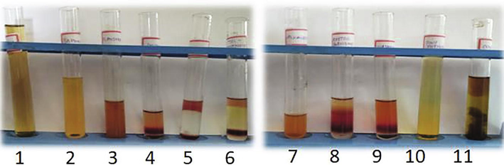
Methanolic extract of Thespesia populnea bark. Phytochemical analysis. 1. Tannin, 2. Saponin, 3. Flavonoids, 4. Steroids, 5. Terpenoids, 6. Triterpenoids, 7. Alkaloids, 8. Anthraquinone, 9. Polyphenol, 10. Glycosides and 11. Coumarins.
3.2 Quantitative analysis of phytochemicals screening
Table 2 shows the quantitative analysis of T. populnea bark aqueous extract which contains high amount of polyphenol (30.1%) and low concentration of flavonoids (2%), and terpenoids (4%). Those compounds were used as a reducing agent to synthesize CuONPs by the reduction process. The total polyphenol constituents were higher when compared to flavonoids and terpenoids. Methanol extracts of T. populnea seeds contains 4.5% of polyphenol and 2.0% alkaloids (Ganga Rao et al., 2011). Medicinal plants have been relatively high amounts of antimicrobial agents. In Nigeria, a variety of herbal plants are used to treat microbial infections (Sofowora, 1986). Plant compounds have been used as many potent drugs worldwide (Iwu et al., 1999).
Photochemical
Concentration (%)
Flavonoids
2.0
Phenols
30.1
Terpenoids
4.0
3.3 Histochemical analysis of bark powder
Histochemical analysis of the T. populnea bark powder corroborates the availability of natural phytocompounds, similar to aqueous and methanol extracts of the park (Table 3 and Fig. 3). John Peter Paul, (2014) reported the histochemical and fluorescence analysis of Turbinaria ornata by light microscopy and UV lamp. Histochemical study used to evaluate the presence of phytochemical constituents in the powder extracts of plant materials (Krishnan et al., 2001). In this present study, histochemical screening of various reagents revealed the presence of terpenoids, flavonoids, polyphenol, tannins, alkaloid, and glycosides in methanol extract. Whereas alkaloids is not found in the water extract.
S. No
Photochemical
Results
1
Terpenoids
(++)
2
Flavonoids
(++)
3
Polyphenol
(++)
4
Tannin
(++)
5
Alkaloids
(+)
6
Glycoside
(++)
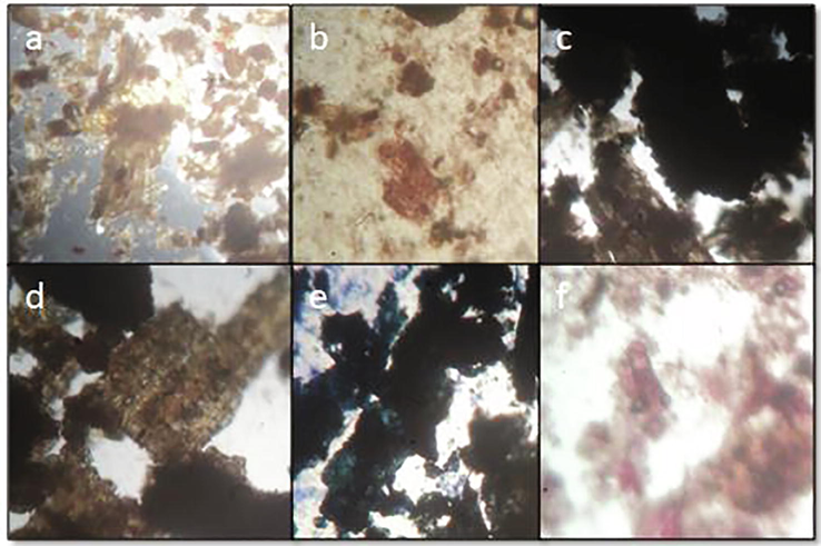
Histochemical analysis of Thespesia populnea bark powder showed tannin, flavonoids, alkaloids, terpenoids, polyphenol, and glycoside. This study indicates that further confirmation of phytochemicals under the light microscope. a) Tannin (black), b) Flavonoids (yellow), c) Alkaloids (yellow), d) Terpenoids (orange), e) Polyphenol (red and blue), f) Glycoside (brown).
3.4 Synthesize of CuO nanoparticles
The aqueous bark extract of Thespesia populnea was used to achieve synthesize of CuO NPs. Throughout the visual appearance, copper acetate and bark extract stirred magnetically, which showed the blue to the green mixture, after 20 min at room temperature. The changes of the blue color of copper ions to green color were apparent for the development of water-soluble mono dispersed CuONPs. We confirmed the efficiency of Thespesia populnea bark extract in the rapid synthesize of copper oxide nanoparticles, due to its numerous organizations of phytochemicals such as phenolics, flavonoids, polyphenols, tannin, anthraquinones, terpenoids and saponin. These phytochemical constituents found in plant extracts function as both reducing and stabilizing agents. Rezaie et al., 2017 and Akintelu et al. (2020), also reported that the biomolecules such as flavonoids, proteins, tannins, phenols and terpenoids are good reducing and stabilizing agents for CuONPs synthesis (Fig. 4).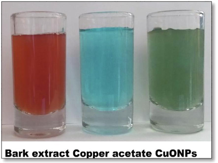
Color changes indicated the synthesize of copper oxide nanoparticles from Thespesia populnea bark extract.
3.5 Characterization of CuO nanoparticles
3.5.1 UV–Vis spectroscopic analysis
UV–Visible spectroscopy is a very useful technique for analyzing the metal nanoparticles because the peak positions and shapes are sensitive to particle size. The surface plasmon resonance peak is the signature of the formation of nanoparticles. According to Akintelu et al., (2020) this wave length is the surface plasmon resonance range for CuONPs. The UV–visible absorption spectrum of copper oxide nanoparticle was scanned from 300 nm to 700 nm and showed the characteristics surface Plasmon resonance (SPR) with absorbance at approximately 390 nm which can be ascribed to the formation of copper oxide nanoparticle (Samuel et al., 2021). Similarly, the present study UV–Vis spectra recorded from the reaction medium after 5 h. The response mixture of copper acetate solution with Thespesia populnea bark extract within the UV–Vis spectra, the peak became found at 400 nm, that confirmed the presence of CuONPs which could be synthesized with the aid of Thespesia populnea bark extract (Fig. 5).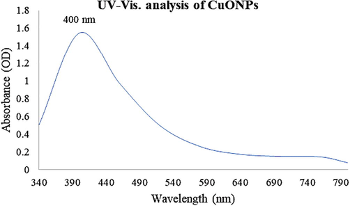
UV–Vis. analysis of CuONPs using Thespesia populnea bark aqueous extract.
3.5.2 Scanning electron microscopy
Scanning electron microscope evaluation had been used to know the size and morphology of the CuO nanoparticles, which showed the high-density poly-dispersed spherical form of copper oxide nanoparticles with variable sizes that ranged from 61 to 69 nm.
SEM examination with an average size of 38.5–48.5 nm was used to validate the production of Cu NPs. The microscope, as shown in, revealed all of the conceivable spherical and irregular forms of Cu NPs. However, the nanocrystals formed having a dimension of 40–60 nm with rod-like morphology (Bhattacharya et al., 2019). The average size of CuNPs was determined from the SEM as 65, 41, 52, 64, 55 and 43 nm corresponding to E. camaldulensis, A. indica, M. koenigii, A. marina, R. rubiginosa and D. stramonium, respectively. The SEM images further confirm the formation of a high density of CuNPs produced by various plant leaves extracts. The dissimilarity distribution in size was due to availability of various naturally derived constituents with different redox properties prevailing in plant (Muhammad Asif Asghar and Muhammad Arif Asghar, 2020) (Fig. 6).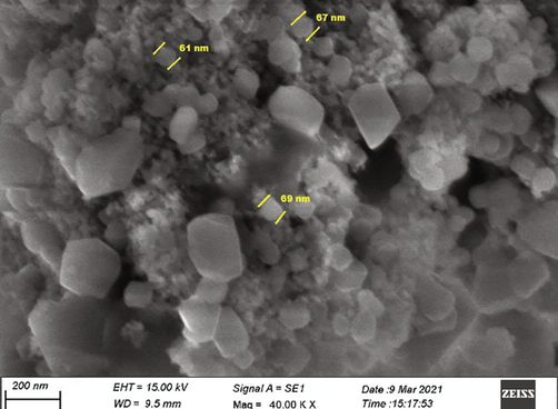
SEM analysis of copper oxide nanoparticles.
3.5.3 Particle size distribution
The particle size distribution study showed the overall size of the nanoparticles ranges from 51 to 145 nm. But, CuONPs were highly distributed between 50 and 80 nm in size. The average size of the CuONPs was found to be 81.23 ± 2.88 nm. In the present study, similarly, the particle size of the nanoparticles was found out as the average diameter of these CuONPs was calculated to be 6.50 ± 1.50 nm by (Joy et al., 2020) (Fig. 7).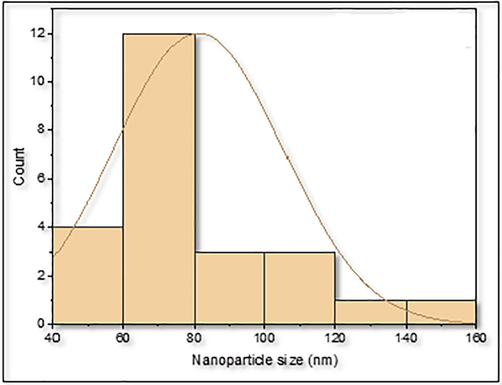
Show the graph Histogram showing the particle size distribution of CuONPs.
3.5.4 Energy-dispersive X-ray spectroscopy (EDX)
EDX was used to discover the composition of the elements from the nanoparticles. EDX analysis of CuONPs confirmed the presence of pure copper (Cu 48.33%) and a very little quantity of chloride (0.95%). Copper was a major constituent element in CuONPs and proved that a higher percentage of copper in CuONPs. The EDX pattern of CuNPs obtained from plant extract, which indicates the presence of Cu and small amount of oxygen. Energy dispersive X-ray spectroscopy (EDX) analysis revealed that pure copper (39.16%) was present in CuNPs (Aher et al., 2019) (Fig. 8).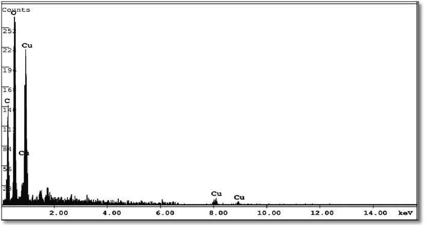
EDX analysis of copper oxide nanoparticles.
3.5.5 Zeta potential
Zeta potential determination is a significant characterization technique of nanoparticles to estimate the surface charge, which can be employed for understanding the physical stability of nanosuspensions. A large positive or negative value of zeta potential of nanoparticles indicate good physical stability of nanosuspensions due to electrostatic repulsion of individual particles.
In the present study, CuONPs showed negatively surface charges with a value of −3.50 mV were stable without aggregate for long period (Sarkar et al., 2011). The stability of the aqueous solution of CuONPs indicated that dispersed nanoparticle capped by negatively charged ions proving CuONPs was stable (Fig. 9).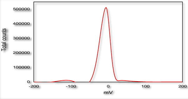
Zeta potential analysis of CuONPs.
3.5.6 Fourier Transform infra-red spectral analysis (FTIR)
FTIR used to examine the potential bioactive compounds, were capped around the nanoparticles. Generally, the phytochemical compounds would act as a capping agent from bark extract by the reduction process. In this study, the peaks showed bioactive compounds which capped around the nanoparticles. The components were identified on the frequency of 3447.28 (Alcohols, Phenols), 2980.63 (Alkanes), 2865.47 (Alkanes), 1638.90 (1° amine), 1453.86 (Alkanes), Aliphatic amines (1053.11, 1032.69), and 693.38 (Alkynes), the presence of flavonoids and phenol adsorbed at the surface of CuONPs. This study revealed that the presence of phytochemical compounds acted as a key to synthesizing nanoparticles by the reduction process and stabilization of Cu ions (Thit et al., 2013) (Fig. 10).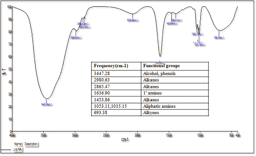
Fourier Transform Infra-Red spectral analysis of CuONPs.
3.6 Antimicrobial activity of CuONPs by disc diffusion method
Synthesize of copper oxide nanoparticles from Thespesia populnea (bark) and copper acetate reaction mixture against skin infection-causing microbes was studied. Evaluation of antimicrobial activity against the skin disease-causing bacteria such as Pseudomonas aeruginosa, Staphylococcus aureus, and Streptococcus pyogenes and the fungal strains of Candida albicans and Trichophyton rubrum (Table 4). *Standard antibiotics, chloramphenicol 10 μg/ml for bacteria; Fluconazole 10 μg/ml for fungal pathogens.
Microbial Strains
Inhibition zone (mm)
Copper acetate
Bark extract
CuONPs
Standard Antibiotic*
Pseudomonas aeruginosa
6.00
6.50
8.50
9.00
Staphylococcus aureus
3.30
4.10
6.70
8.90
Streptococcus pyogenes
5.00
6.50
8.00
8.50
Candida albicans
5.00
6.00
9.00
9.00
Trichophyton rubrum
7.00
7.50
9.50
10.00
In this study, the antimicrobial activity of copper acetate, bark extract, and CuONPs were evaluated. High active properties of CuONPs were reported to compare with copper acetate and bark extract in all microbial strains. Overall, CuONPs had a high zone of inhibition on Pseudomonas aeruginosa bacterial strains, which was achieved when compared with other bacterial strains. The elevation of inhibition rate of CuONPs was equivalently effective with the same concentration of standard antibiotics. From the data obtained from this study, the effect of CuONPs exhibit 1.5 fold times than the copper acetate and bark extracts. The expression of the effect may increases in the occurrence of smaller particle size of nanoparticles. The high zone of inhibition on the Trichophyton rubrum fungal strain gave when compared to Candida albicans. Skin and subcutaneous tissues could be subjected to infection of bacteria and fungi cause dermatitis, acne, and so on. These infections are present all over the world treated by a wide range of antibiotics. Multi-drug resistant strains could be non-sensitive to antibiotics. The discovery of new fighting strategies to control the microbes is essential (Sieradzki et al., 1999). Existing natural products are now grown because of a valuable source of drugs, and novel molecules and chemicals are found in natural products (Fig. 11).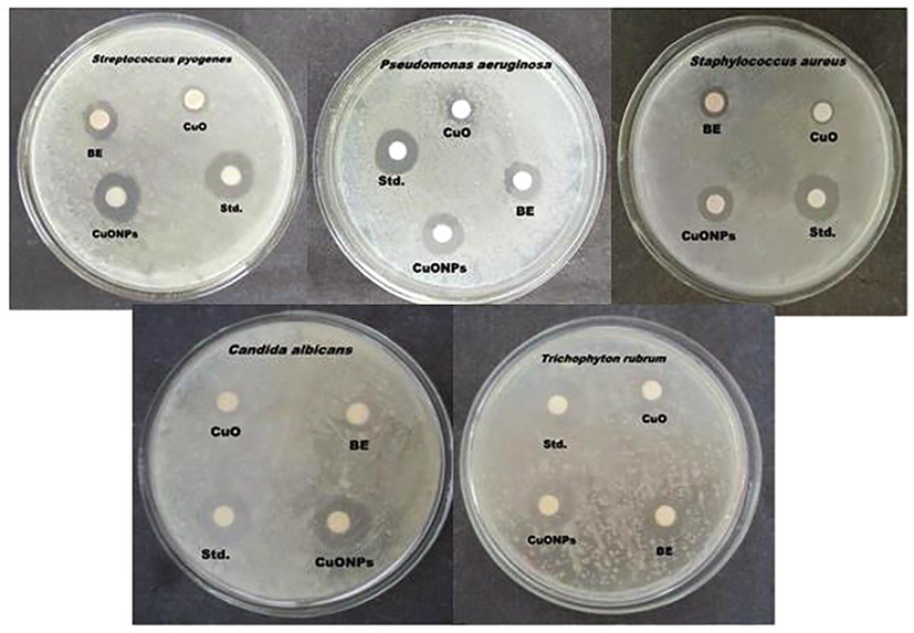
Evaluation of the antimicrobial activity of copper acetate, Bark extract, and CuONPs against the skin causing microbes.
4 Conclusion
The green synthesized CuONPs from Thespesia populnea bark and the copper acetate reaction mixture was treated against skin infection-causing microbes. Various qualitative and quantitative analyses of bark extract and histochemical tests of bark powder revealed the presence of phytochemicals in the extracted plant bark. UV–Vis spectra showed the presence of copper oxide nanoparticles at 400 nm. The particle size distribution showed the average size of the nanoparticles about 81.23 ± 2.88 nm. The zeta potential value of −3.50 mV indicated the negative charge around the nanoparticles proved the CuONPs were stable. SEM analysis showed the higher density poly-dispersed spherical CuONPs at various size ranges from 61 to 69 nm. The disc diffusion method resulted that the green synthesized CuONPs effective against various skin disease-causing bacteria and fungi. The retrieved results from this study showed that the CuONPs are very stable, and have high antimicrobial activity. Further study, such as copper nanoparticle-based ointment to treat skin infection and Copper oxide nanoparticles should check with MDR skin infection causing pathogen and wound healing process.
Conflict of interest
All the authors report no any conflicts of interest.
Acknowledgements
Princess Nourah bint Abdulrahman University Researchers, Supporting Project number (PNURSP2022R82), Princess Nourah bint Abdulrahman University, Riyadh, Saudi Arabia.
References
- Green synthesis of copper nanoparticles using Syzygium cumin, leaf extract, characterization and antimicrobial activity. Chem. Sci. Trans.. 2019;8(1):1-6.
- [Google Scholar]
- Ali, I., Wani, W.A., Saleem, K. Cancer state of affairs in India with future views most cancers therapy. 2011, 8:56-70.
- Green synthesis of copper oxide nanoparticles for biomedical application and environmental remediation. Heliyon. 2020;6(7):e04508.
- [Google Scholar]
- Production of biogenic silver nanoparticles using Boswellia ovalifoliolata stem bark. Digest Mag. Nanomater. Biostruct.. 2010;5(2):369-372.
- [Google Scholar]
- Phytochemical screening and in vitro bioactivity of Cnidoscolus aconitifolius (Euphorbiaceae) J. Med. Plants Res.. 2007;1(3):63-65.
- [Google Scholar]
- Disinfection of drinking water via algae mediated green synthesized copper oxide nanoparticles and its toxicity evaluation. J. Environ. Chem. Eng.. 2019;7(1)
- [Google Scholar]
- Flavonoids and condensed tannins from leaves of Hawaiian Vaccinium vaticulatum and V. calycinium. Pacific Sci.. 1994;48:458-463.
- [Google Scholar]
- Pharmacognostic standardisation and physicochemical evaluations of Amaranthus spinosus Linn. leaves: a review. Inventi Rapid: Ethnopharmacology. 2010;1(2)
- [Google Scholar]
- Phytochemical substances of some Nigerian medicinal plants. African Mag. Biotechnol... 2005;4(7):685-688.
- [Google Scholar]
- A Textual Content-e-Book of Pharmacognosy. New Delhi: Mac Milan agency; 1956. p. :191.
- Studies on phytochemical constituents, quantification of total phenolic, alkaloid content and in-vitro anti-oxidant activity of Thespesia populnea seeds. Free Radicals Antioxid.. 2011;1(4):56-61.
- [Google Scholar]
- Gersbach, S. P., Wyllie, V. G., Sarafis, V. Histochemical method for Localization of the website online of Monoterpene Phenol Accumulation in Plant Secretory systems, 2001.
- Green synthesis of copper oxide nanoparticles using Pucinia granatum peels extract: effect on green peach Aphid. Environ. Nanotechnol. Monit. Manage.. 2016;6:95-98.
- [Google Scholar]
- Harborne J. B. Phytochemical Strategies; A Guide to Fashionable Techniques of Plant Evaluation. 2d version, London New York, 1973.
- New antimicrobials of plant origin. In: Perspective on New Crops and New Uses. Alexandria, VA: ASHS press; 1999. p. :457-462.
- [Google Scholar]
- Histochemistry and fluorescence evaluation of Turbinaria Ornata (Turner) J.Ag. An important Brown Seaweed (Phaeophyceae) Indian J. Plant Sci.. 2014;3(1):40-44.
- [Google Scholar]
- Joy, S., Nilanjan, C., Arindam, C., Avisek, B., Disha, D., Krishnendu, A. Green Synthesized Copper Oxide Nanoparticles Ameliorate Defence and Antioxidant Enzymes in Lens culinaris. Nanomaterials. 2020, 10, 312
- Structural, optical and magnetic investigation of Gd implanted CeO2 nanocrystals. Nuclear Instr. Methods Phys. Res. Sect. B. 2017;409(409):147-152.
- [Google Scholar]
- Investigation of structural and photoluminescence properties of gas and metal ions doped zinc oxide single crystals. J. Alloy. Compd.. 2014;616:614-617.
- [Google Scholar]
- Histochemical localization of storage components in Caryopsis of rice (Oryza sativa L.) Curr. Sci.. 2001;80:567-571.
- [Google Scholar]
- The antidiabetic capacity of Lantana aculeata root extract in alloxan-prompted diabetic rats. Int J Phytomed.. 2010;2:299-303.
- [Google Scholar]
- Green synthesized and characterized copper nanoparticles using various new plants extracts aggravate microbial cell membrane damage after interaction with lipopolysaccharide. Int. J. Biol. Macromol.. 2020;160:1168-1176.
- [Google Scholar]
- Microstructural, electrical and magnetic properties of erbium doped zinc oxide single crystals. Electron. Mater. Lett.. 2015;11(6):998-1002.
- [Google Scholar]
- Phytochemical analysis in the root and leaf of Thespesia populnea (Linn) Soland ex correa. J. Pharmacognosy Phytochem.. 2018;7(1):414-417.
- [Google Scholar]
- NCCLS. Country-wide Committee for scientific Laboratory requirements. performance requirements for antimicrobial disc susceptibility checks. 1993, PA: NCCLS courses 25.
- An evaluation of recent research on tomato nutrients, breeding, and post-harvest technology concerning fruit great. Eur. J. Plant Technol. Biotechnol.. 2007;1(1):1-21.
- [Google Scholar]
- Phytochemical profiling, polyphenol composition, and antioxidant activity of the leaf extract from the medicinal halophyte Thespesia populnea reveal a potential source of bioactive compounds and nutraceuticals. J. Food Biochem.. 2019;43(2):e12731
- [Google Scholar]
- Photo and biocatalytic activities along with UV protection properties on polyester fabric through green in-situ synthesis of cauliflower-like CuO nanoparticles. J. Photochem. Photobiol., B. 2017;176:100-111.
- [Google Scholar]
- Biological synthesis and characterization of copper oxide nanoparticles using aqueous Psidium guajava leave extract and study of antibacterial activity of the copper oxide nanoparticles on Escherichia coli and Staphylococcus aureus. World J. Adv. Res. Rev.. 2021;09(01):114-120.
- [Google Scholar]
- A review on plants extract mediated synthesis of silver nanoparticles for antimicrobial applications: a green expertise. J. Adv. Res.. 2016;7:17-28.
- [Google Scholar]
- Anti-psoriatic and phytochemical evaluation of Thespesia populnea bark extracts. Int. J. Pharm. Pharm. Sci.. 2009;1:176-185.
- [Google Scholar]
- The development of vancomycin resistance in patient with methicillin resistant Staphylococcus aureus infection. N. Engl. J. Med.. 1999;340(7):517-523.
- [Google Scholar]
- A review on phytochemical and pharmacological houses of Holy basil (Ocimum sanctum L.) Ind. Crops Prod.. 2018;118:367-382.
- [Google Scholar]
- Medicinal Plants and Conventional Medicinal Drugs in Africa. Ibadan, Nigeria: Spectrum Books Ltd; 1993. p. :289.
- Toxicity of CuO nanoparticles and Cu ions to tight epithelial cells from Xenopus, cell cycle progression and cell death. Toxicol. In Vitro. 2013;27(5):1596-1601.
- [Google Scholar]
- Trease, G. E., Evans, W.C. Phenols and phenolic glycosides. In: Textbook of Pharmacognosy. Balliese, Tindall and Co Publishers, London pp. 1989, 12, 343-383.
- Pharmacological actions of Thespesia populnea relevant to Alzheimer's disease. Phytomedicine. 2006;13(9–10):677-687.
- [Google Scholar]
- Anthropomorphism in god concepts: the role of narrative. In: Origins of Religion, Cognition and Culture. Routledge; 2014. p. :410-428.
- [Google Scholar]
- Inorganic nanoparticles as carriers for efficient cellular delivery. Chem. Eng. Sci.. 2006;61:1027-1040.
- [Google Scholar]


