

Translate this page into:
Chemical composition, antioxidant, anti-inflammatory and cytotoxic effects of Chondrus crispus species of red algae collected from the Red Sea along the shores of Jeddah city
-
Received: ,
Accepted: ,
This article was originally published by Elsevier and was migrated to Scientific Scholar after the change of Publisher.
Peer review under responsibility of King Saud University.
Abstract
The Red Sea coast is rich in different types of seaweeds, which are suggested to possess medicinal properties. In this study, methanolic extract of Chondrus crispus (MEC), a species of red algae collected from the southeast coast of Jeddah, Saudi Arabia was prepared and analysed for its chemical composition, antioxidant, anti-inflammatory and cytotoxic properties. HPLC analysis revealed that flavonoids were found to be predominant constituents of MEC followed by phenols and tannins. Among phenols, catechin, gallic and p-coumaric acids were found to be major components of MEC. The MEC at 200 µg/mL concentration significantly decreased the levels of free radicals including 2,2-diphenyl-1-picryl-hydrazyl-hydrate (DPPH) and 2,2′-azino-bis (3-ethylbenzothiazoline-6-sulfonic acid (ABTS), and displayed significant total antioxidant capacity (TAC). The MEC also significantly inhibited the denaturation of bovine serum albumin (BSA), indicating its anti-inflammatory potential. Furthermore, hepatocellular carcinoma (HepG2) and adenocarcinoma human alveolar (A549) cells treated with MEC exhibited 81.9% and 71.8% cytotoxic cell death respectively as compared to 69% cell death found in cells treated with reference drug sorafinib. Collectively, the data revealed that flavonoids, phenols and tannins are the major chemical constituents of chondrus crispus species of red algae. Further, the methanolic extract of this species was found to exhibit significant antioxidant, anti-inflammatory and cytotoxic properties in several human cancer cell lines. Thus, red algae commonly found along the shore of Red Sea can be utilized for its medicinal value.
Keywords
Seaweeds
Phenolic and flavonoid
Antioxidant
Anti-inflammatory
Anticancer

- DPPH
-
2,2-diphenyl-1-picryl-hydrazyl-hydrate (DPPH)
- ABTS
-
2,2′-azino-bis (3-ethylbenzothiazoline-6-sulfonic acid
- HPLC
-
high performance liquid chromatography
- HepG2
-
hepatocellular carcinoma cells
- MCF7
-
human breast cancer cells
- CaCo2
-
colon cancer cells
- A549
-
human alveolar adenocarcinoma cells
- DPPH
-
2,2-diphenyl-1-picryl-hydrazyl-hydrate
- MEC
-
methanolic extract of crispus
- BSA
-
bovine serum albumin
- TAC
-
total antioxidant capacity
Abbreviations
1 Introduction
The coastal region of Red Sea along the Jeddah city in KSA is a rich source of diverse types of seaweeds. A collective group of nonflowering plants, acts as a crew of photosynthetic, nonflowering plant-like organic entities called macroalgae are abundant in the ocean. Nearly 500 species of algae have been recorded in the Red Sea (Garcia-Vaquero et al., 2017). Seaweeds are utilized as source of food by humans in various civilisations (Bezerra and Marinho-Soriano, 2010). Seaweeds are also major source of secondary compounds including polysaccharides, proteins and phenols and used as animal feed, prescription drugs and fertilizers (Aziz et al., 2020; Kazłowski et al., 2012). The marine nonflowering growth at the coast of Saudi Arabia warranted comprehensive and concentrated investigation (Garcia-Vaquero et al., 2017). The Red Sea acquired its algal population from the nearby African and Arabian coasts (mainly the Indian Ocean), which has been occupied in particular by sufficiently sized, tropical Indo-Pacific marine flora. Different species occupy exclusive habitats that are based on the depth, degree of exposure to wave action, shore beds and different environmental elements (Sales and Ballesteros, 2009). Benthic algae have been regarded an indicator of marine biodiversity in coastal areas (Kazłowski et al., 2012) because they adopt the colour and content of the marine environment in optically clear, shallow coastal and reef waters (Phinn et al., 2005; Kutser and Jupp, 2006). Lively water flow is important for several species, but dense algal growth occurs on unconsolidated substrates, thus affecting the stability of the environment. Seaweeds or macroalgae belong to three classes: purple (Rhodophyta), brown (Phaeophyta) and inexperienced (Chlorophyta) (McHugh, 2003). Red algae are regarded the principal group and blankets some species that develop deeper in the Red Sea than anywhere else because of water transparency and increasing energy-conserving patterns. Red algae are rich in bioactive compounds, such as proteins, peptides, amino acids, lipids, fibres, colors, polyphenols and polysaccharides (Kumar et al., 2008), and have potential health effects as edible crimson algae against tumour cells; this type of algae also contains beta-carotene and lutein, which can protect against mutagenicity and inflammation (Pulz and Gross, 2004). Due to abundancy of red algae along the shores of red sea and the reported economical and medicinal values, the present study investigated its chemical nature and its effects on major biomarkers of oxidative stress, inflammation and cell survival utilizing three human cancer cell lines.
2 Materials and methods
2.1 Collection of seaweed
Reddish brown seaweed or macroalgae were collected from the Red Sea in the Abhur area of Jeddah City, KSA (Fig. 1) and algal specimens were handpicked and washed completely to eliminate all impurities, such as sand particles and epiphytes.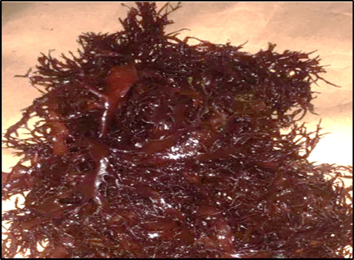
Reddish Brown Macroalgae.
2.2 Preparation of the methanolic extract of condrus crispus (MEC)
The dried algal powder (10 g) was steeped in 100 cc of 80% methanol and was incubated at room temperature under shaking condition for 24 h. The extract was purified, and the extraction was renewed more than once. The obtained extract was analysed for its chemical composition antioxidant, cytotoxic and anti-inflammatory properties.
2.3 Measurement of total phenol, tannin and flavonoid content
The phenolic and tannin compounds in the MEC were measured using the Folin–Ciocalteu reagent (Singleton et al., 1999), whereas, total flavonoid content was estimated using aluminium chloride method with quercetin as a standard (Tambe and Bhambar, 2014).
2.4 Determination of phenolic components of MEC
The phenolic components in the MEC were identified by high-performance liquid chromatography (HPLC) following the method described by Gupta et al. (2012).
2.5 Measurement of free radical scavenging activity of MEC
The free radical–scavenging activity of the methanolic extract was determined by measuring the ability of MEC to inhibit free radicals including ABTS and DPPH as described by Ye et al. (2009).
2.6 Estimation of total antioxidant capacity of MEC
The total antioxidant capacity of MEC was estimated by the method detailed by Prieto et al. (1999).
2.7 Determining anti-inflammatory activity of MEC
The anti-inflammatory property of MEC was assessed by its ability to inhibit BSA denaturation following the method described by Rahman et al. (2015). Aspirin was used as a referral drug (standard) to compare with the effect of MEC on BSA denaturation.
2.8 Assessing cytotoxic effects of MEC
The cytotoxic effect of MEC against HepG2, MCF7, Caco-2 and A549 cancer cells was assessed by MTT assay (Mosmann, 1983). Briefly, cells growing in 96 well plate in DMEM culture media were treated with 200 μg/ml concentration of MEC for 24 h followed by incubation with MTT (3-(4,5-dimethylthiazol-2-yl)-2,5-diphenyltetrazolium bromide) at 5 mg/ml concentration for 4 h at 370C in CO2 incubator. Cells were washed with PBS and further incubated in DMSO at room temperature for 1 h and the absorbance of developed purple colour was measured at 540 nm. The % cell viability was calculated from absorbance of MEC treated cells against absorbance of control cells.
2.9 Statistical analysis
Statistical analysis was performed by CoStat Version 6.45 Statistics package. The differences between different groups were analysed by two-way ANOVA followed by Duncan’s multiple range test. The data were expressed as mean ± SE of three replicates. A P < 0.05 was considered to be significant.
3 Results
3.1 Chemical composition of MEC
Collected seaweed was identified to be Chondrus Crispus species of red algae based on colour, morphological structure, ingredients and collection location. The Chemical composition of MEC is provided in Table 1. HPLC analysis of MEC revealed that flavonoids were predominant constituent than phenols and tannins. On the other hand, phenols and tannins were in comparable amounts in MEC (Table 1). aSignificant difference compared to phenols and tannins. No significant difference was observed between phenols and tannins. CoStat Version 6.45 Statistics package and two-way ANOVA followed by Duncan’s multiple range test was used for statistical analysis. Data were expressed as mean ± SD of three independent experiments, at *P < 0.05.
Phenols
Flavonoids
Tannins
Concentration µg/gm
12.38 ± 2.31
202.66 ± 3.05a*
13.64 ± 0.71
LSD
5.29
3.2 Phenolic constituents of MECCompound
The different phenolic constituents present in MEC was determined by HPLC. The data are presented in Table 2. Catechin at 2.335 µg/mL concentration was the dominant constituent among different phenols followed by gallic acid (1.09 µg/mL) and p-coumaric acid (0.581 µg/mL). However, other compounds such as p-hydroxybenzoic acid, gentisic acid, protocatechuic acid and cinnamic acid were detected in smaller concentrations at different retention times (Table 2).
Phenolic constituent
Retention time (min)
Concentration (µg/ml)
Gallic acid
4.413
1.092
Protocatechuic acid
7.593
0.186
p-hydroxybenzoic acid
11.00
0.255
Gentisic acid
11.839
0.199
Cateachin
12.604
2.335
p-coumaric acid
30.865
0.581
Cinnamic acid
41.33
0.050
3.3 Free radical scavenging activity
Free radical scavenging activity of MEC and Trolox & BHT standards are presented in Table 3. The results suggest the strong free radical scavenging activity pf ethanolic extract of Chondrus sp. In inhibiting ABTS and DPPH free radicals. The percentage of inhibition increased gradually with the increasing concentrations of extract. The highest concentration (200 μg) induced 120.29% of inhibition of ABTS compared to 100.25% inhibition by the Trolox standard. On the contrary, the same concentration exerted 84.17% inhibition of DPPH compared with 91.51% in the BHT standard. DPPH: 2,2-diphenyl-1-picryl-hydrazyl-hydrate; ABTS: 2,2′-azino-bis (3-ethylbenzothiazoline-6 sulfonic acid. Trolox is a ABTS free radical scavenging standard and BHT is a DPPH free radical scavenging standard. aSignificantly different compared to 50 μg of MEC, Trolox or BHT, bSignificantly different compared to 100 μg of MEC, Trolox or BHT, cSignificantly different compared to 150 μg of MEC, Trolox or BHT. Data were expressed as mean ± SD of three independent experiments, *p < 0.05. CoStat Version 6.45 Statistics package and two-way ANOVA followed by Duncan’s multiple range test was used for statistical analysis
Free radical
MEC (μg)
Trolox (μg)
50
100
150
200
50
100
150
200
LSD
ABTS (% inhibition)
82.0 ± 2.6
98.0 ± 2.0a*
108.9 ± 1.9ab*
120.3 ± 0.3abc*
41.9 ± 2.1
81.6 ± 0.8a*
94.07 ± 3.0b*
100.2 ± 0.4c*
2.63
MEC (μg)
BHT (μg)
50
100
150
200
50
100
150
200
DPPH (%inhibition)
25.0 ± 2.6
37.8 ± 1.0a*
47.3 ± 2.6ab*
84.2 ± 3.7abc*
29.3 ± 0.6
45.8 ± 4.6a*
84.2 ± 4.4ab*
91.5 ± 1.5abc*
4.7
3.4 Total antioxidant capacity
The data on total antioxidant capacity of MEC and vitamin C standard are shown in Table 4. Consistent with its free radical scavenging activity, the MEC displayed a significant total antioxidant capacity. Moreover, a dose dependent response was observed as the total antioxidant capacity increased with the increasing concentration of MEC. Highest total antioxidant capacity was observed with the 400 µg concentration of extract which was near equal to the total antioxidant capacity of standard at equivalent concentration (Table 4). MEC, methanolic extract of Chondrus Crispus, TAC, total antioxidant capacity, aSignificantly different compared to 100 μg MEC or standard, bSignificantly different compared to 200 μg MEC or standard, cSignificantly different compared to 300 μg MEC or standard. Data were expressed as mean ± SD of three independent experiments. *P < 0.05. CoStat Version 6.45 Statistics package and two-way ANOVA followed by Duncan’s multiple range test was used for statistical analysis
MEC Concentration(µg)
100
200
300
400
TAC
88.48 ± 1.81
94.77 ± 1.7a*
136.88 ± 2.99b*
235.81 ± 3.20*c
Standard (vitamin C)
TAC
127.97 ± 8.93
147.37 ± 12.15a
230.14 ± 6.20b*
243.46 ± 3.56c*
LSD
11.06
3.5 Anti-inflammatory capacity
The data on anti-inflammatory effects of MEC and standard are provided in Table 5. The anti-inflammatory effect of MEC was assessed by its ability to prevent denaturation of BSA. The SEC significantly inhibited the denaturation of BSA in a dose dependent manner as protein concentration of non-denatured BSA increased with increasing concentration of MEC indicating that less amount of BSA is denatured and more amount of non-denatured BSA remained in the reaction mixture resulting in an increase in protein concentration. At 200 μg concentration of MEC, the amount of non-denatured BSA was 80.78 μg/g of total BSA, which was comparable to 80.48 μg of non-denatured BSA per gram of total BSA found with aspirin (standard). Data were expressed as mean ± SD of three replicates. a,b letters expressed significant difference between concentrations at P<> 0.01. CoStat Version 6.45 Statistics package and two-way ANOVA followed by Duncan’s multiple range test was used for statistical analysis
MEC Concentration (μg) (µg /gm)
100
150
200
BSA (μg/gm)
81.74 ± 7.22 a
85.50 ± 7.23 a
88.48 ± 6.22a
Aspirin concentration (μg)
BSA (μg/g)
33.44 ± 2.12b
56.20 ± 3.52b
80.79 ± 6.85a
Lsd
8.77
3.6 Cytotoxic effects of MEC
The cytotoxic effects of MEC on different cancer cell lines are presented in Table 6. As the data suggest significant cytotoxic effects were noted in all the studied cancer cell lines treated with extract. The MEC at different concentrations induced gradual growth inhibition in HepG2, MCF7, Caco-2 and A549cells. At 100 µg concentration 81.9% and 71.8% growth inhibitions were noted in HepG2 and A549 cells respectively compared 69.1% and 70.6% growth inhibition by sorafinib drug in these two cell lines. On the contrary, at the same concentration (100 µg) MEC induced relatively lower growth inhibition in Caco-2 (64.5%) and MCF7 (47.8%) cells compared to reference drug. MEC: Methanolic extract of Chondrus Crispus, HepG2: hepatic tumour cell line; MCF7: Brest cancer cells; Caco-2: colorectal adenocarcinoma and A549: adenocarcinoma human alveolar cells. Data were expressed as mean ± SD of three independent experiments. CoStat Version 6.45 Statistics package and two-way ANOVA followed by Duncan’s multiple range test was used for statistical analysis
Concentration of MEC/Sorafinib
IC50 µg/ml
100 µg/ml
10 µg/ml
1.0 µg/ml
0.1 µg/ml
0.01 µg/ml
% inhibition of HepG2 cells
1.32 ± 0.07
81.88 ± 5.23
66.02 ± 3.56
44.36 ± 3.69
29.13 ± 1.03
18.72 ± 1.00
MEC
2.23 ± 0.07
69.1 ± 2.67
59.2 ± 1.84
44.4 ± 1.52
34.7 ± 1.42
21.5 ± 1.09
Sorafinib
% inhibition of MCF7 cells
179 ± 8.66
47.82 ± 2.46
34.65 ± 2.35
22.07 ± 1.23
7.71 ± 0.52
0.35 ± 0.012
MEC
4.0 ± 0.33
64.6 ± 1.78
60.7 ± 2.41
59.50 ± 2.76
42.27 ± 0.25
26.10 ± 0.72
Sorafinib
% inhibition of CaCo2 cells
8.24 ± 0.31
64.52 ± 5.63
52.79 ± 3.56
35.82 ± 1.58
23.66 ± 2.01
9.50 ± 0.35
MEC
2.88 ± 0.14
65.6 ± 2.78
56.7 ± 2.41
46.0 ± 1.76
31.8 ± 1.25
25.4 ± 0.85
Sorafinib
% inhibition of A549 cells
7.90 ± 0.33
71.83 ± 4.58
50.00 ± 3.66
31.39 ± 2.35
19.02 ± 1.23
10.25 ± 0.63
MEC
2.55 ± 0.55
70.6 ± 1.78
66.7 ± 2.41
49.50 ± 2.76
32.27 ± 0.25
23.10 ± 0.92
Sorafenib
4 Discussion
Seaweeds are a valuable source of bioactive compounds with pharmacological properties and therefore warrant in depth research for their possible utilization in the treatment and prevention of chronic diseases. Seagrass comprises of enormous species of algae, which have been shown to contain biologically active compounds such as polyphenols, flavonoids and tannins (Ismail et al., 2019). In the present study Chondrus crispus species of red algae picked from the red sea coast in Jeddah was investigated for its bioactive compounds, properties and cellular effects. The extract prepared from the collected Chronrus crispus was found to be abundant in flavonoid content and tannins and simple phenolic compounds in lesser amounts. The dominant phenolic compounds detected by HPLC in the MEC were catechin, gallic aicd and p-coumaric acid. However, other compounds such as p-hydroxybenzoic acid, gentisic acid and protocatechuic acid were detected in smaller amount.
Recently it is shown that lectin from the marine red algae, Bryothamnion seaforthii, inhibited oxidative stress in rats with streptozotocin-induced diabetes (Alves et al., 2020). Similarly, 3-Bromo-4,5-Dihydroxybenzaldehyde from Polysiphonia morrowii red algae attenuated hydrogen peroxide-induced oxidative stress in vitro and in vivo (Cho et al., 2019). The red algae, Porphyra yezoensis, inhibited oxalate-induced oxidative stress in renal epithelial cells (Sun et al., 2019). In an in vitro study Eucheuma cottonii red algae extract displayed significant antioxidant activity in mice (Teo et al., 2020). Consistently, with the above experimental data, a significant antioxidant activity was observed in the present study with the methanolic extract prepared from Chondrus crispus species of red algae as it effectively scavenged ABTS and DPPH free radicals in a dose dependent manner. Additionally, the extract showed remarkable total antioxidant capacity. These data demonstrate the antioxidant properties of Chondrus crispus species of red algae and substantiates previous data. Mechanistically, the capability of the flavonoids constituents in inhibiting the active free radicals depends on the type of reaction between the assay reagent and the sample and in the manner of dimerization and polymerisation during the reaction (Zhang et al., 2019; Jelínek et al., 2015) The antioxidant properties of different species of red algae are attributed to presence of flavonoids and polyphenols and the strength of antioxidant ability depended on the concentration of these bioactive compounds (Zhang et al., 2019; Melpha et al., 2014; Jiang et al., 2008; Meepagala et al., 2005). Since Chondrus crispus extract also found to possess flavonoid and phenols, the antioxidant properties demonstrated by the extract in the present study could be ascribed to these bioactive constituents.
Inflammation can be beneficial as it is the auto immune response to harmful stimulations. However, acute inflammation developing into chronic inflammation may result in the development of inflammation mediated disorders (Peng et al., 2018). Oxidative stress mediated inflammation plays a major role in the onset and pathogenesis of number of chronic diseases (Schramm et al., 2012; Rosanna and Salvatore, 2012; Kim et al., 2012; Hulsmans et al., 2012). The beneficial effects of several marine natural products in the prevention of oxidative stress mediated inflammation are well known (Abad et al., 2008). For example, phytoprostanes and phytofurans from Gracilaria longissima red algae have shown to exert anti-inflammatory effects in endothelial cells (Martínez Sánchez et al., 2020). A dietary polysaccharide from Eucheuma cottonii red algae downregulated proinflammatory cytokines and ameliorates osteoarthritis-associated cartilage degradation in obese rats (Sudirman et al., 2019). In an in vivo study, sulfated polysaccharides from the marine algae Gracilaria caudata prevented tissue damage by blocking inflammation (da Silva et al., 2019). Likewise, brown algae prevented oxidative stress induced inflammation in LPS-stimulated cells (Kim and Kim, 2010). Clearly above data highlights the antiinflammatory capacity of different species of marine red algae. In line with above observations, the MEC in the present study demonstrated a remarkable anti-inflammatory effect in preventing the denaturation of BSA. In most cases inflammation is driven by cellular oxidative stress and is the result of body’s immune response. Therefore, these anti-inflammatory effects of MEC were presumably achieved by blocking oxidative stress through free radical scavenging.
In the present study MEC significantly inhibited cell growth in four different human cancer cell lines treated at varying concentrations. Moreover, at higher concentrations the growth inhibitory effects were comparable to those found when cells treated with referral drug. This clearly indicates the antitumor property of MEC. Moreover, inhibition of cell growth in different cancer cell types underscores the versatility of this extract as an antitumor agent. These findings are consistent with the previous studies where similar antitumor effects of different species of algae were reported. For instance, bioactive lipids from green, brown and red algae inhibited the growth of MDA-MB-231 breast cancer cells (Lopes et al., 2020). Heteropolysaccharide produced by the red alga Porphyridium sordidum are shown to exert cytotoxic effects on MCF7 and MDA-MB-231 breast cancer cells (Nikolova et al., 2019). Porphyrans from Pyropia yezoensis Chonsoo2 red algae had no toxicity in normal human liver cells (HL-7702) but showed antitumor effects on Hep3B, HeLa and MDA-MB-231 cancer cells (He et al., 2019). Likewise, cytotoxic and antitumor algal activities of several algal species against a number cancer cell linesbeen documented (Zandi et al., 2010). Several studies have attributed the antitumor effect of algae to their polyphenols and flavonoids content (Aravindan et al., 2015; Gutiérrez-Rodríguez et al., 2018). Accordingly, antitumor effects of MEC against cancer cells possibly mediated by its flavonoid and phenolic contents. Moreover, these antitumorproperties of polyphenols are shown to be due to their ability to downmodulate oxidative stress which plays an important role in carcinogenesis (Pejčić et al., 2019; Liu et al., 2019; Lee et al., 2012).
5 Conclusion
The methanolic extract of Chondrus crispus species of red algae obtained from the Red sea was found to contain several flavonoids, polyphenols and tannins. The algal extract also displayed remarkable antioxidant, anti-inflammatory and antitumor properties. Therefore, Chondrus crispus extract can be further studied for its pharmacological application in the prevention and treatment of chronic diseases particularly human cancers.
6 Compliance with ethical standards
Not applicable
7 Human and animal rights
This manuscript does not contain any experiments involving human or animals.
Funding
This research did not receive any specific grant from funding agencies in the public, commercial, or not-for-profit sectors.
Declaration of Competing Interest
The authors declare that they have no known competing financial interests or personal relationships that could have appeared to influence the work reported in this paper.
References
- Natural marine anti-inflammatory products. Mini Rev. Med. Chem.. 2008;8(8):740-754.
- [Google Scholar]
- Antihyperglycemic and antioxidant activities of a lectin from the marine red algae, Bryothamnion seaforthii, in rats with streptozotocin-induced diabetes. Int. J. Biol. Macromol.. 2020;30(158):773-780.
- [Google Scholar]
- Polyphenols from marine brown algae target radiotherapy-coordinated EMT and stemness-maintenance in residual pancreatic cancer. Stem Cell Res. Ther.. 2015;6(1):182.
- [Google Scholar]
- An overview on red algae bioactive compounds and their pharmaceutical applications. J. Complement Integr. Med. 2020
- [CrossRef] [Google Scholar]
- Cultivation of the red seaweed Gracilaria birdiae (Gracilariales, Rhodophyta) in tropical waters of northeast Brazil. Biomass Bioenergy. 2010;34(12):1813-1817.
- [Google Scholar]
- Protective Effect of 3-Bromo-4,5-Dihydroxybenzaldehyde from Polysiphonia morrowii harvey against hydrogen peroxide-induced oxidative stress in vitro and in vivo. Microbiol. Biotechnol.. 2019;29(8):1193-1203.
- [Google Scholar]
- Sulfated polysaccharides from the marine algae Gracilaria caudata prevent tissue damage caused by ligature-induced periodontitis. Int. J. Biol. Macromol.. 2019;132:1-8.
- [Google Scholar]
- Polysaccharides from macroalgae: recent advances, innovative technologies and challenges in extraction and purification. Food Res. Inter.. 2017;99:1011-1020.
- [Google Scholar]
- HPLC profiles of standard phenolic compounds present in medicinal plants. IJPPR. 2012;4(3):162-167.
- [Google Scholar]
- Tong H.J Antitumor bioactivity of porphyran extracted from Pyropia yezoensis Chonsoo2 on human cancer cell lines. Sci. Food Agric.. 2019;99(15):6722-6730.
- [Google Scholar]
- Mitochondrial reactive oxygen species and risk of atherosclerosis. Curr. Atherosclerosis Rep.. 2012;14(3):264-276.
- [Google Scholar]
- In vitro potential activity of some seaweeds as antioxidants and inhibitors of diabetic enzymes. Food Sci. Technol. 2019
- [Google Scholar]
- Chlorella vulgaris biomass enriched by biosorption of polyphenols. Algal Res.. 2015;10:1-7.
- [Google Scholar]
- Antioxidant activities of extracts and flavonoid compounds from Oxytropis falcate Bunge. Nat. prod. Res.. 2008;22(18):1650-1656.
- [Google Scholar]
- Prevention of Japanese encephalitis virus infections by low-degree-polymerisation sulfated saccharides from Gracilaria sp. and Monostroma nitidum. Food Chem.. 2012;133(3):866-874.
- [Google Scholar]
- Effect of phloroglucinol on oxidative stress and inflammation. Food Chem. Toxicol.. 2010;48(10):2925-2933.
- [Google Scholar]
- Oxidative stress in inflammation-based gastrointestinal tract diseases: challenges and opportunities. J. Gastroenterol. Hepatol.. 2012;27(6):1004-1010.
- [Google Scholar]
- Seaweeds as a source of nutritionally beneficial compounds-a review. J. Food Sci. Technol.. 2008;45(1):1-13.
- [Google Scholar]
- On the possibility of mapping living corals to the species level based on their optical signatures. Estuar. Coast. Shelf Sci.. 2006;69(3–4):607-614.
- [Google Scholar]
- Oxidative stress and metal carcinogenesis. Free Radical Biol. Med.. 2012;53(4):742-757.
- [Google Scholar]
- Exploring the antioxidant effects and periodic regulation of cancer cells by polyphenols produced by the fermentation of grape skin by Lactobacillus plantarum KFY02. Biomolecules.. 2019;9(10):575.
- [Google Scholar]
- Domingues MR valuing bioactive lipids from green, red and brown macroalgae from aquaculture, to foster functionality and biotechnological applications. Molecules. 2020;25(17):E3883.
- [Google Scholar]
- Bioavailable phytoprostanes and phytofurans from Gracilaria longissima have anti-inflammatory effects in endothelial cells. Food Funct.. 2020;11(6):5166-5178.
- [Google Scholar]
- A guide to the seaweed industry FAO Fisheries Technical Paper 441. Rome: Food and Agriculture Organization of the United Nations; 2003.
- Algicidal and antifungal compounds from the roots of Ruta graveolens and synthesis of their analogs. Phytochemistry. 2005;66:2689-2695.
- [Google Scholar]
- Phytochemical evaluation of two brown seaweeds from Muttom and Rasthacaud coasts of Tamil Nadu, India. J. Chem. Pharm. Res.. 2014;6(10):566-569.
- [Google Scholar]
- Rapid colorimetric assay for cellular growth and survival: application to proliferation and cytotoxicity assays. J. Immunol. Methods. 1983;65(1–2):55-63.
- [Google Scholar]
- Characterization and potential antitumor effect of a heteropolysaccharide produced by the red alga Porphyridium sordidum. Eng. Life Sci.. 2019;19(12):978-985.
- [Google Scholar]
- The polyphenols as potential agents in prevention and therapy of prostate diseases. Molecules. 2019;24(21):3982.
- [Google Scholar]
- Screening of cytotoxicity and anti-inflammatory properties of Feijoa Extracts Using Genetically Modified Cell Models Targeting TLR2, TLR4 and NOD2 pathways, and the implication for inflammatory bowel disease. Nutrients. 2018;10(9):1188.
- [CrossRef] [Google Scholar]
- Mapping water quality and substrate cover in optically complex coastal and reef waters: an integrated approach. Mar. Pollut. Bull.. 2005;51(1–4):459-469.
- [Google Scholar]
- Spectrophotometric quantitation of antioxidant capacity through the formation of a phosphomolybdenum complex: specific application to the determination of vitamin E. Anal. Biochem.. 1999;269(2):337-341.
- [Google Scholar]
- Valuable products from biotechnology of microalgae. Appl. Microbiol. Biotechnol.. 2004;65:635-648.
- [Google Scholar]
- In-vitro antiinflammatory and anti-arthritic activity of oryza sativa var. Joha rice (an aromatic indigenous rice of assam) Am. Eurasian J. Agric. Environ. Sci.. 2015;15:115-121.
- [Google Scholar]
- Reactive oxygen species, inflammation, and lung diseases. Curr. Pharm. Des.. 2012;18(26):3889-3900.
- [Google Scholar]
- Shallow Cystoseira (Fucales: Ochrophyta) assemblages thriving in sheltered areas from Menorca (NW Mediterranean): relationships with environmental factors and anthropogenic pressures. Estuar. Coast. Shelf Sci.. 2009;84(4):476-482.
- [Google Scholar]
- Targeting NADPH oxidases in vascular pharmacology. Vasc. Pharmacol.. 2012;56(5–6):216-231.
- [Google Scholar]
- Analysis of total phenols and other oxidation substrates and antioxidants by means of folin-ciocalteu reagent. In: Methods in Enzymology. Vol Vol. 299. Academic press; 1999. p. :152-178.
- [Google Scholar]
- A dietary polysaccharide from Eucheuma cottonii downregulates proinflammatory cytokines and ameliorates osteoarthritis-associated cartilage degradation in obese rats. Food Funct.. 2019;10(9):5697-5706.
- [Google Scholar]
- Repair activity and crystal adhesion inhibition of polysaccharides with different molecular weights from red algae Porphyra yezoensis against oxalate-induced oxidative damage in renal epithelial cells. JM. Food Funct.. 2019;10(7):3851-3867.
- [Google Scholar]
- Estimation of total phenol, tannin, alkaloid and flavonoid in Hibiscus tiliaceus Linn. wood extracts. Res. Rev. J. Pharmacog. Phytochem.. 2014;2(4):41-47.
- [Google Scholar]
- In vitro evaluation of antioxidant and antibacterial activities of Eucheuma cottonii extract and its in vivo evaluation of the wound-healing activity in mice. J. Cosmet. Dermatol. 2020
- [CrossRef] [Google Scholar]
- Antioxidant activities in vitro of ethanol extract from brown seaweed Sargassum pallidum. Eur. Food Res. Technol.. 2009;230(1):101.
- [Google Scholar]
- In vitro antitumor activity of Gracilaria corticata (a red alga) against Jurkat and molt-4 human cancer cell lines. Afr. J. Biotechnol.. 2010;9(40):6787-6790.
- [Google Scholar]
- Effects of different pretreatments on flavonoids and antioxidant activity of Dryopteris erythrosora leave. PLoS One. 2019;14(1):e0200174
- [CrossRef] [Google Scholar]
Appendix A
Supplementary data
Supplementary data to this article can be found online at https://doi.org/10.1016/j.jksus.2020.10.007.
Appendix A
Supplementary data
The following are the Supplementary data to this article:


