

Translate this page into:
Phyto-biologic bimetallic nanoparticles bearing antibacterial activity against human pathogens
⁎Corresponding author at: Department of Biotechnology, Sri Jayachamarajendra College of Engineering, JSS Science and Technology University, JSS Technical Institutional Campus, Mysore 570006, India. npmicro8@yahoo.com (M.N. Nagendra Prasad) mnnagendraprasad@sjce.ac.in (M.N. Nagendra Prasad)
-
Received: ,
Accepted: ,
This article was originally published by Elsevier and was migrated to Scientific Scholar after the change of Publisher.
Peer review under responsibility of King Saud University.
Abstract
Use of nanomaterials as antimicrobial agents is gaining tremendous importance. In the present investigation, bimetallic nanoparticles were synthesized using an aqueous extract of Annona squamosa L. Initial confirmation was attenuated with a change in color of the reaction mixture and further confirmation was achieved with UV–Visible spectrophotometry which displayed absorbance peak between 500 and 700 nm. The possible role of phyto-components present in aqueous extract to mediate and stabilize nanoparticles was studied using Fourier transform infrared (FTIR) analysis which predicted the presence of hydroxyl, carboxyl and amide functional groups. The X-ray diffraction (XRD) analysis revealed the crystalline nature of bimetallic nanoparticles. The morphological characteristics of bimetallic nanoparticles were studied using Transmission electron microscopy (TEM) which depicted the polydispersity of nanoparticles with myriad size and shapes. The antibacterial activity was assessed with well diffusion, minimal inhibitory concentration. The bimetallic nanoparticles were effective against tested human pathogens and Bacillus subtilis (MTCC 121) followed by Staphylococcus aureus (MTCC 7443), Escherichia coli (MTCC 7410) and Salmonella typhi (MTCC 7407). The obtained preliminary investigation is interesting for future studies to elucidate the mechanism of synthesis and the possible mode of action of bimetallic nanoparticles against the test pathogens.
Keywords
Annona squamosa L.
Bimetallic nanoparticles
Antibacterial activity
Human pathogens

1 Introduction
The intersection of nano-science has led to a new era of nano-revolution (Björnmalm et al., 2016). The field of nanotechnology deals with the particles maneuvering at nano-scale which tends to possess unique physicochemical properties which render different applicative properties (Lin et al., 2014). Nanoparticles are defined as particles bearing size less than 100 nm. These nanoparticles are considered to be particles of the century owing to its extraordinary properties compared to its bulk material (Syed et al., 2017a). The use of nanoparticles in different fields are constantly being explored (Kavitha et al., 2013). Some of the predominant applications of nanoparticles are in textile industries, used in biosensing, targeted drug delivery, nano-agroparticles in agriculture, development of dressing materials and their application as potent antimicrobial agents (Baker and Satish, 2012). Use of nanomaterials as antimicrobial agents has envisioned significant progress in recent years (Baker et al., 2016). The paucity of potent antibiotics has led to a new era of drug resistance (Fair and Tor, 2014). Ever since the first antibiotic was introduced, there is a parallel mode of resistance being developed among the microbial pathogens (Davies and Davies, 2010). In recent decades, there has been rapid expansion of drug resistant bacteria which has posed a serious threat to all form of lives (Ventola, 2015). Hence scientific communities are engaged in developing potent antimicrobial agent with multiple modes of actions (Baker and Satish, 2012). The use of nanoparticles as antimicrobial agents can be attributed to its size and large surface area coupled with suitable surface tunable properties (Zhang et al., 2016). The nanoparticles impart different mode of actions on pathogens for instance, binding to the cell wall followed by depolarizing the contents of the cell wall membrane which results in the loss of cellular contents (Syed et al., 2016). Studies also highlight that nanoparticles interact with the vital components of pathogens and influence the metabolic and genetic pathways, which leads to inactivation of enzymes and prevent replication (Baker and Satish, 2012). Scientific studies report different nanoparticles bearing antimicrobial potential for instance silver, gold, zinc, copper, magnesium, platinum etc. (Baker et al., 2013). One of the important aspects of nanotechnology includes the synthesis of nanoparticles (Kavitha et al., 2013). There are different approaches to synthesize nanoparticles which are broadly grouped into top-down and bottom-up processes (Baker et al., 2013). Majority of these methods are bound with one or more limitations which have resulted in a gradual shift towards developing facile and eco-friendly processes to synthesize nanoparticles via biogenic sources such as plants and microorganisms (Kavitha et al., 2013). The plant-mediated synthesis of nanoparticles is simple, cost-effective and one-pot synthesis process wherein the phyto-components present in plants act as reducing agents and synthesize desire class of nanoparticles (Iravani, 2011). Based on these facts and consideration, the present study reports the synthesis of bimetallic nanoparticles from Annona squamosa L. and their evaluation as antibacterial agents against selected pathogens. Studies pertaining to nanoparticles as antimicrobial agents are well documented but scanty reports are available on bimetallic nanoparticles hence in the present investigation attempt has been made to synthesize bimetallic nanoparticles. The selected plant as a subject of interest was carried out based on its therapeutic index and prior study on bimetallic synthesis. To best of our knowledge, scanty exploratory study has been conducted for synthesis of bimetallic nanoparticles from this plant.
2 Material and methods
2.1 Preparation of plant extract
The Annona squamosa plant material was collected from the abundant growing region of Mysore, Karnataka, India. Plant materials were washed to remove the adhering soil particles and chopped into small segments. Further, 20 g of plant segments was ground using pestle and mortar and the mixture was boiled in a 250 ml Erlenmeyer flask with 200 ml water for 30 min. The aqueous extract was filtered and evaluated for the synthesis of nanoparticles (Syed et al., 2017b).
2.2 Synthesis of bimetallic nanoparticles
The aqueous extract was treated with a mixture of 1 mM silver nitrate and 1 mM gold chloroaurate (1:1 v/v). The initial synthesis was monitored with a change in colour of reaction mixture and further confirmation was achieved with UV–Visible spectrophotometry by recording the spectra between 200 and 700 nm using Shimadzu double beam spectrophotometer.
2.3 Characterization of bimetallic nanoparticles
The synthesized nanoparticles were centrifuged at 10,000×g for 10 min using Eppendorf 5810R Refrigerated Centrifuge. The pellet obtained was air dried under vacuum with a strict aseptic condition and later subjected to characterization using hyphenated techniques. Initial the synthesized was confirmed with UV–visible spectroscopy which recorded the spectral range between 200 and 800 nm. The possible role of phyto-components in mediating the synthesis was confirmed with FTIR analysis using JASCO FT-IR 4100 instrument at room temperature with a resolution of 4 cm−1. The crystalline nature of bimetallic nanoparticles was studied using XRD analysis. The bimetallic nanoparticles were centrifuged and the pellet was lyophilized to remove the liquid phase. Later adequate amount of sample was coated onto XRD grid and spectra were recorded between 2 theta angle by Rigaku Miniflex-II Desktop X-ray diffractometer instrument operating operated at 30 kV. The morphological characteristics were studied using transmission electron microscopy using TECHNAI-T12 JEOL JEM-2100 operated at a voltage of 120 kV with Bioten objective lens.
2.4 Bactericidal property of bimetallic nanoparticles
To examine and determine the bactericidal activity, Muller Hinton media was selected to grow the pathogens. Approximately 106 CFU were grown in media seeded with different bimetallic nanoparticles concentration of 0–100 µg/ml and plates were incubated at 37 °C for 24 h. The number of colonies and growth pattern of the individual pathogens were determined. Further, growth pattern of test pathogens, broth dilution assay was determined in presence of different bimetallic nanoparticles concentration as described earlier and incubated at 37 °C for 24 h. The activity was measured as optical density at 600 nm which determined the antibacterial activity of bimetallic nanoparticles. The activity was further confirmed with well diffusion assay according to the protocol described by Baker et al., (2015). Initially, for well diffusion assay, Muller-Hinton agar plates were seeded with 106 CFU (colony forming unit) suspensions of test organism which was uniformly swabbed followed by punching well of 10 mm diameter and 50 µl of nanoparticles sample was added and incubated at 37 °C for 24 h. The zone across the well was measured and recorded to compare with standard gentamicin. The minimal inhibitory concentration was determined by the protocol described by Sarker et al. (2007) with slight modification. Resazurin dye was used as a growth indicator to check the efficacy of nanoparticles against the test organisms. Gentamicin at concentration 1 mg/ml was used as positive control and bacterial growth in the plate was inspected visually as well as ELISA microtitre plate reader. All the antibacterial tests were determined in accordance to the standard protocols, in each assay positive and negative controls were maintained and validated with values of triplicates.
3 Results and discussion
The present study attributes towards synthesizing bimetallic nanoparticles from Annona squamosa L. and their evaluation for bactericidal properties against significant test pathogens. The selected plant is well documented for its significant therapeutic values. Leaves extract of Annona squamosa L. is reported to have anti-diabetic, anticancerous, anti-hyperthyroidism, antifertility, antitumor, antimutagenic, anthelmintic, scavenging, antidiabetic, licicidal, hepatoprotective, antithyroid, antigenotoxic, antiplasmodial, molluscicidal and antimicrobial properties (Vanitha et al., 2011). According to previous studies on this plant reports the synthesize of silver nanoparticles but scanty reports are available on synthesis of bimetallic nanoparticles which is one of the interesting facet in the present investigation. The synthesis and use of bimetallic nanoparticles have opened new avenues in different fields and implementation of its usage as antimicrobial agents can be very useful during the post antimicrobial resistant era. In the present investigation, bimetallic nanoparticles were synthesized by treating with mixture of silver nitrate and gold chloroaurate. The synthesis was rapid which was monitored with change in the color of reaction mixture. The synthesis was initiated within 5 min and completed by 20 min of incubation time. After 20 min no further synthesis was observed which was confirmed with UV–visible spectrometer. In order to achieve maximum synthesis, different parameters were studied wherein intrinsic factors like alkaline pH 8 and temperature at 80 °C influenced the synthesis. The results justifies with earlier report which confirms the influence of intrinsic factors influencing the maximum synthesis of nanoparticles (Qian et al., 2013). The reduction of metal salts to produce nanoparticles was further confirmed with UV–Visible spectrophotometry with maximum absorption peak attenuating between 500 and 700 nm (Fig. 1). The absorption peaks might be due to increase in the surface plasmon resonance of nanoparticles as per the earlier reports (Song et al., 2009). The UV–visible absorbance of bimetallic exhibited maximum absorbance at 541 nm with broad SPR band instead of two distinct SPR bands indicating the formation of bimetallic nanoparticles. During the synthesis of nanoparticles, there was change in color of reaction mixture with brown and finally dark violet which indicated the synthesis of bimetallic nanoparticles. These results obtained were in agreement with the reports of bimetallic alloys (Sheny et al., 2011; Tamuly et al., 2013). The possible role of phyto-components present in aqueous plant extract responsible for mediating and stabilizing the nanoparticles was depicted using FTIR analysis. Perusal of scientific studies report FTIR as one of the ideal tool to predict the functional moieties. In the present investigation, vibrational stretch occurring at 3339 corresponds to NH stretching (Devi and Gayathri, 2010), 1638 corresponds to C⚌C stretching (Urbaniak-Domalaga, 2012) and 663 corresponds to C-OH (Fan and Dai, 2012) as cited in the Fig. 2. Scientific studies on FTIR analysis of plant mediated nanoparticles reports that different functional moieties like hydroxyl, carboxyl and amide are responsible for reduction of metal ions to produce nanoparticles (Elavazhagan and Arunachalam, 2011; Kavitha et al.,2013). The obtained result of FTIR analysis is in accordance with previous findings (Dauthal and Mukhopadhyay, 2016). Interestingly, in plant-mediated synthesis of nanoparticles, the phyto-components also play important role in stabilization of nanoparticles which is very crucial for rendering its applicative properties. These results also coincide with reports of earlier findings (Syed et al., 2017; Awwad et al., 2013). The XRD analysis of bimetallic nanoparticles exhibited brag intensities which corresponds to face centric cube of nanoparticles which displayed the crystalline nature of the nanoparticles (Fig. 2) the obtained results are in agreement with earlier reports. (Baker et al., 2015). The TEM microgram revealed the morphological characteristics of bimetallic nanoparticles with different size and shapes with majority of them ranging between 30 and 50 nm (Fig. 3). The size distribution pattern of nanoparticles was constructed by counting 100 nanoparticles and histogram was constructed. The obtained results are in accordance with previous studies. Interestingly, TEM analysis revealed that nanoparticles were not well separated which indicated the formation of bimetallic association (Elavazhagan and Arunachalam, 2011). Based on the reductive potential it can be predicted that gold nanoparticles might have formed first followed by silver nanoparticles to form bimetallic nanoparticles (Baker et al., 2015). The possible mechanisms for synthesis is yet to be completely elucidated, scientific studies highlights that the phyto-components interacts with metal salts resulting in reduction of metal ions followed by nucleation growth to attenuate particle size and obtain the thermal stability (Keat et al., 2015). Interestingly, the size of the nanoparticles act as boon to predict the applicative properties of nanoparticles. Especially, for antimicrobial properties of nanoparticles, as the size reduces the bactericidal activity increases. It can be referred that as the size decrease, it can easily penetrate into pathogenic cell and interrupt the metabolism of the pathogen by initiating ROS uptake and early phagocytosis (Baker and Satish, 2012). The antibacterial property of bimetallic nanoparticles determined via CFU colony forming units resulted in decrease in the viable cells of test pathogens as the increase in the concentration of nanoparticles. The growth pattern of test pathogens in presence of bimetallic nanoparticles resulted in decrease in the optical density recorded at 600 nm confirming that, as the concentration of nanoparticles were increased, there was gradual decrease in the optical density of the test pathogens (Fig. 4). In addition, well diffusion assay resulted in bactericidal activity against all the test pathogens which was measured as zone of inhibition across the well. In the present investigation, bimetallic nanoparticles were more effective against Bacillus subtilis (MTCC 121) with 14.66 ± 0.57 mm zone of inhibition in comparison with other test pathogens. Moderate activity was expressed against Staphylococcus aureus (MTCC 7443) with 13.66 ± 0.57 mm, Escherichia coli (MTCC 7410) with 11.00 ± 1.00 mm and least activity was obtained against Salmonella typhi (MTCC 7407) with 09.33 ± 1.52 mm as described in Table 1. In order to determine the minimal concentration of bimetallic nanoparticles to suppress the growth of test pathogens, minimal inhibitory concentration was determined based on the colorimetric measurement. The concentration of nanoparticles was serially diluted in liquid media and test pathogens were seeded into the liquid broth. The activity was measured against all the test pathogens varied from 31.25 to 250 µg/ml. During the MIC, both positive and negative control was maintained in order to compare the results obtained. Interestingly, results with minimal inhibitory concentration were in accordance with broth dilution and well diffusion assay. The results obtained with bactericidal property justifies with the previous scientific reports (Banerjee et al., 2011; Baker et al., 2016). The use of resazurin as growth indicator becomes one of the most viable tool in order to probe the antimicrobial activity via MIC. The cell growth is measured based on the reduction of blue non-fluorescent dye to resorufin by enzyme oxidoreductase present in the cell. Further, resorufin is reduced hydroresorufin (Sarker et al., 2007). In addition to these antibacterial assays, growth pattern of test pathogens on solid media was determined by incorporating different concentration of nanoparticles into the media onto which the test bacteria was smeared. The activity was measured as colony forming unit and growth onto the media. The results displayed the significant activity of bimetallic nanoparticles against Bacillus subtilis (MTCC 121). All the antibacterial assay was carried out in triplicates and the results were compared with gentamicin as standard antibiotics. Theobtained results are in accordance with earlier reports on effective treatment of nanoparticles against array of test pathogenic bacteria (Syed et al., 2017b). The obtained antibacterial activity of bimetallic nanoparticles clearly suggests its potential towards exploiting as one of the alternative against drug-resistant pathogens. Perusal of scientific literatures suggests majority of studies carried out using silver, gold, and other nanoparticles against different test pathogens and to best of our knowledge scanty reports are available on bimetallic nanoparticles and their evaluation against selected pathogens. The results of the present investigation are promising enough to report as brief findings. Future investigation is highly desirable in order to reveal the exact mechanism responsible for synthesis of bimetallic nanoparticles and the mode of action of bimetallic nanoparticles against test pathogens with its comparison with array of antibiotics and other nanoparticles.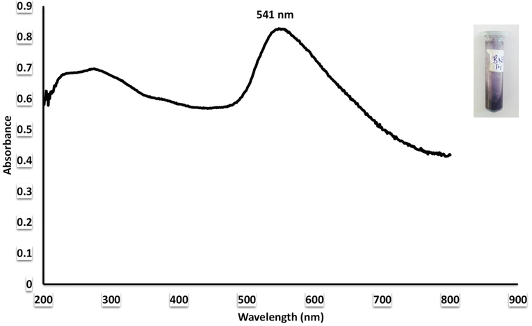
UV–Visible analysis of bimetallic nanoparticles.
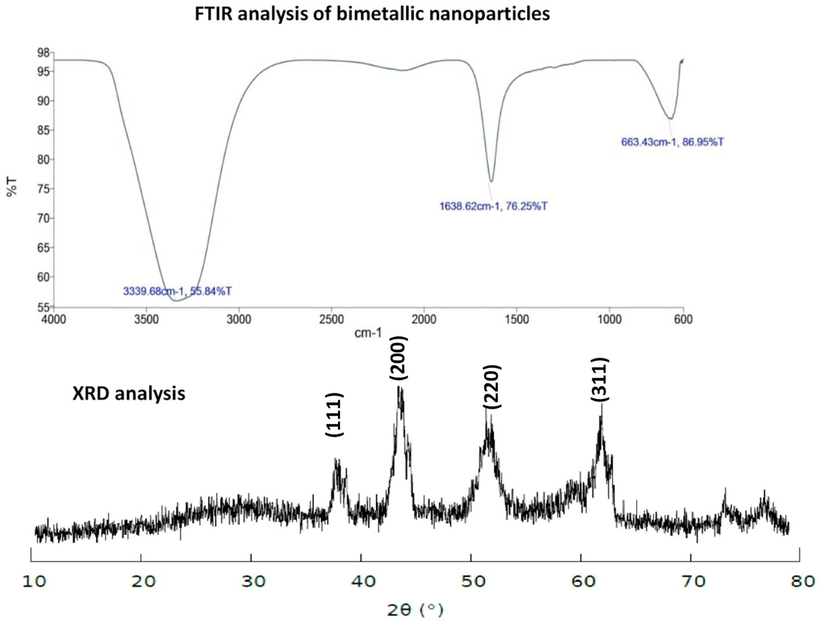
FTIR and XRD analysis of bimetallic nanoparticles.
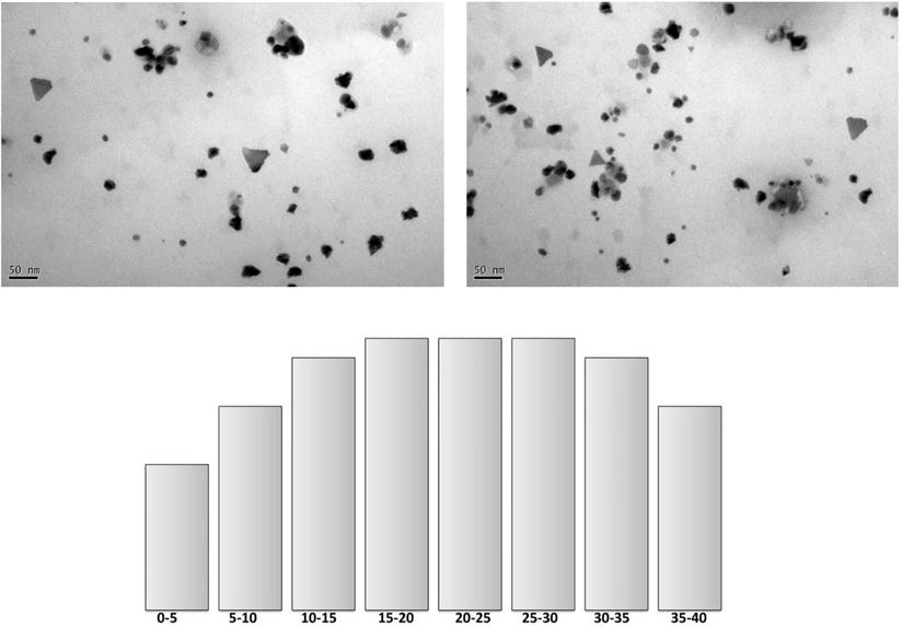
TEM analysis and histogram for size of bimetallic nanoparticles.
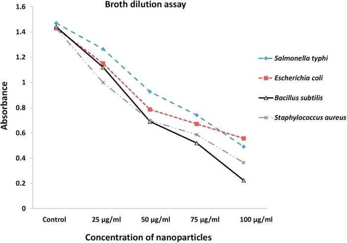
Inhibitory activity of bimetallic nanoparticles via broth dilution assay.
Pathogens
Bimetallic nanoparticles
Gentamicin
Bacillus subtilis (MTCC 121)
14.66 ± 0.57
23.00 ± 1.00
Escherichia coli (MTCC 7410)
11.00 ± 1.00
24.66 ± 0.57
Salmonella typhi (MTCC 7407)
09.33 ± 1.52
14 0.33 ± 0.57
Staphylococcus aureus (MTCC 7443)
13.66 ± 0.57
22.33 ± 1.52
4 Conclusion
The present study attributes towards the synthesis of plant-mediated bimetallic nanoparticles as one of the viable substitutes for combating drug-resistant pathogens. The results obtained in the present investigation are promising enough for future study to elucidate the exact mechanisms responsible for synthesis and antibacterial activity.
Conflict of interest
All authors declare no conflict of interest with regards to publications
Acknowledgments
Authors thank ICMR for financial assistance and Management of SJCE-JSS university technical campus for providing all the necessary facility. Authors are grateful to collaborative institute Siberian Federal University and thankful Ministry of Education and Science of the Russian Federation - Russia for providing funding under 5-100 – Russian academic excellence project.
References
- Green synthesis of silver nanoparticles using carob leaf extract and its antibacterial activity. Int. J. Indus. Chem.. 2013;1:1-6.
- [Google Scholar]
- Plants: emerging as nanofactories towards facile route in synthesis of nanoparticles. Bioimpacts. 2013;3:111-117.
- [Google Scholar]
- Biogenic nanoparticles bearing antibacterial activity and their synergistic effect with broad spectrum antibiotics: emerging strategy to combat drug resistant pathogens, Saudi. Pharm. J.; 2015. 10.1016/j.jsps.2015.06.011
- Synthesis and characterization of silver nanobactericides produced by Aneurinibacillus migulanus 141, a novel endophyte inhabiting Mimosa pudica L. Arab. J. Chem. 2016
- [CrossRef] [Google Scholar]
- Endophytes: toward a vision in synthesis of nanoparticle for future therapeutic agents. Int. J. Bio-Inorg. Hybrid Nanomater.. 2012;2:67-77.
- [Google Scholar]
- Enhanced antibacterial activity of bimetallic gold-silver core-shell nanoparticles at low silver concentration. Nanoscale. 2011;3:5120-5125.
- [Google Scholar]
- Increasing the impact of materials in and beyond bio-nano science. J. Am. Chem. Soc.. 2016;138:13449-13456.
- [Google Scholar]
- Noble metal nanoparticles: plant-mediated synthesis, mechanistic aspects of synthesis, and applications. Ind. Eng. Chem. Res.. 2016;55:9557-9577.
- [Google Scholar]
- Origins and evolution of antibiotic resistance. Microbiol. Mol. Biol. Rev.. 2010;74:417-433.
- [Google Scholar]
- FTIR and FT-Raman spectral analysis of paclitaxel drugs. Int. J. Pharm. Sci. Rev. Res.. 2010;2:106-110.
- [Google Scholar]
- Memecylon edule leaf extract mediated green synthesis of silver and gold nanoparticles. Int. J. Nanomed.. 2011;6:1265-1278.
- [Google Scholar]
- Antibiotics and bacterial resistance in the 21st century. Perspect. Med. Chem.. 2014;6:25-64.
- [Google Scholar]
- Fourier transform infrared spectroscopy for natural fibres. In: Salih Salih Mohammed, ed. Fourier Transform – Materials Analysis. InTech; 2012.
- [CrossRef] [Google Scholar]
- Green synthesis of metal nanoparticles using plants. Green. Chem.. 2011;13:2638-2650.
- [Google Scholar]
- Plants as green source towards synthesis of nanoparticles. Int. Res. J. Biol. Sci.. 2013;2:66-76.
- [Google Scholar]
- Biosynthesis of nanoparticles and silver nanoparticles. Bioresour. Bioprocess.. 2015;2:47.
- [Google Scholar]
- Techniques for physicochemical characterization of nanomaterials. Biotechnol. Adv.. 2014;3:711-726.
- [Google Scholar]
- Biosynthesis of silver nanoparticles by the endophytic fungus Epicoccum nigrum and their activity against pathogenic fungi. Bioprocess Biosyst. Eng.. 2013;36:1613-1619.
- [Google Scholar]
- Microtitre plate-based antibacterial assay incorporating resazurin as an indicator of cell growth, and its application in the in vitro antibacterial screening of phytochemicals. Methods. 2007;42:321-324.
- [Google Scholar]
- Phytosynthesis of Au, Ag and Au–Ag bimetallic nanoparticles using aqueous extract and dried leaf of Anacardium occidentale. Spectrochim. Acta Part A. 2011;79:254-262.
- [Google Scholar]
- Rapid biological synthesis of silver nanoparticles using plant leaf extracts. Bioprocess. Biosyst. Eng.. 2009;32:79-84.
- [Google Scholar]
- Synthesis of silver nanoparticles by endosymbiont Pseudomonas fluorescens CA 417 and their bactericidal activity. Enzyme Microb. Technol.. 2016;95:128-136.
- [Google Scholar]
- Phytogenic synthesis of nanoparticles from Rhizophora mangle and their bactericidal potential with DNA damage activity. Nano-Structures & Nano-Objects.. 2017;10:112-115.
- [Google Scholar]
- Endo-symbiont mediated synthesis of gold nanobactericides and their activity against human pathogenic bacteria. Environ. Toxicol. Pharmacol.. 2017;52:143-149.
- [Google Scholar]
- In situ biosynthesis of Ag, Au and bimetallic nanoparticles using Piper pedicellatum C.DC: green chemistry approach. Colloids Surf. B. 2013;102:627-634.
- [Google Scholar]
- The Use of Spectrometric Technique FTIR-ATR to Examine Polymer Surface. Advance Aspects of Spectroscopy. Rijeka (Croatia): Intech Open Science; 2012.
- Determination of bioactive components of Annona squamosa L. leaf by GC-MS analysis. Int. J. Pharm. Sci. Drug. Res.. 2011;4:309-312.
- [Google Scholar]
- The antibiotic resistance crisis: part 1: causes and threats. Pharm. Ther.. 2015;40:277-283.
- [Google Scholar]
- Silver nanoparticles: synthesis, characterization, properties, applications, and therapeutic approaches. Int. J. Mol. Sci.. 2016;17:1534.
- [Google Scholar]


