Translate this page into:
Spectroscopic studies on Permian plant fossils in the Pedra de Fogo Formation from the Parnaíba Basin, Brazil
⁎Corresponding author at: Institution: Faculdade de Educação Ciências e Letras do Sertão Central, Universidade Estadual do Ceará-Quixadá, Brazil. gilberto.saraiva@uece.br (G.D. Saraiva)
-
Received: ,
Accepted: ,
This article was originally published by Elsevier and was migrated to Scientific Scholar after the change of Publisher.
Peer review under responsibility of King Saud University.
Abstract
The Pedra de Fogo Formation dated from the early Permian (approximately 280 Mya), belongs to the sedimentary Parnaíba Basin, located in the northeastern region of Brazil. It is recognized by their well preserved plant fossil contents and it is notable for having several fossilized trunks in the life position. This study presents physical and/or chemical properties of the fossilized plants through vibrational spectroscopies, SEM/EDS spectroscopy and X-ray diffraction techniques. Specimens from different localities were selected for the purpose of identifying and characterizing compounds related to the fossilized materials. These different techniques allowed obtaining information from the molecular spectra in organic and inorganic substances, which are present in these stated fossils and in the atomic elements, as well as the crystalline phases. Based on the results duly obtained, we were able to identify the presence of silica and confirm that the dominant process of the fossilized specimens investigated has occurred through quartz silicification with the contribution of persistence from amorphous carbon.
Keywords
Vibrational spectroscopies
Fossil plants
Permian Period
1 Introduction
The study of fossils allows to know the evolution (emergence, development and decline) of ancient (mostly extinct) biological species, and to understand the complex interactions verified between organisms and the physical environments in the past. Fossil are also important as an aid for inferring the age of strata, the evolution of climate, and elucidating the position of continents in the past. In brief, fossils are essential tools for assembling the history of Earth.
The processes of preservation of vascular plants in the fossil record, especially those related to wood, have attracted the attention of many scientists since the last century (e.g., Murata, 1940; Buurman, 1972; Mitchell and Tufts, 1973; Leo and Barghoorn, 1976; Stein, 1982; Scurfield and Segnit, 1984), and a series of hypotheses on the mechanisms of silicification that permitted the preservation of cellular structures and on the origin and nature of the silicification agents in these fossils have been raised.
Cellular permineralizations (fossilization through incorporation of minerals at the cellular level) can be considered as the most frequent and scientifically important type of fossil plant preservation (Witke et al., 2004). Through the analysis of permineralization (mainly silicification) of fossil plant tissues, relatively important information is obtained, especially when preservation has maintained the three-dimensional structure of the plant, and there are still cellular tissues that may help in paleoenvironmental and paleoecological interpretations (Tavares, 2011). In this sense, the physico-chemical analysis of fossil plants has brought relevant information about the diagenetic processes involved in the permineralization of plant parts or organs, especially woods. However, despite of a significant number of researches on this topic, particularly in the last years (Dietrich et al., 2001;Witke et al., 2004; Mustoe, 2008; Matysová et al., 2010; Zodrow et al., 2010; Silva et al., 2013; Alencar et al., 2015), there is still much questioning and lack of consensus on the actual conditions involved in the processes of permineralization of fossil plants over geological time (Buurman, 1972; Dietrich et al., 2001, Mustoe, 2008; Alencar et al., 2015).
In this way, the main goal of this study is to infer the possible processes involved in the fossilization of Permian woods from the Parnaíba Sedimentary Basin (Santos and Carvalho, 2004), in Northeast Brazil. Several physic-chemical techniques were employed (sensu Silva et al., 2013), in order to determine the composition of these fossilized woods, therefore, inferring the sequence of chemical events and the main mechanisms of the processes that produced these fossil specimens.
2 Experimental
2.1 Samples
The four fossils analyzed in the present study are shown in the optical image in Fig. 1, where the four kinds of trunk fossil samples appear surrounded by their sediments. The identification of the four log fossils with the taxonomy and the location where these samples were collected are listed on Table 1. These samples are identified as gymnosperms and tree-ferns, which came from the outcrops belonging to the Pedra de Fogo Formation in the eastern margin of Sedimentary Parnaíba Basin. This basin is located in the northeastern region of Brazil, the studied fossil samples were collected in the municipalities of Teresina and Monsenhor Gil (State of Piauí) and in Duque Bacelar (State of Maranhão). MA = Maranhão State; PI = Piauí State.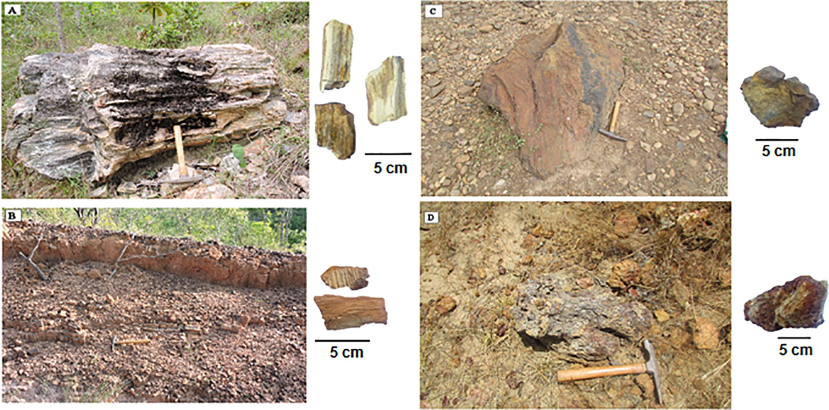
Stem fossil samples analyzed herein. A and B: The gymnorsperm and pteridophyte, respectively. Both were collected from the Duque Bacelar municipality; C and D: The gymnosperm and pteridophyte, respectively. Both were collected from the city of Teresina and Monsenhor Gil municipality.
Samples
Taxonomic identification
Locations
A
Gymnosperm
APA of the Morros Garapenses, Duque Bacelar, MA
Geographical Coordinates: 4°10′1.28″S 42°58′52.41″O
B
Pteridophyte
APA of the Morros Garapenses, Duque Bacelar, MA
4°10′1.28″S 42°58′52.41″O
C
Gymnosperm
The fossil florest of the river Poti, Teresina, PI
Geographica lCoordinates: 5° 6′51.42″S 42°43′48.24″O
D
Pteridophyte
The Monsenhor Gil, PI
Geographical Coordinates: 5°36′56.45″S 42°30′20.48″O
The sample A corresponds to the fossil of a gymnosperm, collected from Duque Bacelar municipality. The sample B corresponds to a tree-fern fossil also collected from Duque Bacelar. The sample C belongs to a gymnosperm fossil from Teresina municipality and, finally, the sample D, originated from Monsenhor Gil municipality, corresponds to a tree-fern fossil specimen. The sample A and C belong to gymnosperms based on the internal organization of the trunks in concentric bands of permineralized tissues, here interpreted as possible growth rings of the secondary xylem. In addition, the external structure of the trunk does not present bundles of rachises and/or adventitious roots as seen in the stems that belong to tree-ferns and found in the same areas.
The sample B and D belongs to tree-fern. Although detailed histological descriptions have not been carried out on these specimens, is possible to visualize bundles of rachises and adventitious roots typically found in tree-fern stems. This specific characteristic is insufficient for taxonomic identification in the generic level but it is enough to confirm a botanical affinity with the ferns.
2.2 Characterization
The X-ray diffraction (XRD) patterns were obtained using a Rigaku powder diffractometer with the Bragg–Brentano geometry. The Co–Kα radiation was used and operated at 40 kV and 25 mA. The XRD measurements were taken in the 2θ range of 10–100°, using the step scan procedures (0.02°) in counting times of 5 s. To perform the XRD measurements, we used 1 g of powder samples and for the data treatment, we used the Xpert High score software with the powder diffraction files (PDFs) included. The Fourier transform infrared spectroscopy (FTIR) measurements were performed using a Bruker spectrometer, model Vertex 70. The spectral region analyzed spanned from 400 up to 4000 cm−1. The attenuated total reflectance (ATR) technique was used in the measurements of the sample. The energy dispersive spectroscopy (EDS) and scanning electron microscopy (SEM) analysis were performed using an Hitachi TM-3000 tabletop equipment, increasing up to 30,000 times at 15 kV with an EDS coupled Swifft ED 3000 with a solid state detector. The Fourier transform (FT)-Raman spectra, were collected with a Bruker RAM II FT-Raman module coupled with the VERTEX FT-IR spectrometer and with a liquid nitrogen cooled in a high-sensitivity Ge detector with the samples excited through the 1064-nm line of an Nd: YAG laser. The resolution was 4 cm−1 with an accumulation of 60 scans per spectra, as well as a nominal laser power of 120 mW.
3 Results and discussion
3.1 Analytical data
Fig. 2 shows the X-ray diffractograms for the four samples. According to the analysis of the XRD diffractograms (Fig. 2), all aforementioned fossilized samples presented quartz as the major crystalline constituent. This compound exhibits a hexagonal crystal structure with space group of the type P3121. According to X-ray diffractogram all peaks show the presence quartz is being the main component with chemical formula SiO2. The presence of the quartz is confirmed by the infrared and the Raman spectroscopic studies, as well as the small quantities of the amorphous phase, which was not detected by the XRD.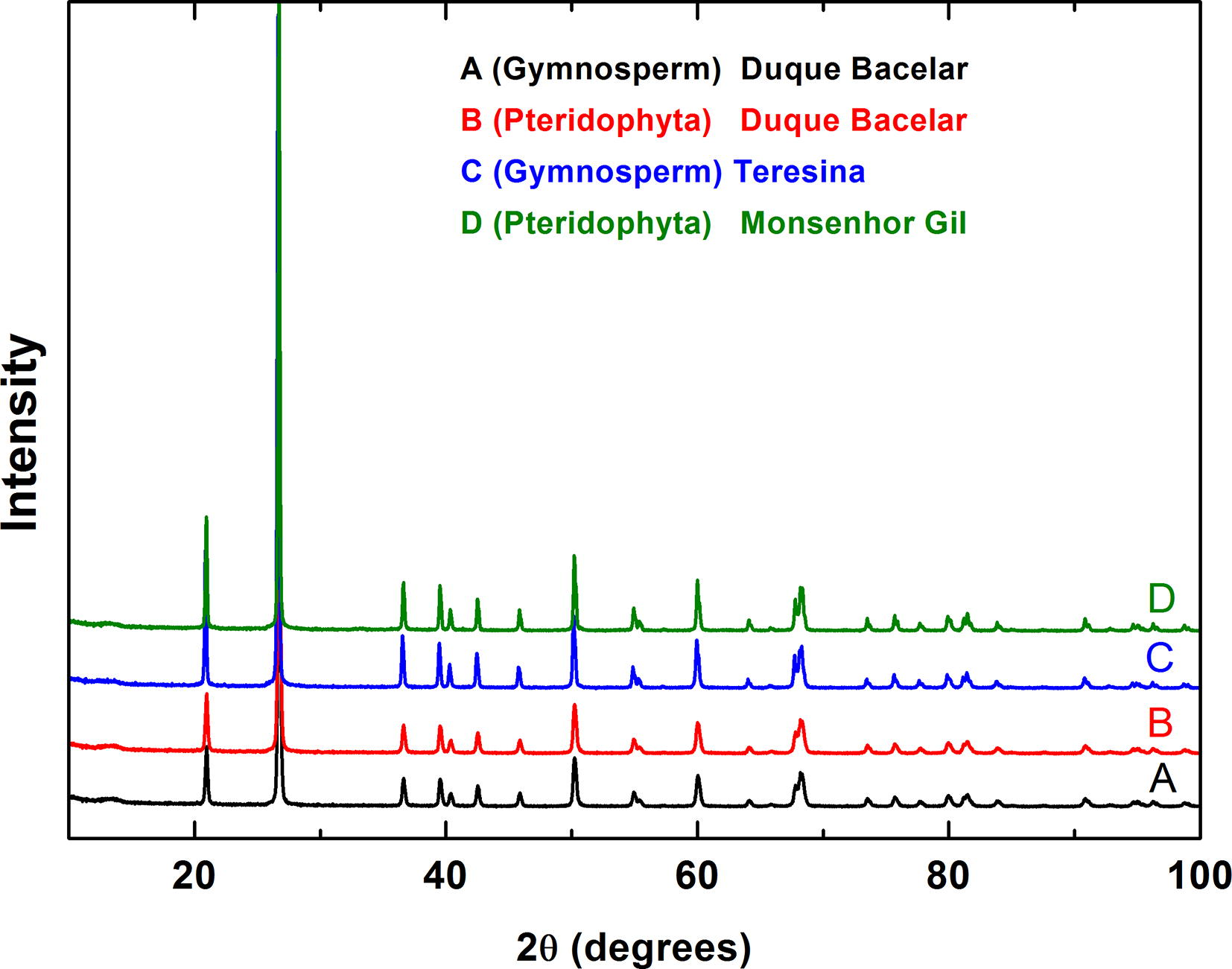
X-ray diffractograms of the samples A (in black), B (in red), C (in blue) and D (in green) shows the crystalline phase in the samples.
Fig. 3 shows the FTIR spectra in the four specimens of fossil trunks (samples A, B, C and D). Regarding the profile in the FTIR spectra of these fossils, the presence of SiO2 vibrations associated with the quartz (Lippincott et al., 1958), confirming the previous results shown by the XRD results. The characteristics of the infrared bands associated with the quartz, are described on Table 2. The bands located at 462 cm−1 and 517 cm−1, are due to the bending vibrations of the Si-O, whereas the bands observed at 694, 779, can be assigned as Si – Si stretching and the bands at 1085 and 1164 cm−1, corresponding to the stretching vibration of the Si-O bonds (Lippincott et al., 1958; Saikia et al., 2008). The doublet peaks near 800 cm−1, confirmed the presence of quartz in relation to other polymorphs of silica, such as the cristobalite and the tridymite (Russell and Fraser, 1994).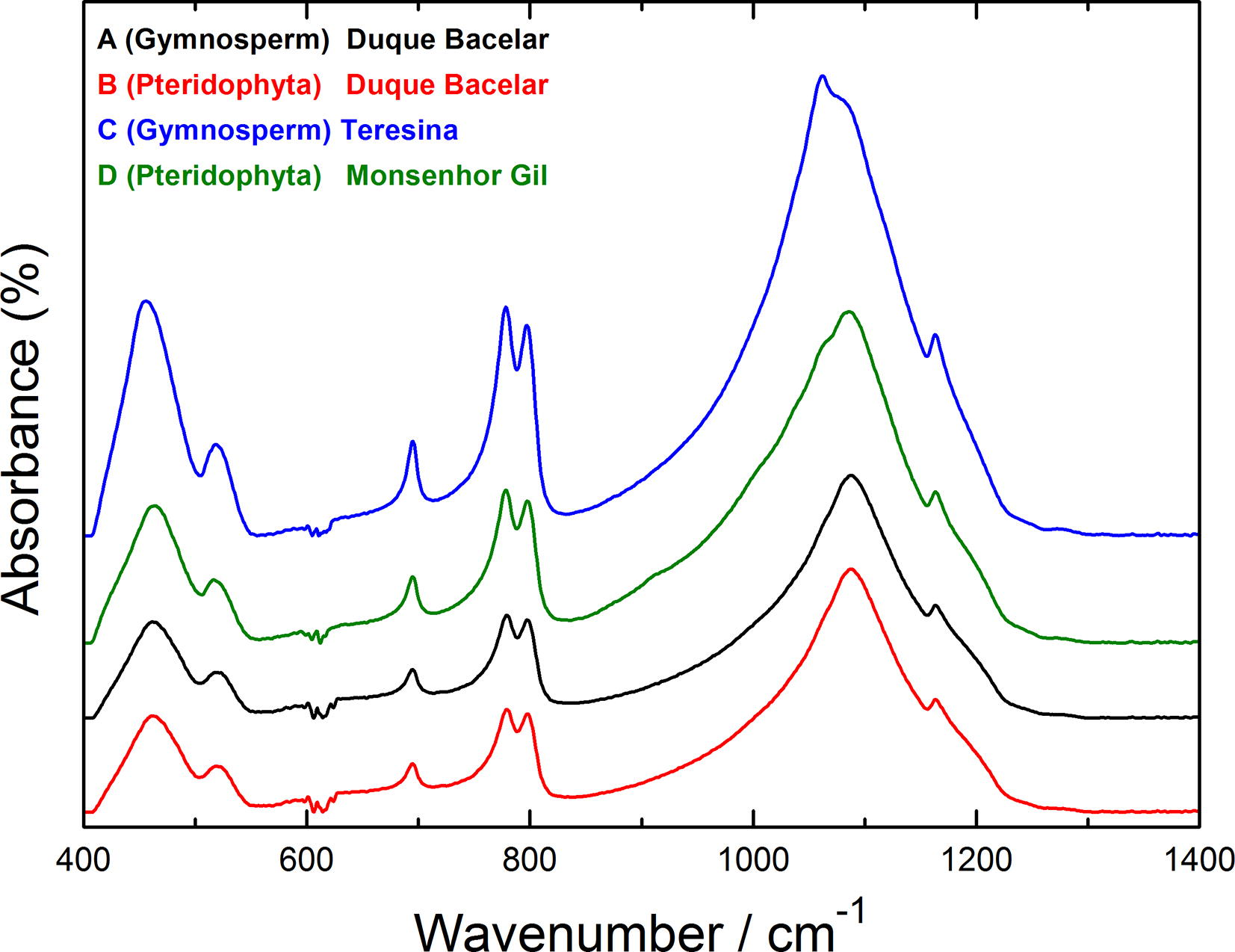
Infrared spectra of the fossilized logs in the spectral range from 400 to 1400 cm−1: A (in black), B (in red), C (in blue) and D (in green).
IR modes
Samples
A
B
C
D
Assignment
461
462
455
462
Si-O bending
520
502
518
517
Si-O bending
694
694
694
694
Si stretching
778
779
778
778
Si stretching
797
797
797
797
Si stretching
1087
1087
1085
1087
Si-O stretching
1164
1164
1164
1164
Si-O stretching
Raman modes
Sample
A
B
C
D
Assignment
201
Si-O stretching
Wavenumber (cm−1)
464
464
464
462
Si-O bending
1347
1356
–
–
Carbon vibration
1575
1577
–
–
Carbon vibration
–
1595
1604
–
Carbon vibration
Fig. 4 shows the Raman spectra of the fossils corresponding to the samples A, B, C and D. We also observed two intense bands around 201 cm−1 and 464 cm−1 in the spectrum of sample D, which were assigned, respectively as Si–O stretching/O–Si–O, bending/Si–O torsion and Si – O stretching/–Si–O bending from the quartz compound (Schmidt and Ziemann, 2000), and yet, in relation to the Raman spectra, three samples (A, B and C) presented the band centered at 201 cm−1 with a low intensity, however, the band at 464 cm−1 remained relatively intense. It is interesting to note that the peaks associated with the quartz have large bandwidths which were supposed to be related with the distortion of the quartz crystal structures due to the changes during the process of the fossilization (Legodi and Waal, 2007). Furthermore, two other broad bands observed around 1350 and 1600 cm−1 also appeared in the Raman spectra of the three (A, B and C) said samples. These are very intense bands and can be associated with the carbon of carbonaceous material present in the fossil (hybridizations sp3 and sp2, respectively). These bands can be assigned as well, respectively as D and G bands corresponding to the amorphous carbon (Ferrari and Robertson, 2001). It is interesting also to note that in a previous study, the D and G bands were observed in the Raman spectrum of a wood fossil collected from the Crato Formation of Northeastern Brazil, belonging to the Cretaceous Period, being associated with the amorphous carbon, contained in the sample as well (Lippincott et al., 1958). In the fossil from the “Crato” Formation, the presence of the amorphous carbon and the additional presence of oxygen were interpreted as being a consequence of a natural fire. Additionally, the presence of carbon was interpreted as originated from the plant itself (highly altered remains from cellulose or lignin from the living plant) (Alencar et al., 2015) in fossil plants from the Parnaíba Basin. It is probable that this last interpretation may explain the presence of carbon in the fossils analyzed in the present study.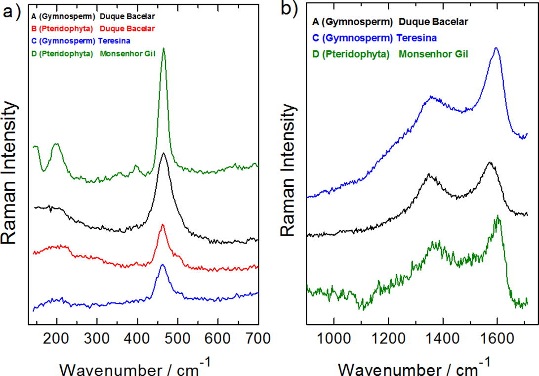
Raman spectra of the fossils A, B, C and D for the spectral ranges (a) from 150 to 700 cm−1 and (b) from 950 to 1650 cm−1. The identification of the four fossils in reference, are given on Table 2.
Fig. 5(a–d) shows the scanning electron microscopy images of the four samples (A – D), previously analyzed by the infrared and Raman spectroscopy and the X-ray diffraction (a complete set of images is given in the Supplementary Materials). The analysis of the EDS spectroscopy for the different points on the sample surfaces was used to give a semiquantitative determination of the chemical composition. The use of this technique allowed us to recognize that the most of the samples are not perfectly homogeneous. For instance, in Fig. 5(a) from the white region studied, we noted the presence of 39% weight of Si and 49% of oxygen, whereas in the black region (see the Supplementary Material), the silicon corresponded to 46% and oxygen, 53%. In relation to the sample B in Fig. 5(b), it is possible to note a certain homogeneity regarding silicon and oxygen in different parts of the fossil. However, if we look at the concentration of iron, for example, a difference in 3% of weight in different regions of the sample is verified. Regarding the sample C appearing in Fig. 5(c), an impressive non–homogeneity was observed: the quantity of silicon varies from 24% to 45% and the quantity of oxygen varies from 53% to 62% of weight. Similarly, the sample D in Fig. 5(d) presents silicon varying from 20% to 26% in weight and oxygen varying from 38% to 54%. In addition to this, the sample D analysis showed iron with a weight of 5% in certain parts and up to 35% in other parts. Such non-homogeneity is not exclusive of the fossils studied herein, e.g. the Brazilian fossil plant Brachyphyllum castilhoi (Araucariaceae) from the Cretaceous Period (Crato Formation), evinced different concentrations of pyrite at different points of the sample (Sousa Filho et al., 2011).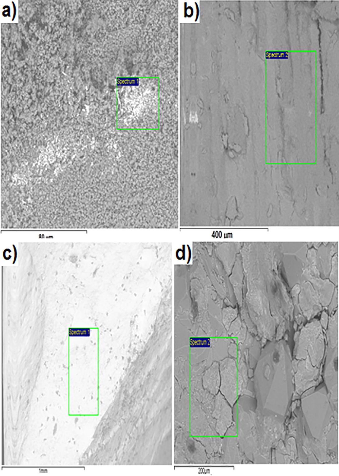
The SEM images of the following four trunk fossil samples: (a) fossil A, (b) fossil B, (c) fossil C, (d) fossil D (shown on the text), which demonstrated the locations where the EDS spectra were performed.
The analysis of the micromorphological patterns are characterized by the microporous on the surface of the samples. The images (Fig. 5) showed the silica occurring primarily as masses corresponding to the microgranular quartz (Landmesser, 1984). Additionally, it was noted the presence of other chemical elements found in smaller quantities, such as aluminum, iron (except for sample D) and potassium.
3.2 Fossilization process and silica source
The silica involved in the fossilization process of the plant remains within the Pedra de Fogo Formation, could be interpreted as being originated from an external source, comprised of terrigenous alkaline materials present in typical arid climate conditions. This could promote the solubility of the silica and its transference to the deposited areas (Faria JR, 1979). Still, it is worth mentioning that processes related to volcanism are particularly well known as a source of silica (Leo and Barghoorn, 1976). Regarding the quartz found in a previous study, we observed large crystals formats, contained in the fine-grained matrix or glass. In these cases, we found combined crystalline forms, constituted of bipyramidal prisms (Tröger, 1979).
An important parameter to be considered during the silicification is the pH value, as this can present substantial changes during the process (Leo and Barghoorn, 1976). It is generally accepted that in order to occur silicification of wood, it must be rapidly buried in sediments that preclude it from being exposed to oxygen, avoiding, in this way, decomposition by bacteria and fungi. The environment where this occurs is generally a fluvial one with water pH varying from 6 to 9. If the ambient is acid, fungi can be developed, and if the ambient is excessively basic, it can destroy the molecules of the wood itself. Ideally, dissolved silica must be found in the environment, which is readily available where volcanic ash was present (Landmesser, 1984, Faria JR, 1979, Leo and Barghoorn, 1976, Tröger, 1979; Landmesser, 1994). It is believed that volcanic ash in glass or amorphous forms absorbs water, thus, releasing silica and producing a solution with monosilicic acid [Si(OH)4]. According to Leo and Barghoorn (1976), when a concentration of the solution increases, the monosilicic acid polymerizes, with the consequent production of siloxane bonding and releasing water. The pivotal point in the process is that the polymerized silicic acid leads to form hydrogen bonds with organic molecules in the tissue of the wood. As consequence, layers of the silicic acid are formed in the woody tissue. In the process of the polymerization, a coating of silica gel is produced on the cells (or filling voids of the woods). When the gel looses water, it solidifies in the amorphous opal [SiO2.nH2O]. Successive crystallizations and losses of water form the cristobalite, trydimite, chalcedony and finally the quartz. Therefore, an eventual silification process is a complex event extending over many years. The other chemical elements found in smaller quantities, especially the enrichment by iron (in sample D), can be interpreted as consequence of the replacement of original silica mostly by iron oxides due to processes driven by chemical weathering. Silicate dissolution and neoformation of minerals such as kaolinites, iron oxides (goethite, hematite), aluminum oxides-hydroxides (gibbsite, boehmite) which are stable at low temperature and pressure (25 °C, 1 atm) is quite common in the pedogenesis of tropical soils (Formoso, 2006). Taking into account that the area of Parnaíba Basin was under tropical climate regimes since end of Paleozoic-to- early Mesozoic, the dissolution and remobilization (including leaching) of silica, and the increase in iron-containing compounds, is expected in silica-permineralized fossils according they gradually approach the surface (entering the alteration cover) due to the upwelling of the deposits and subsequent erosion of the strata over time. However, this hypothesis needs to be better investigated in future work.
The spectroscopic analysis used in this study showed a high prevalence of silica levels in all fossil plant samples duly checked. These results corroborate with the study done by Alencar et al. (2015) on wood fossils originated from other sites within the same geological unit, e.g. Pedra de Fogo Formation. Therefore, it is possible to infer that the fossilization process that acted on the phytofossil records in the eastern is the same as the northern margin of the basin. However, the source of silica present in these stated fossils is still a matter of debate. In the Pedra de Fogo Formation, as previously commented, the silica source ought to be external Faria JR (1979). In addition, Matysová et al. (2010), suggested a similar climatic-driven process to explain the silicification of plant stems recovered from the overlying Motuca Formation strata, located in Filadélfia municipality in the Tocantins State (Permian, southwestern Parnaíba Basin). For these authors, the presumed silica source for the initial stage of silicification was the weathering of labile minerals, mostly feldspars in the alluvium. Thus, they excluded the volcanic influence on the silicification mode in the plant stems from the Motuca Formation and interpreted it as a process occurred in fluvial sediments from the basin fills, under a seasonally dry and warm climate, perhaps during a relatively fast tectonic basinal development.
In the case of phytofossiliferous outcrops present in the northeastern and eastern margins of the basin, some of them are associated with autochthonous assemblages, i.e., fossil woods in a living position (Caldas et al., 1989; Conceição et al., 2016a,b). It is worth noting that in the Pedra de Fogo Formation, some trunks in a vertical position reach up to 2.3 meters of length, whereas other trunks in a horizontal position have more than 180 cm in diameter (Conceição et al., 2016a,b). Trees preserved in living position imply that there was a rapid burial of these plant communities (Gastaldo and Staub, 1999; DiMichele and Gastaldo, 2008; Gastaldo and Demko, 2010), which can be occasioned by some catastrophic event. As proposed by Alencar et al. (2015), the phenomenon which enabled the rapid permineralization of these macrophytofossils could have been one or more pyroclastic depositional events in the presence of aqueous streams. Some examples of petrified forests with elements in living-position are known from the Paleozoic and are considered to be associated with events of pyroclastic depositions: Permian petrified forests in Chemnitz, Germany (Rossler, 2001, 2006); a Middle Pennsylvanian (Bolsovian) peat-forming forest preserved in situ in volcanic ash of the Whetstone Horizon (Opluštil et al., 2009); a Late Pennsylvanian coniferopsid forest in growth position, near Socorro, New Mexico, U.S.A (Falcon-Lang et al., 2016).
However, on the other hand, Matysová et al. (2010) noted, based on observations in the Australian modern environments, that trees from the riparian vegetation, are prone to survive in semiarid climatic conditions, therefore, they are predisposed to be rapidly buried after extreme seasonally river discharges and later silicified. As a consequence, some of the trunks at these mentioned deposits are found not too far from their likely sites of growth, consequently these must have also belonged to the original riparian vegetation (Matysová et al. 2010). The aforementioned flash fluvial regimes could be easily considered as catastrophic events. It has to be noted, however, that the fossil trees from the Motuca Formation in Tocantins, in the southwestern margin of the Parnaíba Basin, have never been found in living position (Dias Brito et al., 2007). This situation contrasts with vertical stance of the some fossil trees from the Pedra de Fogo Formation considered in this study, located at the northeastern margin. Although both the Motuca and Pedra de Fogo Formations are of Permian age, the former unit is generally considered slightly younger (Santos and Carvalho, 2004; Dias Brito et al., 2007), consequently, the fossil layers that bear trees in vertical situation within the Pedra de Fogo Formation might be older and could have been fossilized under different circumstances than those in the Motuca Formation in Tocantins.
4 Conclusions
This study reported the characterization of four fossil trunks by X-ray diffraction measurements, SEM-EDS spectroscopy, as well as Raman and infrared spectroscopy. The samples from the trunk fossils were obtained from the sedimentary Parnaíba Basin, in the Permian Pedra de Fogo Formation. These aforesaid results suggest that during the fossilization process, the fossils in question were affected by the environment and also suffered a process of permineralization with a partial substitution of its original composition by quartz. Therefore, the results indicated that all plant remains have undergone a similar diagenetic process, which would be expected if they belong to the same lithostratigraphic unit. The quartz is the most abundant mineral in all the analyzed samples. However, the origin of the significant amount of silica, available in the depositional settings that gave the origin to microcrystalline quartz available in the original depositional settings, is still controversial. Furthermore, an analysis must be carried out in order to characterize signatures of other materials that are present in a silicified plant stems and also in the surrounding rocky matrixes. Nevertheless, a limited detail study of the fossils and the associated sedimentary deposits, may eventually clarify the source of the silica, and consequently, explaining the initial event taking place with the fossilization process in the plant remains of the Permian strata from the Parnaíba Basin.
Acknowledgments
G.D. Saraiva, Ph.D, acknowledges the support from the MCTI/CNPq/Universal 14/2014 (Grants#449471/2014-4) and the PQ – 2014 (Grants#306631/2014-8): R. Iannuzzi thanks the Brazilian National Council for Scientific and Technological Development (CNPq) for the grants (PQ 309211/2013-1) and PTCF thanks the CNPq. Acknowledgements are due to Francisco Carlos from the APA dos Morros Garapenses in Duque Bacelar. The authors also wish to thank the following Brazilian institutions, such as CAPES, FAPESPI, CNPq and FUNCAP and J. H. da Silva acknowledges grant from BPI - Notice n° 09/2015.
References
- Spectroscopic analysis and X-ray diffraction of trunk fossils from the Parnaíba Basin, Northeast Brazil. Spectrochim. Acta Part A Mol. Biomol. Spectrosc.. 2015;135:1052-1058.
- [Google Scholar]
- Nota sobre a ocorrência de uma floresta petrificada de idade permiana em Teresina, Piauí. Boletim IG-USP. Publicação Especial.. 1989;7:69-87.
- [Google Scholar]
- New petrified forest in Maranhão, Permian (Cisuralian) of the ParnaíbaBasin, Brazil. J. S. Am. Earth Sci.. 2016;70:308-323.
- [Google Scholar]
- Novo registro de uma Floresta Petrificada em Altos, Piauí: relevância e estratégias para geoconservação. Pesquisas em Geociências. 2016;43:311-324.
- [Google Scholar]
- Analytical x-ray microscopy on Psaronius sp. - a contribution to permineralization process studies. Microchim. Acta.. 2001;133:279-283.
- [Google Scholar]
- Floresta petrificada do Tocantins Setentrional - O mais exuberante e importante registro florístico tropical-subtropical permiano no Hemisfério Sul. Sítios Geológicos e Paleontológicos do Brasil. Brasília: CPRM; 2007. p. :337-354.
- Faria JR, L.E.C., 1979. Estudo Sedimentológico da Formação Pedra de Fogo – Permiano da Bacia do Maranhão. Dissertação de Mestrado 70f. Universidade Federal do Pará.
- A Late Pennsylvanian coniferopsid forest in growth position, near Socorro, New Mexico, USA: tree systematics and palaeoclimatic significance. Rev. Palaeobotany Palynol.. 2016;225:67-83.
- [Google Scholar]
- Resonant Raman spectroscopy of disordered, amorphous, and diamondlike carbon. Phys. Rev. B.. 2001;64:75414-75423.
- [Google Scholar]
- Some topics on geochemistry of weathering: a review. Anais da Academia Brasileira de Ciências. 2006;78(4):809-820.
- [Google Scholar]
- A mechanism to explain the preservation of leaf litters in coals derived from raised mires. Palaeogeogr. Palaeoclimatol. Palaeoecol.. 1999;149:1-14.
- [Google Scholar]
- Long term hydrology controls the plant fossil record. In: Allison P.A., Bottjer D.J., eds. Taphonomy: Processes and Bias Through Time. 32. Topics in Geobiology; 2010. p. :249-286.
- [Google Scholar]
- Raman spectroscopic study of ancient South African domestic clay pottery. Spectrochimica Acta Part A Mol. Biomol. Spectrosc. Spectrosc. Acta A. 2007;66:135-142.
- [Google Scholar]
- Silicification of wood: Harvard University Botanical Museum Leaflets. 1976;25(1):1-47.
- Infrared studies of polymorphs of silicon dioxide and germanium dioxide. J. Res. Natl. Bur. Stand.. 1958;61:61-70.
- [Google Scholar]
- Volcanic ash as a source of silica for the silicification of wood. Am. J. Sci.. 1940;238:586-596.
- [Google Scholar]
- Alluvial and volcanic pathways to silicified plant stems (Upper Carboniferous–Triassic) and their taphonomic and palaeoenvironmental meaning. Palaeogeogr. Palaeoclimatol. Palaeoecol.. 2010;292:127-143.
- [Google Scholar]
- Mineralogy and geochemistry of late Eocene silicified wood from Florissant fossil beds National Monument, Colorado. Geol. Soc. Am. Spec. Pap.. 2008;435:127-140.
- [Google Scholar]
- A Middle Pennsylvanian (Bolsovian) peat-forming forest preserved in situ in volcanic ash of the Whetstone Horizon in theRadnice Basin, Czech Republic. Rev. Palaeobot. Palynol.. 2009;155:234-274.
- [Google Scholar]
- Der versteinerte Wald von Chemnitz.KatalogzurAusstellungSterzeleanum. Chemnitz: Museum fürNaturkunde; 2001.
- Sphenopsids of the Permian. 1: the largest known anatomically preserved calamite, an exceptional find from the petrified forest of Chemnitz, Germany. Rev. Palaeobotany Palynol.. 2006;140:145-162.
- [Google Scholar]
- Infrared methods. Clay Mineralogy: Spectroscopic and Chemical Determinative Methods. London: Springer, Netherlands; 1994. p. :11-67.
- Fourier transform infrared spectroscopic estimation of crystallinity in SiO2 based rocks. Bull. Mater. Sci. Indian Acad. Sci.. 2008;31:775-779.
- [Google Scholar]
- Paleontologia das Bacias do Parnaíba, Grajaú e São Luís: Reconstituições Paleobiológicas. Rio de Janeiro: CPRM/ Serviços Geológicos do Brasil; 2004.
- Silica recrystallization in petrified wood. J. Sediment. Petrol.. 1982;52:1277-1282.
- [Google Scholar]
- Studies of wood fossils from the Crato Formation, Cretaceous Period. Spectrochimica Acta Part A Mol. Biomol. Spectrosc.. 2013;115:324-329.
- [Google Scholar]
- In-situ Raman spectroscopy of quartz: a pressure sensor for hydrothermal diamond-anvil cell experiments at elevated temperatures. Am. Mineral.. 2000;85:1725-1734.
- [Google Scholar]
- Combination of Raman, Infrared, and X-Ray Energy-Dispersion Spectroscopies and X-Ray Diffraction to Study a Fossilization Process. Braz. J. Phys.. 2011;41:275-280.
- [Google Scholar]
- Raman and cathodoluminescence spectroscopic investigations on Permian fossil wood from Chemnitz — a contribution to the study of the permineralization process. Spectrochimica Acta A. 2004;60:2903-2912.
- [Google Scholar]
- Tavares, T.M.V., 2011. Estudo de Marattiales da “Floresta Petrificada do Tocantins Setentrional” (Permiano, Bacia do Parnaíba). (Ph.D. Thesis). Instituto de Geociências e Ciências Exatas - Universidade Estadual Paulista “Júlio de Mesquita Filho”, Rio Claro, p. 184.
- Optical determination of rock-forming minerals. Part 1, determination tables. Stuttgart, Germany: E. Content Free Trial; 1979. p. :188.
- Raman and cathodoluminescence spectroscopic investigations on Permian fossil wood from Chemnitz: a contribution to the study of the permineralisation process. Spectrochimica Acta A. 2004;60:2903-2912.
- [Google Scholar]
- Medullosan fusain trunk from the roof rocks of a coal seam: Insight from FTIR and NMR (Pennsylvanian Sydney Coal Field, Canada) Int. J. Coal Geol.. 2010;82:116-124.
- [Google Scholar]
Appendix A
Supplementary data
Supplementary data associated with this article can be found, in the online version, at https://doi.org/10.1016/j.jksus.2018.05.019.
Appendix A
Supplementary data


