

The effects of melatonin and vitamin D3 on the gene expression of BCl-2 and BAX in MCF-7 breast cancer cell line
⁎Corresponding author at: Chair for Biomarkers of Chronic Diseases, Biochemistry Department, College of Science, King Saud University, PO Box, 2455, Riyadh 11451, Saudi Arabia. ndaghri@ksu.edu.sa (Nasser M. Al-Daghri)
-
Received: ,
Accepted: ,
This article was originally published by Elsevier and was migrated to Scientific Scholar after the change of Publisher.
Peer review under responsibility of King Saud University.
Abstract
Abstract
Objective
Compelling evidence from epidemiological and experimental studies indicate that the disruption of circadian rhythm and decreased melatonin synthesis are risk factors for breast cancer. With the ever-increasing rate of breast cancer cases globally, there is a strong need for safer, effective, and alternate treatment strategies with lesser side effects. This study aimed to investigate the effects of melatonin and vitamin D3 on the gene expression of two apoptotic factors, BCL-2 and Bax, in the MCF-7 cancer cell line.
Methods
Cell viability (MCF-7) was studied through MTT reduction assay in response to melatonin (1 nM, 5 nM and 10 nM) and vitamin D3 (0.5 nM, 1 nM and 10 NM). The optimum dose of melatonin (5 nM) and vitamin D3 (0.5 nM) were selected for their combination and Bax/BCL-2 ratio determination. Changes on the expression of genes BCL-2 and Bax, were determined through mRNA expression while Western blot analysis was used to detect changes in the expression of BCL-2 and Bax at the protein level.
Results
There was an upregulation of Bax gene expression and protein expression with the treatments of melatonin, vitamin D3, and their combination, with concomitant downregulation of Bcl-2 gene and protein expression. The Bax/BCL-2 ratio was increased significantly (p < 0.001) suggesting potential therapeutic treatments.
Conclusion
The results suggest that treatment with melatonin and vitamin D3 inhibits the proliferation and induced apoptosis in breast cancer cells. We report here for the first time that this combination has effectively activated Bax and downregulated BCL-2 at the DNA and protein level.
Keywords
MCF-7 breast cancer cell line
BCL-2
Bax
Melatonin
Vitamin D3
Apoptosis

1 Introduction
Standard treatment options for breast cancer (BC) include chemotherapy, radiation, surgery and endocrine therapy (employing antiestrogen and/or progesterone treatment). Selective estrogen receptor modulators (SERMs) including tamoxifen (estrogen receptor antagonist), raloxifene, (an aromatase inhibitor) and GnRH (gonadotrophic releasing hormone) agonists, are clinical agents being used for the estrogen receptor-positive (ER + ) tumors (Lumachi et al., 2011). There are many side effects and risks associated with their use including increased risk of uterine cancer, osteoporosis, stroke, pulmonary embolism, and vision problem (Fisher et al., 2005). These undesirable side effects and the ability of the cancer cells to become resistant to the drug, unfortunately, made these agents ineffective in the fight against breast cancer. Due to the growing number of breast cancer cases globally, the need of natural agents with better safety as adjuvant treatment option cannot be underestimated.
Melatonin is a non-steroidal lipophilic hormone secreted by the pineal gland, and is associated with a wide range of physiological functions including anti-inflammatory, antioxidant, onco-statin, and regulation of circadian rhythm. There is a consistent increase in the number of reports exploring the role of melatonin in the etiology of cancer during past two decades and several studies related to the disruption of melatonin's circadian profile by exposure to light at night has been described to play an important role in the initiation, promotion, and progression of breast cancer (Touitou et al., 2017; Van Dycke et al., 2015). Many studies have shown that reduced melatonin levels linked to a greater risk of breast cancers might be an outcome of increased estrogen production, altered estrogen receptor (ER) functions, by repressing the activity of various enzymes such as aromatase, sulfatase and aldo–keto-reductases. (Dauchy et al., 2009; Li et al., 2017; Nooshinfar et al., 2016).
Calcitriol (1, 25 dihydroxy vitamin D3) is the active form of vitamin D produced in the liver and kidney. Anticancer effects of vitamin D3 are believed to be the result of apoptosis, inhibition of cell cycle progression, and regulation of estrogen receptor signaling (Abd-Elsalam et al., 2015; Estébanez et al., 2018; O'Brien et al., 2018; Ansari et al., 2020). Multiple epidemiological studies indicated that vitamin D is not only important for bone growth and skeletal integrity but its deficiency is linked to some metabolic and autoimmune disorders, besides several other malignancies (Al-Daghri et al., 2014, 2020; Jeon and Shin, 2018; Zhu et al., 2019). The evidence for the chemo-preventive role of vitamin D3 and for association of low vitamin D3 levels with increased breast cancer risk have been reported in recent years (Yao et al., 2011; Wang et al., 2013). An epidemiological analysis was conducted in Saudi Arabia by Yousef et al (2013) highlighting the inverse association between serum levels of 25(OH)D (vitamin D) and risk of breast cancer.
Several clinical trials were undertaken to assess the benefits of melatonin and vitamin D3 either as single or in combination (as adjuvant therapy) in breast cancer patients. In one such clinical trial with small sample size (n = 14) the use of melatonin before tamoxifen (a SERM) resulted in partial response (28%), reduced anxiety and toxicity. A more recent phase II trial in breast cancer patients has shown that melatonin improved the quality of life with better quality and improved sleep time, reduced fatigue and improved cognitive functioning (Lissoni et al., 1995; Innominato et al., 2016). However, results are inconsistent regarding the relationship between breast cancer risk and melatonin levels due to different methods, small cohorts, different times of melatonin sample collection and differences in menopausal status (Li et al., 2017, Estébanez et al., 2018).
The disturbance in apoptotic molecular signaling pathways was thought to be involved in all stages of carcinogenesis (Hassan et al., 2014). BCL-2 family of proteins is the hallmark of apoptosis, and its role in the regulation of apoptotic pathways is related to the cancer pathophysiology and resistant to conventional chemotherapy. BCL-2 prevents BAX/BAK oligomerization, which leads to the cascade of various caspases release from the mitochondria resulting in apoptosis (Reyna et al., 2017).
The current research was aimed at investigating the effects of melatonin and vitamin D3 treatments on the transcription (mRNA) as well protein expression of two apoptotic factors, BCL-2 and Bax of MCF-7 cells.
2 Materials and methods
2.1 Cell culture
The breast cancer cell line used in this study was MCF-7 (ATCC, USA). Cells were maintained in Dulbecco's Modified, Eagle Medium (DMEM) supplemented with 10% fetal bovine serum (FBS) and 1% of streptomycin and penicillin. Cells were grown as an adherent monolayer in T-25 or T-75 culture flasks and 6-well plates or 96-well plates (according to the type of the experiment) at 37 °C in humidified environment (5% CO2). The medium was changed every third day. At confluence, cells were sub-cultured after removal with 0.05% trypsin-EDTA. Cell viability was assessed with trypan blue.
2.2 MTT reduction assay
To find the optimum concentration of melatonin and vitamin D3 for growth inhibition of breast cancer cell line MCF-7, cells were plated onto 96 well plates and exposed to different concentrations of melatonin (1, 5 and 10 nM) and vitamin D3 (0.5, 1 and 10 nM) for different periods (24 and 48 h) at 37 °C (5% CO2 incubator). 10 µl of MTT reagent (5 mg/mL in PBS) was added to each well and further incubated for 3–4 h at 37 °C. After that, the medium was removed and the formed formazan crystals were solubilized by adding 100 µl of isopropanol in 0.04HCL per well for 30 min. The absorbance was measured at 540 nm (using the BioTek ELX 800 absorbance microplate reader). The percentage of cell viability was calculated using the following formula:
Cell viability (%) = (OD of experimental samples/OD of experimental control sample) × 100.
2.3 RNA extraction and cDNA synthesis
Total RNA was isolated after 48 h treatment of cells with 0.5 nM vitamin D3, 5 nM melatonin, and their combination (0.5 nM vitaminD3 + 5 nM melatonin) using the Trizol reagent RNA easy kit (Qiagen, Germany) according to the manufacturer’s protocol. Reverse transcription (RT) was carried out by 2 µg of total RNA using the SuperScript Vilo cDNA Synthesis Kit (Thermo-Fisher, USA) for first-strand complementary deoxyribonucleic acid (cDNA).
2.4 Quantification of mRNA expression by Real-Time polymerase chain reaction (RT-PCR)
Quantitative analysis of specific mRNA expression was performed by RT-PCR by subjecting the resulting cDNA to PCR amplification using a 96-well PCR plate in CFX 96 TM Real-Time System (BIO-RAD, USA) using Kapa Sybr Fast Universal qPCR Kit (KAPA BIOSYSTEM, USA). The primers sequences for both the target genes (Bax and BCL-2) and the housekeeping gene (GAPDH) are given in Table 1. The standard deviation in samples was calculated using results from at least three independent experiments.
| Forward | Reverse | |
|---|---|---|
| GAPDH | 5′-CTTTTGCGTCGCCAGGTGAA-3′ | 5′-AGGCGCCCAATACGACCAAA-3′ |
| BAX | 5′-GCGACTGATGTCCCTGTCTCC-3′ | 5′-AAAGATGGTCACGGTCTGCCA-3′ |
| BCL-2 | 5′-GAACTGTACGGCCCCAGCAT-3′ | 5′-GGGGCAGGCATGTTGACTTC-3′ |
2.5 Preparation of cellular extracts and western blot analysis
MCF-7 cells were treated with melatonin (5 nM) or vitamin D3 alone (0.5 nM) and in combination (5 nM melatonin + 0.5 nM vitamin D3) for 48 h. After that, cells were washed with PBS and scraped in RIPA buffer supplemented with protease inhibitors. The protein content of cell lysate was determined using the Bicinchoninic Acid Protein Assay Kit (Sigma-Aldrich, USA). For immunoblot analysis, cell lysate was separated on 10% SDS-PAGE gels. Protein from the gel was electrophoretically transferred to the PVDF membrane in a 1X transfer buffer containing Tris-HCl, glycine, methanol, and 20% SDS at 13 V for 27 min. Protein blots were blocked overnight at 4 °C in a solution containing 5% non-fat dry milk powder in Tris-buffered saline (TBS) solution (NaCl/Tris base pH 7.6). The blocking solution was removed, and the blots were washed in a wash buffer (TBS) followed by 2–3 h incubation at 4 °C with monoclonal primary mouse antibody against tubulin and BCL-2 and Bax. The primary antibody was removed, and blots were rinsed three times with a wash buffer TBST (NaCl/Tris-base + 0.1% Tween pH 7.6), followed by incubation with peroxidase-conjugated goat anti-mouse IgG (1:1000) at room temperature for 2 h. The bands were visualized (by LI-COR Bioscience, Lincoln, Nebraska, USA) using the enhanced chemo-luminescence method according to the manufacturer’s instruction. Individual bands were quantified using Image J software and normalized with standard tubulin bands.
2.6 Statistical analysis
Results were presented as mean ± SD and analyzed using Two-way ANOVA. Microsoft excel 2010 and GraphPad Prism 6 software (GraphPad Software, San Diego, CA, US) were used for the densitometric analysis of immunoblots.
3 Result
3.1 Effects of vitamin D3 and melatonin on cell viability
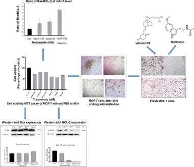
The cell viability of MCF-7 cells was detected through MTT reduction assay at different concentrations of vitamin D3 (0.5 µM, 1 µM, and10 µM) and melatonin (1 µM, 5 µM, and 10 µM). We found 0.5 nM of vitamin D3 (79.7% cell survival) and 5 nM of melatonin (64.1% cell survival) as most effective dose for growth inhibition of MCF-7 cells after 48 h (Fig. 1).
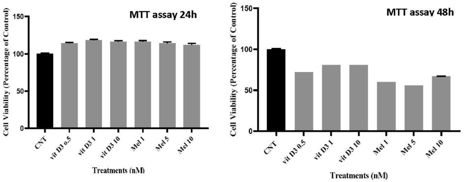
- Effect of Melatonin and vitamin D3 on MCF-7 cell viability. Percentage cell viability as compared to the control was measured by MTT assay as described in the methods sections after 24 h and 48 h of exposure to melatonin and vitamin D3.
3.2 Effects of vitamin D3 and melatonin on the mRNA expression level of Bax and BCL-2
The relative mRNA expression levels of Bax and BCL-2 were measured in the MCF-7 cells treated with 0.5 nM of vitamin D3, 5 nM of melatonin, and their combination (Fig. 2). There was an increase in mRNA expression of Bax (1.6045) with vitamin D3 and melatonin treatment (1.6882), however their combined treatment had a significant (1.8657; p < 0.01) effect on upregulating the mRNA expression of Bax (Fig. 2). When the mRNA expression of anti-apoptotic gene BCL-2 was measured, down-regulations of BCL-2, from 0.9917 (control) to 0.8648 (vit D3) and 0.7955 (melatonin), was the result of single agents. The significant effect, however, resulted from the combination treatment (0.4700) (Fig. 3). This was further echoed by looking at the ratio of Bax/BCL-2. As shown in (Fig. 4), the ratio was more significantly increased (p < 0.001) as compared to the control in the case of their combined treatments (Vitamin D3 + melatonin).
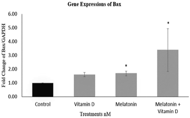
- Relative gene expression (mRNA) levels of BAX. The relative gene expression for BAX using quantitative RT-PCR analysis. Results are expressed as the mean of fold change as compared to control GAPDH (±SD for three independent experiments). The expression of mRNA of BAX in Vitamin D3 samples was 1.6-fold higher than the control and 1.69-fold increased with melatonin. The MCF-7 cells treated with both melatonin and vitamin D3 showed a 3.39-fold increase. * indicate significant (p < 0.05) values.
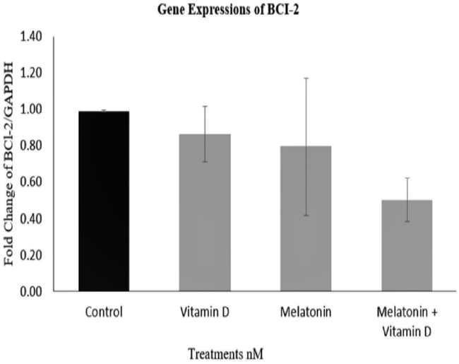
- Relative expression of BCL-2 mRNA. The relative mRNA expression of BCL-2 with the vitamin D3 treatments was 0.86-fold less than the control and about 0.8-fold less than the control in the presence of melatonin. The cells treated with both melatonin and vitamin D3 showed 0.5 less than the control. The overall difference was not significant.
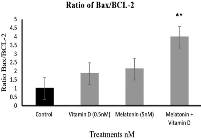
- The ratio of BAX/BCL-2 ratio. The ratio of BAX/BCL-2 was calculated and found to be highly significant (**p < 0.001) for the combined treatments with melatonin and vitamin D3.
3.3 Effects of melatonin and vitamin D3 on the protein expression of Bax and BCL-2 on MCF-7
In this experiment, the effects of most effective dose of melatonin and vitamin D3 (0.5 nM of vitamin D3 and 5 nM of melatonin) were investigated on the protein expression of Bax and BCL-2 through western blot analysis. These results agreed with the mRNA gene expression study, as an upregulation of the Bax protein expression (Fig. 5A and C) was also significant on combination treatment of vitamin D3 (0.5 nM) and melatonin (5 nM).
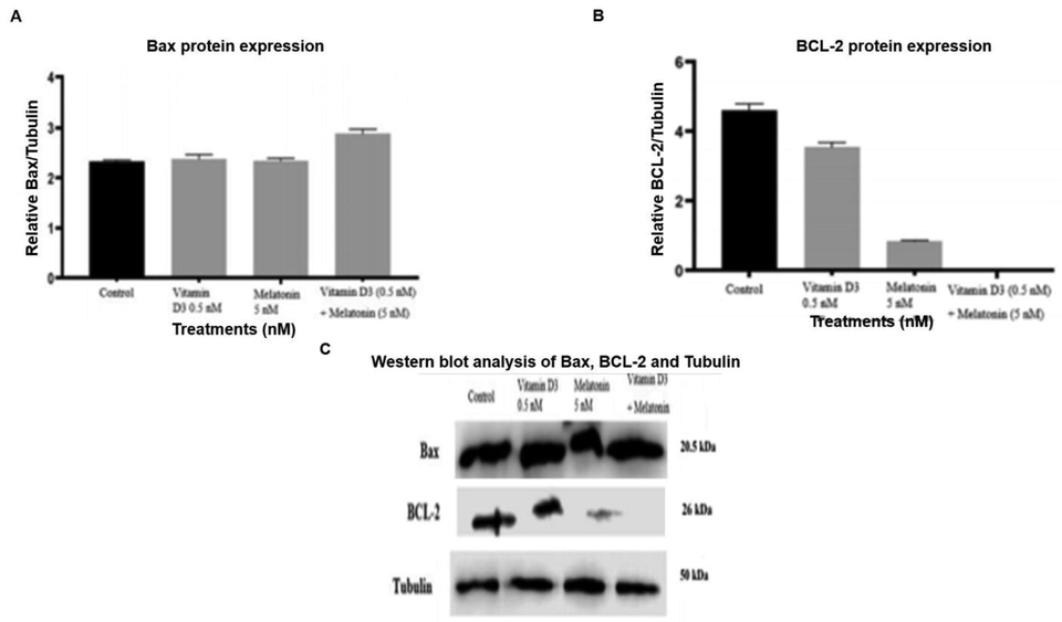
- The effect of vitamin D3 and Melatonin on Protein expression of Bax and BCL-2. The effect on the protein expression of Bax; B. The effect of vitamin D3 and melatonin on the protein expression of BCL-2; C. Immune blot showing bands of standard tubulin, BAX and BCL-2. The relative individual band intensity was quantified using Image J software and each band was normalized and expressed as relative BAX/Tubulin.
The protein expression of BCL-2 clearly showed that there was a downregulation in their expression with vitamin D3 and melatonin treatments, which was again even more obvious with their combined treatments (Fig. 5B and C). The ratio of Bax/BCL-2 showed a significant (p < 0.001) rise with the combined treatments of vitamin D3 and melatonin (Fig. 6).
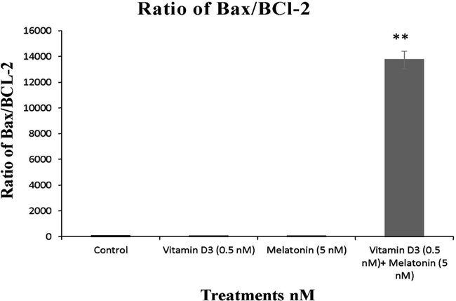
- The ratio of BAX/BCL-2 at the protein level. The relative ratio of the protein expression of BAX /BCL-2 was quantified and compared. The ratio was more significantly increased (** P < 0.001) as compared to the control as a result of their combined treatment of melatonin and vitamin D3.
4 Discussion
Majority of breast cancer patients respond to initial treatment, but unfortunately this progress is followed mostly by development of resistance to first line treatment options. This resistance necessitates the addition of other agents, including chemo and immunotherapeutic agents or their combinations. MCF-7 cell line is a commonly used in-vitro model for studying the drug resistance and treatment strategies in breast cancer. Vitamin D3 is a reputed supplement and is in current use in many experimental and epidemiological ongoing studies for repurposing it as an adjuvant in cancer patients or experimental models, and also in palliative care. The mechanisms involved in its anticancer potential have been reported to be the induction of cell cycle arrest, inhibition of ERα, upregulation of p53, p21 and p27, induction of caspase dependent as well as independent programmed cell death, downregulation of BCL-xL and BCL-2, and inhibition of angiogenesis (Constantinou and Kolokotroni, 2019).
BCL-2 and Bax are two distinct members of a gene family involved in the cellular regulation of apoptosis. Extensive evidence based on the manipulation of BCL-2 and Bax expression in the cultured cells had revealed that BCL-2 protein can be functionally characterized as apoptosis suppressing factor (Hassan et al., 2014; Perlman et al., 1999) whereas Bax is related to apoptosis promotion (Reyna et al., 2017). The intracellular ratio of Bax/BCL-2 is considered as the key indicator in the decision of the cells to respond to apoptotic signals. A cell with a high Bax/BCL-2 ratio will be more sensitive to the given apoptotic signal as compared to a similar cell type with a low Bax/BCL-2 ratio. This ratio was also considered a valuable and reliable biomarker to assess the success in the treatments of various types of cancer. The success of chemotherapy and radiation therapy, as well as resistance to chemotherapy, could all be monitored very accurately using this ratio (Khodapasand et al., 2015). The role of melatonin and vitamin D3 in the prevention of tumor progression has been documented (Benabu et al., 2015; Cannell and Hollis, 2008; Estébanez et al., 2018). The present study aimed to explore in detail the therapeutic effect of melatonin, vitamin D3 and their combined treatments on the gene and protein expression of BCL-2 and Bax.
In this study, the most effective dose of melatonin and vitamin D3 (Fig. 1) treatment after 48 h was used for their combination study (Figs. 2 and 3). The effectiveness of combination was manyfold than individual effects and same trend was reflected in their ratio (Bax/BCL-2), as evident in Figs. 2–4. This finding is an interesting addition in the literature because it highlights that this combination is not only effective through indirect positive effects on patients health, including better quality of life mediated through melatonin and vitamin D3, but the effect is also direct though attenuating the key genes directly.
The transmission of melatonin may be melatonin receptor (MT1 and MT2) mediated as well as through other second-messenger pathways, e.g. estrogen receptor signaling, and thus can directly or indirectly reduce cellular proliferation through estrogen receptor mediated signaling, thus bringing about downregulation/ inhibition of estrogen mediated proliferation of breast cancer cells (Fig. 7). However, remarkable contribution of melatonin towards cancer prevention is not only through attenuating the modulation of circadian rhythm but also through free-radical scavenging and antioxidant potential through multiple cell-protecting signaling cascades (Blask, 2009; Kaczor, 2010; Liu et al., 2018). Reduction in tumor size in breast cancer patients has also been previously (Kaczor, 2010) attributed to the inhibition of insulin-like growth factor.
Overall, the role of melatonin and vitamin D3 on induction of apoptosis and inhibition of cancer cell proliferation is multifaceted, but playing a direct role in key molecular players, Bax and BCL-2. The effects of this combination on other related cascades, e.g estrogen receptor and insulin-lie growth factor receptors should also be investigated. In conclusion, the study supports the use of both melatonin and vitamin D3 as an adjuvant therapy along with conventional chemo- and radiation therapy in breast cancer. The treatment of both melatonin and vitamin D3 had a modest but beneficial effect in the intrinsic pathway of programmed cell death or apoptosis of breast cancer cells, which could potentially be an effective strategy in the future treatment of breast cancer patients.
CRediT authorship contribution statement
Abir A. Alamro: Conceptualization, Writing ‐ original draft, Writing ‐ review & editing. Manal M. Al-Malky: Conceptualization, Writing ‐ original draft, Writing ‐ review & editing. Mohammed G.A. Ansari: Investigation, Writing ‐ original draft, Writing ‐ review & editing. Osama E. Amer: Investigation, Writing ‐ original draft, Writing ‐ review & editing. Abdullah M. Alnaami: Investigation, Writing ‐ original draft, Writing ‐ review & editing. Syed D. Hussain: Formal analysis, Writing ‐ original draft, Writing ‐ review & editing. Tlili A. Barhoumi: Methodology, Writing ‐ original draft, Writing ‐ review & editing. Amani A. Alghamdi: Data curation, Writing ‐ original draft, Writing ‐ review & editing. Samina H. Haq: Supervision, Writing ‐ original draft, Writing ‐ review & editing. Shaun Sabico: Data curation, Writing ‐ original draft, Writing ‐ review & editing. Nasser M. Al-Daghri: Supervision, Resources, Writing ‐ original draft, Writing ‐ review & editing.
Acknowledgements
We are very grateful to the technical support and assistance of the Chair for Biomarkers of Chronic Diseases at King Saud University for carrying out this project. The authors also wish to acknowledge the financial support of the Deanship of Scientific Research at King Saud University for funding this project (RG-1441-534).
Declaration of Competing Interest
The authors declare that they have no known competing financial interests or personal relationships that could have appeared to influence the work reported in this paper.
References
- Vitamin D receptor gene polymorphisms and breast cancer risk among postmenopausal Egyptian women. Tumour Biol.. 2015;36:6425-6431.
- [Google Scholar]
- Association of VDR-gene variants with factors related to the metabolic syndrome, type 2 diabetes and vitamin D deficiency. Gene. 2014;542:129-133.
- [Google Scholar]
- Vitamin D status and its correlation with parathyroid hormone level among population in Riyadh, Saudi Arabia. Journal of King Saud University. Science. 2020;32:2016-2019.
- [Google Scholar]
- Vitamin D supplementation is associated with increased glutathione peroxidase-1 levels in Arab adults with prediabetes. Antioxidants (Basel).. 2020;9:118.
- [Google Scholar]
- Night work, shift work: Breast cancer risk factor? Gynecol Obstet Fertil.. 2015;43:791-799.
- [Google Scholar]
- Constantinou, C& and O. Kolokotroni., 2019. Vitamin D: A multi-faceted role for the prevention, management, survivorship and palliative care of breast cancer. W J Breast Can Res. 2, 1011.
- Antineoplastic effects of melatonin on a rare malignancy of mesenchymal origin: Melatonin receptor-mediated inhibition of signal transduction, linoleic acid metabolism and growth in tissue-isolated human leiomyosarcoma xenografts. J. Pineal Res.. 2009;47:32-42.
- [Google Scholar]
- Vitamin D exposure and Risk of Breast Cancer: A meta-analysis. Sci. Rep.. 2018;8:9039.
- [Google Scholar]
- Tamoxifen for the prevention of breast cancer: current status of the National Surgical Adjuvant Breast and Bowel Project P-1 study. J. Natl. Cancer Inst.. 2005;97:1652-1662.
- [Google Scholar]
- Apoptosis and molecular targeting therapy in cancer. Biomed. Res. Int.. 2014;2014:150845
- [Google Scholar]
- The effect of melatonin on sleep and quality of life in patients with advanced breast cancer. Support Care Cancer.. 2016;24:1097-1105.
- [Google Scholar]
- Is Bax/Bcl-2 ratio considered as a prognostic marker with age and tumor location in colorectal cancer? Iran. Biomed. J.. 2015;19:69-75.
- [Google Scholar]
- Modulation of cancer endocrine therapy by melatonin: A phase II study of tamoxifen plus melatonin in metastatic breast cancer patients progressing under tamoxifen alone. Br. J. Cancer. 1995;71:854-856.
- [Google Scholar]
- Melatonin for the prevention and treatment of cancer. Oncotarget.. 2017;8:39896-39921.
- [Google Scholar]
- Melatonin promotes ATO-induced apoptosis in MCF-7 cells: Proposing novel therapeutic potential for breast cancer. Biomed. Pharmacother.. 2016;83:456-465.
- [Google Scholar]
- An elevated bax/bcl-2 ratio corresponds with the onset of prostate epithelial cell apoptosis. Cell Death Differ.. 1999;6:48-54.
- [Google Scholar]
- Direct activation of BAX by BTSA1 overcomes apoptosis resistance in acute myeloid Leukemia. Cancer Cell.. 2017;32:490-505.e10.
- [Google Scholar]
- Association between light at night, melatonin secretion, sleep deprivation, and the internal clock: Health impacts and mechanisms of circadian disruption. Life Sci.. 2017;173:94-106.
- [Google Scholar]
- Chronically alternating light cycles increase breast cancer risk in mice. Curr. Biol.. 2015;25:1932-1937.
- [Google Scholar]
- Wang, D., Vélez de-la-Paz O.I., Zhai, J., Liu, D.W., 2013. Serum 25-hydroxyvitamin D and breast cancer risk: a meta-analysis of prospective studies. Tumour Biol. 34, 3509-17.
- Pretreatment serum concentrations of 25-hydroxyvitamin D and breast cancer prognostic characteristics: A case-control and a case-series study. PLoS One. 2011;6:e17251 PMID: 21386992; PMCID: PMC3046139
- [CrossRef] [Google Scholar]
- Vitamin D status and breast cancer in Saudi Arabian women: Case-control study. Am. J. Clin. Nutr.. 2013;2013(98):105-110.
- [Google Scholar]
- Association of the vitamin D metabolism gene GC and CYP27B1 polymorphisms with cancer susceptibility: A meta-analysis and trial sequential analysis. Biosci. Rep.. 2019;39
- [Google Scholar]


