

Translate this page into:
Synthesis of silver nanoparticle (Ag NPs) using phytochemical rich medicinal plant Lonicera japonica for improve the cytotoxicity effect in cancer cells
⁎Corresponding author at: State Key Laboratory of Biocontrol, Guangdong Provincial Key Laboratory of Plant Resources and Southern Marine Science and Engineering Guangdong Laboratory (Zhuhai), School of Life Sciences, Sun Yat-Sen University, Guangzhou 510275, PR China. (W.-J. Li) liwenjun3@mail.sysu.edu.cn (Wen-Jun Li)
⁎⁎Corresponding author at: Department of Marine Science, Bharathidasan University, Tiruchirappalli 620024, Tamil Nadu, 620024, India (N. Manoharan) biomano21@yahoo.com (Natesan Manoharan),
-
Received: ,
Accepted: ,
This article was originally published by Elsevier and was migrated to Scientific Scholar after the change of Publisher.
Peer review under responsibility of King Saud University.
Abstract
Initially, the excellent anti-oxidant property of the Ag NPs was synthesized from Chinese medicinal plant of Lonicera japonica, and their spectroscopic evidences were confirmed by UV-spectrometer. The morphology, size and shape of the synthesized Ag NPs were effectively identified by scanning electron microscope and transmission electron microscope. Both the spectroscopic results were more evident, which UV-spectrometer peak was exhibited at 456 nm, scanning electron microscope and transmission electron microscope results were also clearly shown ball like spherical shape. The potential anti-cancer effect of the Ag NPs was revealed, the selected Chinese plant Lonicera japonica was influenced the A549 lung cancer cells through excessive ROS production. The IC50 concentration of 75 µg/mL was shown decreased viability in Ag NPs treated A549 lung cancer cells of MTT assay. Consecutively, the MTT assay result was proved by irregular shape and damaged morphology in A549 lung cancer cells by images of phase contrast microscope. Finally, the result was also clearly stated that the synthesized Ag NPs was very effective against A549 human lung cancer cells and it use for future drug discovery for inhibiting the lung cancer.
Keywords
Medicinal plant
Silver nanoparticle
Anti-oxidant activity
Cancer cells
Cell viability
Morphological damage
Phase contrast microscope

1 Introduction
In modern years, metallic nanoparticles have enormous properties like chemical, physical, biological and etc. (Ghosal et al., 2020). Because, it has extended surface area with variety of application process including catalysis, anti-oxidant, electronic, pharmaceutical, environmental and other (He et al., 2016; Patil et al., 2020). It is used frequently in the entire field for improve the better yield than before standardized materials. Usually, metallic nanoparticles have increase efficiency, greatest potentiality, excellent bioactivities, extended biodegradable efficiency with lowest level of toxicity (Lyu et al., 2020; Mani et al., 2021a,b). Based on the greater ability in entire field, now day nanoparticles research is heightened word wide. Apart from this, the toxicity level is played a major concern in biomedical field, because of the side effect or chance to increase the infections and their virulence (Bolbanabad et al., 2020; Anna et al., 2017). It also leads to advantages some of some related infections. So, syntheses of lower toxicity level of the nanoparticles are very essential to eradicate any kind of infections (Hamedi et al., 2017). Recent years, researchers are concentrated only in the safe method of nanoparticle synthesis that also increased high reproducibility and standard purity. So, all the researchers are focused biogenic nanoparticles with reduced toxicity index for all the applications including soil, agriculture, environment, food, pharmaceutical, biomedical and various other fields (Velázquez-Velázquez et al., 2015; Al-Brahim and Mohammed, 2020; Lakhan et al., 2020; Mani et al., 2021a,b). Also, utilized in large scale production with convenient size and shape of the nanomaterials. So far, most of the researchers are stated that the biogenic mediated nanomaterials are very effective than chemical, physical methods including microbes, plant, algae, and etc (Qais et al., 2020).
Mechanistically, the water soluble bioactive compounds are heightened in the nanoparticle synthesis and act as an effective reducing agent for metal ions especially for metal oxide nanoparticles (Elshaarawy et al., 2020). The Chinese medicinal plant of Lonicera japonica has rich flavonoid derivatives and excellent bioactive compounds have excellent anti-cancer properties against various cancer cells and it has excellent reducing agent for various metal oxide nanoparticles (Shang et al., 2011). Plant extract, bioactive derivatives, chemical molecules and respective synthesized nanoparticles are having tremendous anti-oxidant, anti-malarial, anti-biofilm, anti-microbial, anti-cancer and anti-diabetics (Wang et al., 2016; Rantang et al., 2018). As same as the plant of Lonicera japonica synthesized nanoparticles are also having same kind of activities with higher efficiency (Annapurna, 2015).
Taken to consideration, the current study is focused on synthesis of silver nanoparticles using Lonicera japonica against human lung cancer cells of A549 and it anti-cancer effects of plant extract and silver nanoparticles are performed using various in vitro assays. In addition, this is the first time approach to realize the use of Chinese plant extract and respective silver nanoparticles to perform against A549 lung cancer cells.
2 Materials and methods
2.1 Needed chemicals and materials
2.1.1 Extraction of plant extract
The Chinese medicinal plant of Lonicera japonica leaves were grinded well and taken 1 Kg in sterile distilled water and followed by heat vigorously until the extract was separated from mixture of the samples. Taken Whatman No. 1 filter paper and filtered the liquid portion of the filtrate from extracted samples. Then needed amount of methanol was added into the samples and taken together in soxhlet apparatus. The process was conducted for three hours at 37 °C. After, the sample was put into the hot air oven and maintained at 45 °C until the color changes from green to dark green color in the evaporated samples. In this process, rotary evaporator was used for evaporation process and dark green filtrate for nanoparticles synthesis (Dirar et al., 2019).
2.2 Available chemical derivatives by LC–MS
The available phytochemical compounds, flavonoid, phenols, secondary metabolites and other chemical compounds of the selected plant was analyzed by LC–MS spectroscopy with the help of previously report of Rahul et al. (2014). In LC–MS analysis, the solvent of dichloromethane was used as an injection solution, the split ration, and 1:20 and 70 Ev respectively. All the chemical compound detection, dichloromethane (95%) and phenyl (5%) combination was taken together and performed with the procedure of 40 m × 0.45 mm × 0.25 μm. The temperatures and helium gas of 1 mL/m were used as a format of 40 °C initial and 250 °C extension. Then, maintained in oven at 4 °C, and followed by sample with chloroform at the ratio of 1:10 taken together. After completion of all the procedure, the available chemical derivatives present in the selected plant were used by NIST and report effectively.
2.3 Amount of phenol and flavonoid content
The detection of phenol and flavonoid derivatives detection, the successful method of Folin–Ciocalteu’s was performed to our sample with the followed procedure of Fui et al. (2021). For measurement, gallic acid is act as a standard and compared with standard, the result was interpreted by UV-spectrometer. One in double concentration of our sample and Folin-Ciocalteu’s solution was taken together. Continuously, the sample was taken with 2 mL of Na2CO3 also. All the solutions were mixed tightly for 10 min and maintained at sterile dark place. After incubation, the sample was read at 410 nm using spectrophotometer and compared with standard before conclude the result. As same as 10–100 µg/mL of the gallic acid was acted as a positive control. The result was positive when 1 µg of powder sample to result.
Further, the rich flavonoid content of the selected plant was measured by colorimetric method and universal standard of rutin was also used in this study. In this experiment, 500 µL of extract and 200 µL of sodium nitrate were taken together in test tube. Apart from this, 150 µL of sodium nitrate was taken separately before process start. Then, potassium iodide and aluminium chloride solutions were added gradually into side wall in tubes. Then, the mixture solutions were maintained at room temperature 1 h, whereas without addition of extract containing control also maintained with same procedure. Finally, the O.D value of the control and treated were taken separately and noted based on the 1 mg of flavonoid equal to 1 1 µg gallic acid (Nastaran and Haldi, 2021) (Fig. 1).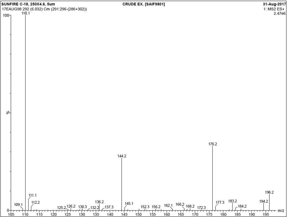
LC–MS analysis of selected plant extract for detection of available chemical derivatives.
2.4 Synthesis and characterization of Ag NPs
The 1 mL of selected plant extract filtrate and 10 mL of silver nitrate were taken for synthesis of silver nanoparticles based on the recent reports of Masum et al., (2019). Briefly, the mentioned amount of the required materials were taken together in a single test tube and heated in heavy sun light 1 h, also 1 N H3PO4 was used as a pH maintenance. After, the color was changed to green to yellow or brown to yellow in the test tube was suggested that the plant extract influenced the Ag NO3 to form silver ions and then converted to silver nanoparticles. Basically, the formed silver nanoparticles were initially proved by UV-spectroscopy and structurally confirmed with size and shape by SEM and TEM (Sahoo et al., 2020) (Fig. 2).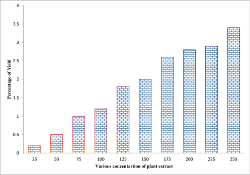
Measurement of available phenolic content percentage in the selected medicinal plant extract of Lonicera japonica.
2.4.1 Anti-cancer activity of synthesized Ag NPs by cell viability assay
The viability of A549 lung cancer cells in the presence of Ag NPs treatment plate was monitored by UV-spectrometer with the help of dimethylthiazol-diphenyltetrazolium bromide (MTT) solution interruption (Krishnamoorthy et al., 2020). The 96-well plate was evenly filled with 24 h cancer cells before filled with complete medium of RPMI and followed necessary procedure. Initially, ∼2 × 10⁴ of cancer cells were diluted into the 96-wells eventually in the 5% CO2 availability incubator for 4 h. In addition, the humidity of 95% was very effective to pure process. Then, 25–250 µg/mL concentration of synthesized Ag NPs was added into all the 96-well containing A549 lung cancer cells without control and blank. The plate was maintained based on the above said procedure for 1 day. Then, MTT solution was added after complete treatment and also control wells of the plate eventually and sealed by using aluminum foil. The plate was clearly monitored until the formation of formazan crystal production, once it formed; the plate was taken to consideration for measurement by UV-spectroscopy at 500 nm of O.D. Experiment was conducted three times without any changes and calculated each and every plate. Then, the result of IC50 value of the tested Ag NPs against A549 lung cancer cells was calculated based on the universal available formula,
3 Result
3.1 Detection of available phytochemical derivatives using LC-MS spectroscopy
In chemical composition of medicinal plant Chinese medicinal plant of Lonicera japonica was shown more than 40 different peaks in LC-MS analysis. Among these peaks, 13 different peaks were observed with phytochemical derivatives of Lonicera japonica and relatively confirmed to previous reported data of Shang et al. (2011). In addition, all these peaks were confirmed by Wiley chemical compounds Library that attached with International Repository of phytochemical derivatives, Sun Yat-Sen University, China. Previously, these 13 peaks were screened based on the retention time, occupied percentage and occupied area and have excellent biomedical inhibition effect. All those peaks were possible to relate with phenolic, flavonoids, bioactive and alkaloids derivatives. Further, the phytochemical rich derivatives of Lonicera japonica were reported with high rate of anti-oxidant property and various biomedical activities (Rantang et al., 2018). Yuke et al., 2020 also documented for anti-microbial and anti-cancer properties of Lonicera japonica. The present result was more similar to previously reported author of Tang et al. (2021) and the Chinese medicinal plant of Lonicera japonica suggested with anti-oxidant and anti-cancer properties. Based on the Wiley Repository Library reports, the list of phytochemicals, bioactive compounds, phenols and flavonoid derivatives were listed below Phenol, 2,4-bis(1,1-dimethylethyl) (206.32), 1-acetyl-3-amino-4-cyano-3-pyrroline, methyl 2-amino-3-methylbutanoate hydrochloride, Pyrrolo[1,2-a]pyrazine-1,4-dione, pyrimido[1,2-a]azepine, 2,3,4,6,7,8,9,10-octahydro-, 1,2,5-oxasilaborolane, 4,4,5-triethyl-2,2,3-trimethyl, hexahydro-3-(2-methylpro (210.27), butanoic acid, 2-hydroxy-3-methyl- (146.18 g/Mol), 5 h,10 h-dipyrrolo[1,2-a:1′,2′-d]pyrazine-5,10-dione, octahydro-, (5as-cis, 4-cyanobenzoic acid, 1-(cyclopentyl)ethyl ester), phenylethyl Alcohol (122.16 g/Mol, formamide, n-(4-[2-(1,1-dimethylethyl)-5-oxo-1,3-dioxolan-4-yl]butyl), benzoic acid, 2-amino (137.14), n,n-diethyl-2-formyl-5-methoxybenzamide, dodecanoic acid. Their total retention time was also covered with 10, 50,000 and their percentages were matched to these percentages. Further, the potential compounds were also closely matched to respective retention time, occupied area and occupied percentages. Similar result evidences were also reported by previous researcher for medicinal plant of Lonicera japonica with increased anti-oxidant activities. Previously similar result was published by Annapurna (2015), and the plant bioactive compounds were identified based on the LC-MS analysis using NIST Wiley Repository Library (Fig. 3).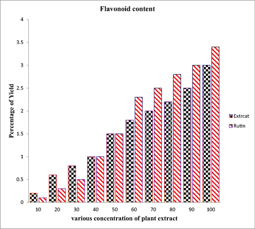
Identification of available flavonoid content percentage in the selected medicinal plant extract of Lonicera japonica.
3.2 Calculation of available photochemical amounts
In result of phenol content amount detection, the result was more favor to Lonicera japonica extract and it confirmed that the Chinese medicinal plant Lonicera japonica has more phenol content and the control of gallic acid contain R2 = 0.95052 was closely related to the extract. After comparison with universal control was revealed that the Chinese medicinal plant Lonicera japonica was very higher and effective. Further, the concentration of 10–100 µg/mL was also revealed that the extract has more phenol content and it was comparatively very high. As same as, the 10–100 µg/mL concentration of extract was also very effective for flavonoid content production. The universal positive control result of R2 = 0.9423 was clearly lower than extract production. In addition, both the result was suggested that the Chinese medicinal plant was efficiently produced phenol and flavonoid contents very high in rate, and it confirmed by after comparison of positive control of rutin. Recently, Tang et al., 2021, Yuke et al., 2020 reported that the high phenol and flavonoid content was extracted from Chinese medicinal plant of Lonicera japonica. In addition, the biological applications like anti-bacterial, anti-fungal, and anti-cancer were very high rate and evidently proved by Annapurna, 2015; Shang et al., 2011. Plant has more nutrients, hormones, nitrogen content, carbon contents and some other related contents for improve their productivity and activity (Wang et al., 2016). As same as, the medicinal plant and their secondary metabolites were eradicated most of the infectious pathogens with the help of excessive nutrients. In the stress environment, the plant produced excessive bioactive metabolites and phytochemical derivative; they are also acted as excellent biological properties and used in various biomedical and pharmaceutical properties Rantang et al., 2018.
3.3 Silver nanoparticle synthesis using Chinese medicinal plant of Lonicera japonica
Based on the MiO theory, the present Ag NPs peak was observed at the nm of 456 and it effectively checked in Fig. 4. In the silver nitrate plus extract containing test tube was slightly changed their original color after addition of Chinese medicinal plant Lonicera japonica extract at 30 min. Later, the color formation was changed to increasing time interval and changed the color from brown to pale yellow and clear nature of the sample was observed at 1 h. The surface plasma resonance between the silver nitrate and extract was clearly stated the synthesized material was Ag NPs and the plant extract was an effective precursor molecule. The color variations of the tube were suggested that the silver nitrate was converted to silver nanoparticles with the help of plant extract at 1 h time duration. Crystalline structure of the biosynthesiszed Ag NPs were carried out by XRD. The Ag nanostructure employing by Lonicera japonica plant extract was additional established by the distinguishing peaks detected in the XRD analysis in Fig. 4b. The four separate diffraction peaks of the 2θ values of 36.3°, 47.5°, 66.4° and 76.9° corresponded to the plane of (1 1 1), (2 0 0), (2 2 0) and (3 1 1) respectively. The designates Ag NPs are fcc and crystalline in nature and exhibited planes were originate to be a faultless match with the JCPDS card number 04-0783 and earlier conclusions also indicated that same JCPDS card number (Suman et al., 2013; Lee et al., 2013).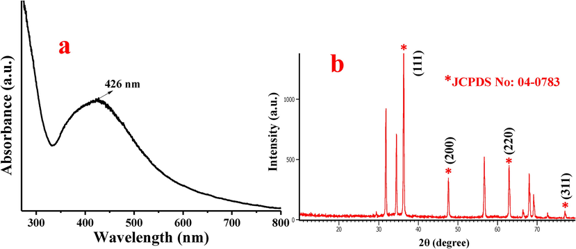
Detection of synthesized Ag NPs by UV-spectroscopy (a) and XRD (b) using medicinal plant extract of Lonicera japonica.
In addition, the SEM result of minute agglomerate structure was clearly formed in the viewed structure and it confirmed the Ag NPs presence in the viewed picture (Fig. 5a). The morphological appearance of present result was agreed by previously reported evidences of Ruíz-Baltazar et al. (2018); Velmurugan et al. (2016), and the morphology was spherical in shape. Supportively, the TEM micrograph picture was clearly delivered the original spherical morphology of Ag NPs. Also, the clear spherical shape of the Ag NPs and their size was clearly zoomed. Both the scanning electron microscopic method and transmission electron microscopic method was clearly mingled each other in the stage of shape and size (Kup et al., 2020). So, present result was accepted that the Chinese medicinal plant Lonicera japonica as an excellent precursor for synthesis of Ag NPs. Similarly Sathiyaseelan et al., 2020; reported that the plant extract is the best choice for synthesis of Ag NPs due to the low cost and environmental free nature. Recently, Wong et al., 2017; Hussain et al., 2019 reported that the biosynthesized Ag NPs was very efficient than chemical and physical method synthesis due to the less toxic nature. This statement was agreed with Que et al. (2019) and reported contrary chemical and physical methods.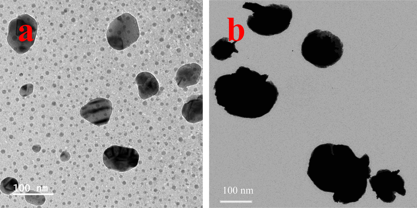
Size and shape morphology of Lonicera japonica mediated Ag NPs confirmed by SEM (a) and TEM (b).
3.4 Inhibition of A549 lung cancer cells by MTT assay
Among the concentrations of 5–75 µg/mL, the result was proved at the concentration of 75 µg/mL with 52% of cell viability. In this concentration, the viability was observed half rate due to the influence of Chinese medicinal plant of Lonicera japonica extract and it may helped to regenerate the ROS when ROS level is decreasing. The apoptosis level was very high at this concentration because of the Ag NPs. Particularly, 5–10 µg/mL concentration of Ag NPs was tried to stabilize the cells in decreased phase and helped to produce the ROS initially. After 50 µg/mL the apoptosis was increased due to the continuous production of ROS level. In addition, the nucleus was collapsed and unformed. Due to this deficient, the cancer cells failed their regeneration process and gene alteration was stimulated. Further, late apoptosis was more severe to the cancer cells and damaged the structural integrity. These evidences were clearly indicated by Jeyaraj et al. (2015); Sangour et al. (2021). In this stage, the cells were not able to produce gene expression and failed to produce the favorable enzymes (Malvin et al., 2020). Chromatin condensation was clearly happened into the cancer cells and it shrinked totally. Ag NPs may enter into the internal membranes and helped to continuous ROS production and leads to death (Ramar et al., 2015). Hence, the concentration of 75 µg/mL Ag NPs was more efficient than other concentration for damaged the A549 lung cancer cells and this concentration was fixed as an IC50. Recently, Almalki and Khalifa (2020), Rajesh Kumar et al. (2018) reported that the plant mediated Ag NPs was very efficient for ROS production and helped to more apoptosis process (Fig. 6).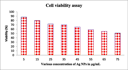
Anti-cancer ability of Ag NPs proved against A549 lung cancer cells by cell viability assay.
4 Conclusion
Based on the above study, the excellent anti-oxidant property of the Chinese medicinal plant of Lonicera japonica was chosen in this study for synthesis of Ag NPs. As per result, the SPR peak of the synthesized Ag NPs was confirmed by UV-spectroscopy and morphological with size and shape of the synthesized Ag NPs were confirmed by microscopic observations. In biomedical process, the result was proved that the selected plant extract has anti-cancer properties against tested A549 lung cancer cells and confirmed by decreased viability formation. The phase contrast images were supported to MTT result, and clearly stated that the synthesized Ag NPs damaged the morphology of A549 human lung cancer cells at very lowest concentration. Finally, the present study was proved that the synthesized Ag NPs were very efficient against A549 lung cancer cells and it will be used in future for best drug choice with complete inhibition of cancer cells.
Acknowledgement
All the authors gratefully acknowledge the National Natural Science Foundation of China (Project Approval Number: 41950410573) and Postdoctoral Science Foundation of China (Project Approval Number: 2019M663213) for financial support for this work. Wen-Jun Li was also supported by Introduction project of high-level talents in Xinjiang Uygur Autonomous Region. The authors extend their appreciation to the Researchers Supporting Project number (RSP-2021/70), King Saud University, Riyadh, Saudi Arabia.
Declaration of Competing Interest
The authors declare that they have no known competing financial interests or personal relationships that could have appeared to influence the work reported in this paper.
References
- Antioxidant, cytotoxic and antibacterial potentials of biosynthesized silver nanoparticles using bee’s honey from two different floral sources in Saudi Arabia. Saudi J. Biol. Sci.. 2020;27(1):363-373.
- [Google Scholar]
- Silver nanoparticles synthesis from Bacillus sp KFU36 and its anticancer effect in breast cancer MCF-7 cells via induction of apoptotic mechanism. J. Photochem. Photobiol. B: Biol.. 2020;204:111786
- [Google Scholar]
- Annapurna M.S., Traditional applications and phytochemical investigations of Lonicera japonica Thunb. 7, 2015, 042-049.
- Development and evaluation of different strategies for the clean synthesis of silver nanoparticles using Yarrowia lipolytica and their antibacterial activity. Process Biochem.. 2020;94:319-328.
- [Google Scholar]
- Effects of extraction solvents on total phenolic and flavonoid contents and biological activities of extracts from Sudanese medicinal plants. S. Afr. J. Bot.. 2019;120:261-267.
- [Google Scholar]
- Inhibitory activity of biofunctionalized silver-capped N-methylated water-soluble chitosan thiomer for microbial and biofilm infections. Int. J. Biol. Macromol.. 2020;152:709-717.
- [Google Scholar]
- The development of callus and cell suspension cultures of Sabah Snake Grass (Clinacanthus nutans) for the production of flavonoids and phenolics. Biocatal. Agric. Biotechnol.. 2021;33:101977
- [Google Scholar]
- Natural polysaccharide derived carbon dot based in situ facile green synthesis of silver nanoparticles: Synergistic effect on breast cancer. Int. J. Biol. Macromol.. 2020;20:34074-34075.
- [Google Scholar]
- Evaluation of the catalytic, antibacterial and anti-biofilm activities of the Convolvulus arvensis extract functionalized silver nanoparticles. J. Photochem. Photobiol. B: Biol.. 2017;167:36-44.
- [Google Scholar]
- Effects of green-synthesized silver nanoparticles on lung cancer cells in vitro and grown as xenograft tumors in vivo. Int. J. Nanomed.. 2016;11:1879-1887.
- [Google Scholar]
- Hybridization and functionalization with biological macromolecules synergistically improve biomedical efficacy of silver nanoparticles: Reconceptualization of in-vitro, in-vivo and clinical studies. J. Drug Delivery Sci. Technol.. 2019;54:101169
- [Google Scholar]
- Biogenic metal nanoformulations induce Bax/Bcl2 and caspase mediated mitochondrial dysfunction in human breast cancer cells (MCF 7) RSC Adv.. 2015;5:2159.
- [Google Scholar]
- Avicennia marina engineered nanoparticles induce apoptosis in adenocarcinoma lung cancer cell line through p53 mediated signaling pathways. Process Biochem.. 2020;94:349-358.
- [Google Scholar]
- Biosynthesis of silver nanoparticles using leaf extract of Aesculus hippocastanum (horse chestnut): Evaluation of their antibacterial, antioxidant and drug release system activities. Mater. Sci. Eng. C. 2020;107:110207
- [Google Scholar]
- Eco-friendly green synthesis of clove buds extract functionalized silver nanoparticles and evaluation of antibacterial and antidiatom activity. J. Microbiol. Methods. 2020;173:105934.
- [Google Scholar]
- Synthesis of silver nanoparticles using cow milk and their antifungal activity against phytopathogens. Mater. Lett.. 2013;105:128-131.
- [Google Scholar]
- Synthesis of silver nanoparticles using oxidized amylose and combination with curcumin for enhanced antibacterial activity. Carbohydr. Polym.. 2020;230:115573.
- [Google Scholar]
- Biosynthasized silver nanoparticle using Bacillus amyloliquefaciens: Application of cytotoxicity effect on A549 cell line and photocatalytic degradation of p-nitrophenol. J. Photochem. Photobiol. B: Biol.. 2020;202:111642
- [Google Scholar]
- Mani M., Okla M.K., Selvaraj S., Ram Kumar A., Kumaresan S., Muthukumaran A., Kaviyarasu K., El-Tayeb M.A., Elbadawi Y.B., Almaary K.S., Mohsen A. Almunqedhi B., Elshikh M.S., 2021. A novel biogenic Allium cepa leaf mediated silver nanoparticles for antimicrobial, antioxidant, and anticancer effects on MCF-7 cell line, Environ. Res., 198, 111199.
- Systematic green synthesis of silver oxide nanoparticles for antimicrobial activity. Environ. Res.. 2021;202:111627.
- [Google Scholar]
- Biogenic synthesis of silver nanoparticles using Phyllanthus emblica fruit extract and its inhibitory action against the pathogen Acidovorax oryzae strain RS-2 of rice bacterial brown stripe. Front. Microbiol.. 2019;10:820.
- [Google Scholar]
- Evaluation of the effect of organic pollutants exposure on the antioxidant activity, total phenolic and total flavonoid content of lettuce (Lactuca sativa L.) using UV–Vis spectrophotometry and chemometrics. Microchem. J.. 2021;170:106632.
- [Google Scholar]
- Synthesis of silver nanoparticles colloids in imidazolium halide ionic liquids and their antibacterial activities for gram-positive and gram-negative bacteria. Chemosphere. 2020;243:125302.
- [Google Scholar]
- Qais F.A., Shafiq A., Ahmad I., Husain F.M., Ahmad Khan R., Hassan I., 2020. Green synthesis of silver nanoparticles using Carum copticum: Assessment of its quorum sensing and biofilm inhibitory potential against gram negative bacterial pathogens, Microbial Pathog., 144, 104172.
- Size dependent anti-invasiveness of silver nanoparticles in lung cancer cells. RSC Adv.. 2019;9:21134-21138.
- [Google Scholar]
- Determination of selected biogenic amines in Acacia rigidula plant materials and dietary supplements using LC–MS/MS methods. J. Pharm. Biomed. Anal.. 2014;88:457-466.
- [Google Scholar]
- Rajesh kumar, S., Venkat Kumar, S., Ramaiah, A., Happy, A., Lakshmi, T., Mohana Roopan, S., 2018. Biosynthesis of Zinc oxide nanoparticle using Mangifera indica leaves and evaluation of their antioxidant and cytotoxic properties in lung cancer (A549) cells. 117, 91-95.
- Synthesis of silver nanoparticles using Solanum trilobatum fruits extract and its antibacterial, cytotoxic activity against human breast cancer cell line MCF 7. Spectrochim. Acta A, Mol. Biomol. Spectrosc.. 2015;140:223-228.
- [Google Scholar]
- Lonicerae Flos: A review of chemical constituents and biological activities. Digital Chinese Med.. 2018;1(2):173-188.
- [Google Scholar]
- Development of noncytotoxic silver–chitosan nanocomposites for efficient control of biofilm forming microbes. RSC Adv.. 2017;7(83):52398-52413.
- [Google Scholar]
- Biosynthesis of Ag nanoparticles using Cynara cardunculus leaf extract: Evaluation of their antibacterial and electrochemical activity. Results Phys.. 2018;11:1142-2114.
- [Google Scholar]
- Biogenic silver nanoparticle synthesis with cyanobacterium Chroococcus minutus isolated from Baliharachandi sea-mouth, Odisha, and in vitro antibacterial activity. S. J. Biol. Sci.. 2020;27(6):1580-1586.
- [Google Scholar]
- Effect of Ag nanoparticles on viability of MCF-7 and Vero cell lines and gene expression of apoptotic genes. Egypt. J. Med. Hum. Genet.. 2021;22:9.
- [CrossRef] [Google Scholar]
- Biocompatible fungal chitosan encapsulated phytogenic silver nanoparticles enhanced antidiabetic, antioxidant and antibacterial activity. Int. J. Biol. Macromol.. 2020;153:63-71.
- [Google Scholar]
- Lonicera japonica Thunb.: Ethnopharmacology, phytochemistry and pharmacology of an important traditional Chinese medicine. J. Ethnopharmacol.. 2011;138:1-21.
- [Google Scholar]
- Biosynthesis, characterization and cytotoxic effect of plant mediated silver nanoparticles using Morinda citrifolia root extract. Colloids Surf., B Biointerfaces. 2013;106:74-78.
- [Google Scholar]
- Potential application of lonicera japonica extracts in animal production: from the perspective of intestinal health. Front. Microbiol.. 2021;12:719877
- [Google Scholar]
- Anti-biofilm and cytotoxicity activity of impregnated dressings with silver nanoparticles. Mater. Sci. Eng. C. 2015;49:604-611.
- [Google Scholar]
- Phyto-crystallization of silver and gold by Erigeron annuus (L.) Pers flower extract and catalytic potential of synthesized and commercial nano silver immobilized on sodium alginate hydrogel. J. Saudi Chem. Soc.. 2016;20:313-320.
- [Google Scholar]
- Research progress on chemical constituents of Lonicerae japonicae flos. Biomed. Res. Int.. 2016;2016:1-18.
- [Google Scholar]
- Incidence and mortality of lung cancer: global trends and association with socioeconomic status. Sci. Rep.. 2017;7:14300.
- [Google Scholar]
- Lonicerae japonicae flos and Lonicerae flos: a systematic review of ethnopharmacology, phytochemistry and pharmacology. Phytochem. Rev.. 2020;19(1):1-61.
- [Google Scholar]


