

Translate this page into:
Synthesis of gold nanoparticles (AuNPs) with improved anti-diabetic, antioxidant and anti-microbial activity from Physalis minima
⁎Corresponding authors. Al-Ansari-myalansari@ksu.edu.sa (Mysoon M. Al-Ansari), amutha1994santhanam@gmail.com (Amutha Santhanam)
-
Received: ,
Accepted: ,
This article was originally published by Elsevier and was migrated to Scientific Scholar after the change of Publisher.
Peer review under responsibility of King Saud University.
Abstract
Background
Gold nanoparticles have come to prominence among noble metals due to their characteristics. Bio-transformed nanoparticles have a low toxicity and are biocompatible and ecologically benign. Physalis minima has been used in traditional medicine for its diuretic, laxative, and anti-inflammatory qualities since ancient times.
Methods
Gold nanoparticles are formed when water gold metal ions interact with an aqueous plant extract of Physalis minima (Au NPs).
Results
In a UV–visible spectrophotometer, the resulting Au NPs formed a broad surface peak at 546 nm. According to the FTIR data, the peaks represent flavonoids, phenols and polyphenols meaning that flavonoids, phenols, and polyphenols were utilised as the reducing agent to create gold nanoparticles. According to the XRD pattern, the synthesised gold nanoparticles are crystalline in nature in a cubic system, with average particle sizes of roughly 36 nm. Gold nanoparticles were evaluated and found to have a zeta potential of −20.5mv, suggesting that they were stable and suitable for biological uses. The morphology of gold nanoparticles was confirmed using microscopic imaging.
Conclusion
This study looked at the anti-oxidant, anti-diabetic, and antibacterial properties of gold nanoparticles (Au NPs) made from Physalis minima, making it exceedingly promising for biological applications.
Keywords
Gold nanoparticles
Physalis minima
Antioxidant
Antidiabetic
Antimicrobial
Biomedical application

1 Introduction
Biosynthesis of metal nanoparticles utilising an environmentally safe and green synthesis process is presently in high demand to reduce unwanted effects in therapeutic and medical applications (Elahi et al., 2018; Kalimuthu et al., 2020; Lee et al., 2020; Hu et al., 2020; Wani, 2017). In contrast to damaging chemical synthesis techniques, biological approaches such as plants and microorganisms offer a wide range of uses in dentistry, imaging, medication administration, sensing, photothermal treatment, and many other domains. In this way, plant-mediated biosynthesis via the bio reduction process has increased anti-diabetic, antioxidant, and anti-microbial capabilities. The bulk of bio converted NPs come from metals like Au, Ag, Cu, Pt, Zn, and Fe that are processed against various organic agents that comprise various portions of a plant material (Qiao and Qi, 2021; Bapat et al., 2020; Siddiqi and Husen, 2017; Patil et al., 2020), due to their phytochemical composition to the bio reduction process.
Gold nanoparticles (AuNPs), for example, have a long history of application in bioimaging, drug delivery vehicles, and other fields. The unique physiochemical and photothermal properties of gold nanoparticles govern the conjugation and distribution of tailored medicines to specific tissues or cells. Au-NPs have several unique features, including surface Plasmon resonance and optical criteria. Biosynthesised metal nanoparticles are biocompatible and environmentally beneficial. Au-NPs have recently been used in imaging and medicinal applications to investigate their unique properties. Engineered NPs may now be labelled with biological, physiological, and biomedical carriers and used in nano-based systems to deliver potent and precise drug delivery in cancer cell imaging zones (Chahardoli et al., 2018; Jat et al., 2020).
Recent research focuses on the various structures of gold nanoparticles such as gold spheres, nanocubes, nanorods, nanostars, and nanocages. In addition, gold nanoparticles are functionalized with probe, polymers, mono clonal antibodies for biomedical applications (Al-Ansari et al., 2021; Vijilvani et al., 2020; Nagaraj et al., 2022). The perennial herb Physalis minima (Native gooseberry) is a member of the Physalis genus and belongs to the Solanaceae family (Grunberger et al., 1988). It is a pantropical annual herb that grows to be 20–50 cm tall when fully grown. P. minima fruit contained 61.4% and 76.7% moisture. There are also sugars, tannins, minerals, protein, pectin, and a good amount of vitamin C (24.45 mg/100 ml juice). The seed, according to reports, contains oil and protein. Palmitic, stearic, oleic, and linoleic fatty acids, as well as minor amounts of hexadecenoic and hydroxy fatty acids, are all present in the oils (Grunberger et al., 1988). Withanone, withaferin A, withanolide A, stigmasterol, and sitosterol are found in the leaves, root, and stem, whereas withanolide A, withanone, withaferin (fruits and flowers), dihydroxyphysalin B2-4, and physalins A, B, and X are found in the fruits and flowers (aerial part). Physalindicanols, withaphysalin E, C, D, physalinicanol A, withaphysalin A, physalin, physalinicanol A, withametelins, withanolide, withangulatins, phygrine, withaphysalin A, B, C, D, withaphysalin, withaphysalin A, B, C, D (Zhu et al., 2002). In rats, the antiulcer characteristics of P. minima aqueous extract (AEPM) were discovered.
The fruits of P. minima insides have a variety of uses, including anticancer which are particularly useful for colon and thyroid cancers (Karpagasundari and Kulothungan, 2014a, 2014b). Alkaloids, steroids, tannins, phenol glycosides, sugars, and anthocyanins were identified in high amounts in the methanolic extract of raw Physalis minima fruit. HPLC is used in conjunction with other detection methods, like as UV and MS, to provide preliminary data on the amount and properties of chemicals discovered in active extracts, especially steroid alkaloids (Patel et al., 2009; Nurhaslina et al., 2019). On the basis of fruit development, the levels of vitamin C, phenol chemicals, and free amino acids were measured.
Based on the scientific benefit of P. minima, this current study is concerned with the synthesis of gold nanoparticles from the herbal plant physalis minima aqueous extract via gold ion bio-reduction due to its phytochemicals. The anti-diabetic, anti-oxidant, and anti-bacterial activities of the biosynthesized gold nanoparticles are thoroughly investigated using various characterisation methods.
2 Materials and methods
2.1 Plant collection
The herb Physalis minima were collected from an agriculture field in the Tiruvallur district region of Tamil Nadu, India, near Chennai. The plant sample were collected from the latitude of 13.264080, longitude of 79.848470 and GPS coordinates: 13° 15′ 50.6880″ N 79° 50′ 54.4920″ E. The plant was identified by Dr. K.R. Jayappriyan at Department of Centre for advanced study in Botany, University of Madras. All of the chemicals used in this investigation were analytical grade and purchased from Hi-media, Sigma Aldrich.
2.2 Bio-reduction of gold nanoparticles
The Physalis minima plant was carefully plucked and cleansed before being cut into thin pieces to make an aqueous extract. The 20 g of the whole plant were boiled in 100 ml of distilled water to create the aqueous extract. After 20 min, the boiling extract was collected using Whatman no.1 filter paper. Three distinct concentrations of 0.1 mM gold chloride solution and aqueous extract of Physalis minima were used to make gold nanoparticles, namely 1:10, 1:5, and 1:3, and the mixture was blended for an hour before being stored in the dark for bio reduction. The purple-colour solution that resulted was saved for additional characterisation experiments.
2.3 Characterizations of biosynthesized AuNPs
The preliminary conformation of gold nanoparticles was measured using a UV–visible spectrophotometer with wavelengths spanning from 200 to 800 nm (PerkinElmer UV Lambda 650 UV/VIS), which conforms gold nanoparticle formation. The size and surface morphology of the generated gold nanoparticles were determined using a scanning electron microscope (ZEISS Gemini SEM 360), which clearly exhibits a tube-like morphology with a flower-like structure. The geometry and morphology of biosynthesized AuNPs were assessed using a Bruker X-flash in energy dispersive X-ray spectroscopy (eZAF). For EDX and imaging, the electron beam's energy was kept at 20 keV. Under FT-IR spectrometers-thermo iS50, the functional groups involved in the production of gold nanoparticles were examined. HORIBA SZ-100 for Windows [Z Type] Ver2.20 was used to test the stability of the produced gold nanoparticles. The stability of the synthesized gold nanoparticles was determined by HORIBA SZ-100 for Windows [Z Type] Ver2.20. The size and structure of the particles were subsequently investigated using TEM (FEI Tecnai G220 S-TWIN), which revealed the size of gold nanoparticle with a morphology. Powder X-Ray diffractometer (D8 Advance Bruker) was used to characterise the crystallinity, phase purity, and crystal systems of the produced gold nanoparticles.
2.4 Antioxidant assay
2.4.1 DPPH (2,2-diphenyl-1-picrylhydrazyl) assay
The free radical scavenging activity of aqueous plant extract and gold nanoparticles was determined using the DPPH radical method. The aqueous plant extract and gold nanoparticles were mixed properly and incubated in the dark at room temperature for 30 min, with the sample's optical density measured at 517 nm. 1.5 ml of 0.1 mM DPPH solution was added, and the final test tube volume was made up to 2 ml using sterile distilled water. The linear regression value was calculated using ascorbic acid as the standard (Nurhaslina et al., 2019).
The proportion of radical scavenging activity was calculated using the formula below (RSA)
2.5 Anti-diabetic assay (α-Amylase inhibition assay)
The 3,5-dinitrosalicylic acid (DNS) technique was used to perform the amylase inhibition assay, as described (Kifle and Enyew, 2022). The gold nanoparticles and aqueous extract were dissolved in sodium phosphate buffer at various concentrations of 100–500 mg/ml. A total of 200U amylase (Units/mL) was combined with various qua µl of the starch solution, which was incubated for 3 min. Adding 200 µl of DNS reagent and boiling for 10 min in 85 °C stopped the process. The absorbance was determined at 540 nm using a UV spectrophotometer after the mixture was blended with 5 ml of distilled water. By substituting the sample in different concentrations in buffer, a blank with 100% enzyme activity was created. In the absence of the enzyme solution, a blank reaction was generated using the sample to be evaluated at each concentration. A positive control sample was made with acarbose (commercial), and the reaction was carried out in the same way as the test sample reaction.
The inhibition of α amylase was stated as a percentage of inhibition, and the following equation was used to compute it:
where Ac is the control's absorbance, Acb is the control blank's absorbance, As is the sample's absorbance, and Asb is the sample blank's absorbance.
2.6 Antimicrobial studies
Staphylococcus aureus (ATCC25923), Pseudomonas aeruginosa (ATCC10231), Streptococcus pneumoniae (ATCC49619), and E. coli (ATCC11229) were used in this study. The bacterial cultures were maintained as a glycerol stock at deep freezer −80 °C in the laboratory. A single colony was revived from the glycerol stock culture and maintained as subculture in the nutrient broth at 4 °C. The antibacterial activity was tested using agar well diffusion (Al-Dahmash et al., 2021). The suspended culture was equally spread on Nutrient agar plates. The solid medium was then gently perforated with a cork borer to produce wells, and each well was filled with 100 µl of synthesized AuNPs. The plates were incubated for 24 h at 37 °C. As a control, the antibiotic amoxicillin was utilized. The diameter of the inhibition zone generated around the well was used to measure antibacterial activity.
2.7 Statistical analysis
All data were presented as the mean ± standard deviation from triplicate runs (n = 3). Data are given as the Mean ± SD; statistics were performed using t-tests and p values less than 5% were considered statistically significant (p < 0.05).
3 Results
3.1 UV visible spectroscopy analysis
The gold nanoparticles that had been synthesised in this study was examined using UV–visible spectroscopy. After adding aqueous plant extract to Au+ ions solution, biotransformation of gold ions into bunches of atoms (AuNPs) was detected. The resulted purple colour is due to Surface Plasmon Resonance (SPR). Colour shifts in phytofabricated AuNPs revealed the presence of excited electrons Fig. 1(a). At wavelengths spanning from 200 to 800 nm, UV spectra were obtained. The presence of AuNps synthesised in three distinct ratios (1:3, 1:5, 1:10), utilising Physalis minima aqueous plant extract was indicated by a robust and broad surface plasmon peak at 546 nm, 544 nm, 555 nm in Fig. 1(b). The absorption band visible around 500–600 nm is the distinctive peak of gold nanoparticles. AuNps have a distinct UV absorbance band due to the excitation mode of their surface plasmons, which varies with nanoparticle size.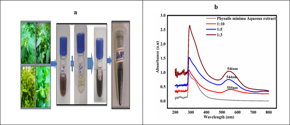
a) Initial screening of synthesis process of AuNPs; b) UV–Visible spectrum of synthesized gold nanoparticles from the aqueous extract of Physalis minima.
3.2 Fourier transform infrared spectroscopy [FTIR]
The biomolecules responsible for bio-reduction, capping, or stabilising the AuNPs synthesised using Physalis minima aqueous extract were identified using FTIR measurements in Fig. 2. FT-IR analysis was performed on Physalis minima aqueous extract and phytosynthesised gold nanoparticles. The spectra of AuNPs was collected in the 500–4000 cm−1 region to produce an excellent signal-to-noise ratio. At 3200 cm−1, 2100 cm−1, and 1600 cm−1, there are notable intensity peaks. Functional groups such as C–H stretch (aromatics), CH3 –R, N–H, C–O–C, and C=O stretching were detected at various wave numbers. The strong band at 3297 cm−1 may be caused by the stretching vibration of OH or NH groups found in carbohydrates or proteins, which is the fundamental cause of bio-reduction.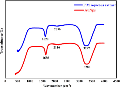
FTIR Spectrum of biosynthesized gold nanoparticles and aqueous extract of Physalis minima.
3.3 X-ray powder diffraction [XRD]
The crystalline size, structure, and Phase purity of the synthesised gold nanoparticles were validated by X-ray diffraction analysis. Fig. 3 shows the XRD pattern of biosynthesized gold nanoparticles. The X-ray diffraction pattern of AuNPs synthesised from Physalis minima aqueous extract showed dominant peaks at 38.089, 44.256,64.379,77.312, 81.412 in the 2 θ range which originated for (1 1 1), (0 0 2), (0 2 2), (1 1 3) and (2 2 2) planes with crystalline cubic lattice of nanogold (JCPDS:98-006-2677).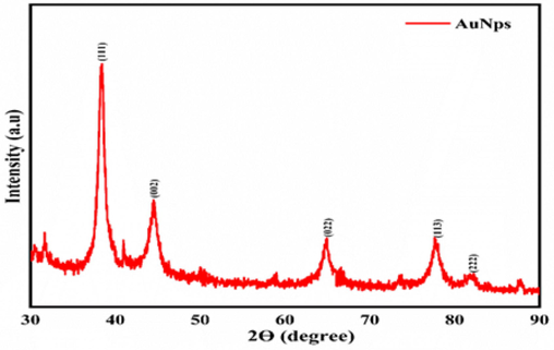
XRD Pattern of synthesized gold nanoparticle from the aqueous extract of Physalis minima.
3.4 Microscopic image analysis
SEM is used to determine the structure and morphology of photosynthesized gold nanoparticles. The structure of the gold nanoparticles obtained here are triangular and cube-like, with irregular and agglomeration flower-like features. The elemental distribution of the material is determined via EDAX analysis; the EDAX spectrum distinctly shows the presence of gold nanoparticle shows in Fig. 4.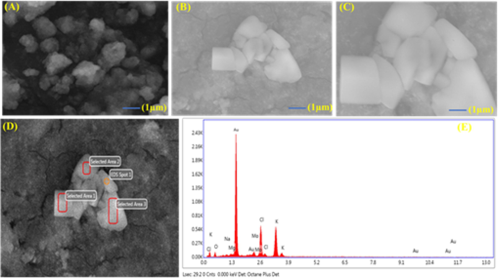
SEM micrographs and EDAX Spectrum of biosynthesized gold nanoparticles.
3.5 High resolution transmission electron microscope (HR-TEM)
HR-TEM is used to examine the size and structural conformation of biosynthesized gold nanoparticles. Fig. 5 depicts the average particle size and structure. The average particle size was 15 nm and the majority of the nanoparticles were spherical in shape. The formation of gold nanoparticles is due to bio-reduction of gold ions with the phytochemical present in the Physalis minima.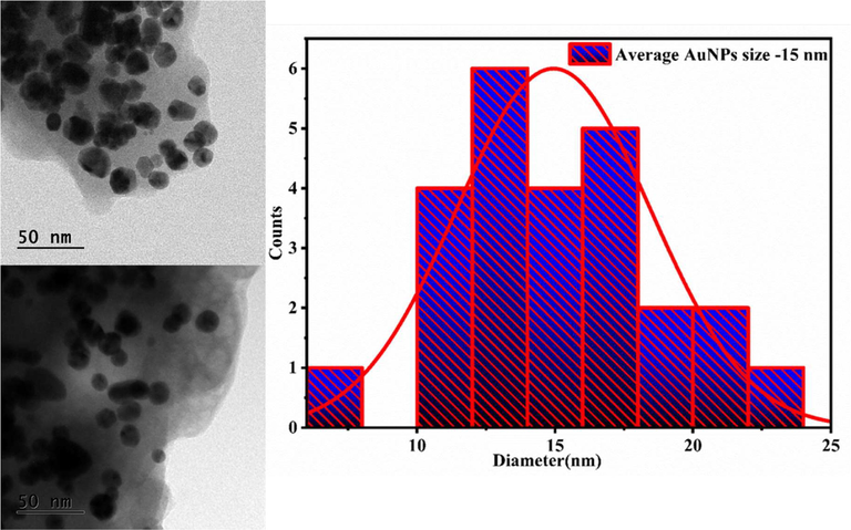
HR-TEM Picture and distribution curve of biosynthesized gold nanoparticles.
3.6 Dynamic light scattering analysis (DLS) and zeta potential
DLS analysis was used to determine the hydrodynamic size of the gold nanoparticles. The hydrodynamic size of the synthesised gold nanoparticles was 65 nm in Fig. 6. The stability of photosynthesized gold nanoparticles was discovered to be around −20.5 mv in Fig. 6. The figures are indicating that they are suitable for biological applications.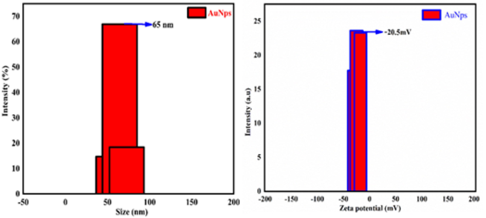
Dynamic light scattering (DLS) and Zeta potential investigation of synthesized gold nanoparticles.
3.7 Antioxidant assay
The free radical scavenging DPPH test was used to examine the antioxidant capabilities of phytochemical substances using a spectrometric technique. The ability of phytofabricated AuNPs and Physalis minima aqueous extract to transport DPPH (free radicals) was analysed at various concentrations (100, 200, 300, 400, and 500 mg/mL). The DPPH free radicals carrying property of the extract, the Phyto generated AuNPs, increased significantly with increasing concentrations, as shown in Fig. 7. The plant extract and AuNPs showed maximum DPPH free radical scavenging activity approximately 86–90% (v/v) and the result shows statistically significant (p < 0.05). The AuNPs had a larger surface area and catalytic capabilities, which could have facilitated their antioxidant properties. Based on the DPPH assay, it was determined that aqueous extract of Physalis minima and Phyto synthesised gold nanoparticles are excellent antioxidant agents.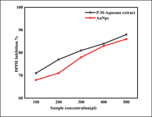
Antioxidant activity of Physalis minima aqueous extract and biosynthesized gold nanoparticles.
3.8 Antidiabetic activity of AuNPs and Physalis minima aqueous extract (α-amylase inhibition assay)
The anti-diabetic potential of Physalis minima aqueous extract and photosynthesized gold NPs were investigated by alpha amylase inhibition assay in Fig. 8. The aqueous extract and Phyto fabricated AuNPs suppressed the most alpha amylase enzyme activity and had a 90–93% (v/v) anti-diabetic effect (p < 0.05). The presence of phytochemicals such as flavonoids and polyphenols have the explanation for Au-NPs' highest amylase inhibitory property which has been confirmed by phytochemical and FTIR studies. The synthesized gold nanoparticles from the plant Physalis minima had an excellent antioxidant because they minimise oxidative stress, and enhanced anti-diabetic activity. Furthermore, the nanoparticles inhibit carbohydrate hydrolysing enzymes. This study shows that Physalis minima aqueous extract and Phyto generated gold nanoparticles are promising anti-oxidant and anti-diabetic agents.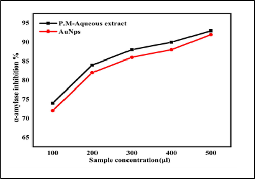
Antidiabetic activity of Physalis minima aqueous extract and synthesized gold nanoparticles.
3.9 Antimicrobial activity
The antimicrobial activity of synthesised gold nanoparticles was investigated using the agar well diffusion assay, in which 100 µl of synthesised gold nanoparticles were added to an agar well and incubated for 12 h, while βlactam antibiotic was used as a positive control in Fig. 9. The zone created around the well was measured after incubation. From this, it was concluded that herbal plant-mediated nanoparticles were found to have the potential to be used as antibacterial carriers (Table1).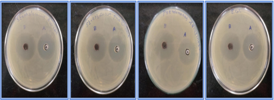
Antimicrobial activity of biosynthesized gold nanoparticles against Staphylococcus aureus (ATCC-25923); Streptococcus pneumoniae (ATCC-49619; Pseudomonas aeruginosa (ATCC-10231), 4-Escherichia coli (ATCC-11229). Zone of A- control, Zone of B- Bacteria.
S.NO
Name of the microorganisms
Zone of inhibition
Gold nanoparticles
Control
1.
Staphylococcus aureus (ATCC-25923)
4.9 mm
4.5 mm
2.
Pseudomonas aeruginosa (ATCC-10231)
5.6 mm
5.2 mm
3.
Streptococcus pneumoniae (ATCC-49619)
6.2 mm
5.8 mm
4.
Escherichia coli (ATCC-11229)
6.6 mm
6.2 mm
4 Discussion
The distinctive peak of gold nanoparticles is the absorption band visible around 500–600 nm. Because of the excitation mode of their surface plasmons, which varies with nanoparticle size, AuNps have a distinct absorbance band. Surface plasmon resonance (SPR) absorption is caused by the simultaneous vibration of metal nanoparticle electrons in resonance with light waves in metal nanoparticles. Nanogold has SPR bands about 530–550 nm, according to earlier findings (Mujeeb et al., 2020; Noruzi 2015). In this study, the SPR peak for Physalis minima was found around 540–560 nm. As a result, the aqueous extract of Physalis minima was discovered to have a significant potential for bio-reduction of Au ions to Au-NPs. Metal ions were bio transformed into NPs in a quick and efficient manner.
The stretching vibration of OH or NH groups found in carbohydrates or proteins might create the strong band at 3297 cm−1, which is the primary reason for the bio-reduction. Phytochemicals are the cause of gold ion reductions, according to FTIR research, therefore other phytochemicals are the cause of gold ion reductions, and other similar studies indicate similar vibrations in the FTIR (Ashwini and Mahalingam 2020; Hadi et al., 2016). Similar XRD studies for other plant-mediated gold nanoparticle production may be found (Khatua et al., 2020; Annamalai et al., 2013). The (1 1 1) plane produced the most powerful peak in Au-NPs biosynthesised from Physalis minima crystals, suggesting the crystals preferred orientation. As a result, Gold NPs were recognised as Phyto fabricated NPs. The present data confirm the distribution of AuNPs, the similar gold nanoparticles made through phytosynthesis have been reported earlier (Shahriari et al., 2019; Gupta and Padmanabhan, 2021). The HR-TEM report of Physalis minima identified the nanoparticles with smaller size and shape, the similar structures were found in TEM analysis in other studies (Shahriari et al., 2019; Gupta and Padmanabhan, 2021; Hammami et al., 2021). The DLS findings of this study are also corroborated by previous research (Wacławek et al., 2018; Uzma et al., 2020). The biosynthesized gold nanoparticles have a good degree of stability and are in the nm range, according to the PDI (0.25) and nanoparticle size measurements.
The ROS activity of gold nanoparticles was measured using DPPH. Many illnesses, including diabetes, high blood pressure, atherosclerosis, cancer, and others, are linked to oxidative stress. The herbal plant Physalis minima has stronger antioxidant and anti-diabetic effects. Antioxidants are organic chemical compounds that, even at low concentrations, can block or slow down a substrate's oxidation reaction (Al-Radadi, 2022). According to comparable studies (Karpagasundari and Kulothungan, 2014a, 2014b), Physalis minima contains antioxidants. Although the antioxidant capacities of synthesised AuNPs have yet to be explored, similar plant-mediated synthesised gold nanoparticles have been reviewed (Veeramani et al., 2021; Ghosh et al., 2020).
Diabetes affects people all across the world and the best therapy is found in nature. The alpha amylase inhibition experiment was performed to show that the anti-diabetic effects of Physalis minima aqueous extract and synthesised gold nanoparticles are different. T2DM (Type 2 Diabetes Mellitus) is a carbohydrate metabolic disorder that results in hyperglycemia and has been linked to cardiovascular and neurological complications. Long-chain complex carbohydrates can be converted to simple glucose by two enzymes: alpha-glucosidase and alpha-amylase. As a result, deactivating these enzymes is a common T2DM therapy. Physalis minima extract and Physalis minima mediated synthesised gold nanoparticles have not been examined for anti-diabetic action so far, however similar plant mediated synthesised gold nanoparticles and aqueous extract have been demonstrated (Banerjee et al., 2017; Veeramani et al., 2021). The antimicrobial assay microorganisms used here are clinically significant pathogens that cause serious infections, and it is critical to combat them with active therapeutic giants. Gram-positive bacteria have a thick peptidoglycan layer, but Gram-negative bacteria have a thin lipopolysaccharide coating that is impermeable to lipophilic substances. However, the exact mechanism of antimicrobial action is not fully understood. It is speculated that nanoparticles cause morphological changes in cell wall membrane structure, disrupting membrane permeability, and ultimately leading to cell death. Free radicals, which interact with proteins, lipids, and DNA, are produced by the nanoparticles and it obviously suggests that the Physalis minima mediated gold nanoparticles have good antimicrobial property. It was also proven that phytosynthesized gold nanoparticles showed enhanced antimicrobial activity (Donga et al., 2020; Durga et al., 2020).
5 Conclusions
When compared to other synthesis techniques, the waste to wealth concept [herbal plant to sustainable material with enhanced biological activity] has an abundant source of raw material, and the synthesis procedure is relatively simple, inexpensive, most significantly eco-friendly. Gold nanoparticles were successfully synthesised from the herbal plant Physalis minima and confirm with different characterization analyses. The particles have a hydrodynamic average particle size of 65 nm, and zeta potential study has shown that they are stable for biomedical use. The photosynthesized gold nanoparticles from physalis minima were exhibited a good activity for antidiabetic, antioxidant, and antibacterial effects. The synthesised Au-NPs generated from Physalis minima extract show superior therapeutic anti-diabetic and anti-microbial properties and can be employed as optimal drug delivery and industrial values in the future Nano biopharma and biomedical industries.
Acknowledgements
The authors extend their appreciation to the Researchers Supporting Project Number (RSP-2021/228), King Saud University, Riyadh Saudi Arabia.
Declaration of Competing Interest
The authors declare that they have no known competing financial interests or personal relationships that could have appeared to influence the work reported in this paper.
References
- Synthesis of silver nanoparticles using gum Arabic: Evaluation of its inhibitory action on Streptococcus mutans causing dental caries and endocarditis. J. Infect. Public Health. 2021;14(3):324-330.
- [CrossRef] [Google Scholar]
- Frankincense, an aromatic medicinal exudate of Boswellia carterii used to mediate silver nanoparticle synthesis: Evaluation of bacterial molecular inhibition and its pathway. J. Drug Delivery Sci. Technol.. 2021;61:102337.
- [Google Scholar]
- Biogenic proficient synthesis of (Au-NPs) via aqueous extract of Red Dragon Pulp and seed oil: Characterization, antioxidant, cytotoxic properties, anti-diabetic anti-inflammatory, anti-Alzheimer and their anti-proliferative potential against cancer cell lines. Saudi J. Biol. Sci.. 2022;29(4)
- [CrossRef] [Google Scholar]
- Green synthesis, characterization and antimicrobial activity of AuNPs using Euphorbiahirta L. leaf extract. Colloids Surf. B Biointerfaces. 2013;108:60-65.
- [CrossRef] [Google Scholar]
- Green synthesized metal nanoparticles, characterization and its anti-diabetic activities- a review. Res. J. Pharm. Technol.. 2020;13:468-474.
- [CrossRef] [Google Scholar]
- Green synthesis of gold NPs using Sumac aqueous extract and their antioxidant activity. Int. J. Curr. Pharm. Res.. 2017;9
- [CrossRef] [Google Scholar]
- Recent advances of gold nanoparticles as biomaterial in dentistry. Int. J. Pharm.. 2020;586:119596.
- [Google Scholar]
- Green approach for synthesis of gold NPs from Nigella arvensis leaf extract and evaluation of their antibacterial, antioxidant, cytotoxicity and catalytic activities. Artif. Cells Nanomed. Biotechnol.. 2018;46:579-588.
- [CrossRef] [Google Scholar]
- Antimicrobial, antioxidant and anticancer activities of gold nanoparticles green synthesized using Mangifera indica seed aqueous extract. Artif. Cells Nanomed. Biotechnol.. 2020;48(1)
- [CrossRef] [Google Scholar]
- Study of phytochemical constituents and antibacterial activity of methanol extract of Physalis minima Linn. Eur. J. Mol. Clin. Med. 2020
- [Google Scholar]
- Recent biomedical applications of gold NPs a review. Talanta 184 2018:537-556.
- [CrossRef] [Google Scholar]
- Biosynthesis of gold NPs using leaf extract of Desmodium gangeticum and their antioxidant activity. Res. J. Pharm. Technol.. 2020;13:2685-2689.
- [CrossRef] [Google Scholar]
- Preferential cytotoxicity on tumour cells by caffeic acid phenethyl ester isolated from Tecoma stans. Cell. Mol. Life Sci.. 1988;44:230-232.
- [CrossRef] [Google Scholar]
- Biogenic synthesis and characterization of gold nanoparticles by a novel marine bacteria marinobacter algicola: progression from nanospheres to various geometrical shapes. Biotechnol. Sci. F 2021:732-737.
- [CrossRef] [Google Scholar]
- Analysis of bioactive chemical compounds of Nigella sativa using gas chromatography-mass spectrometry. J. Pharmacogn. Phytother.. 2016;8:8-24.
- [CrossRef] [Google Scholar]
- Gold nanoparticles: Synthesis properties and applications. J. King Saud Univ. – Sci.. 2021;33(7):101560.
- [CrossRef] [Google Scholar]
- Hu X, Zhang Y, Ding T, Liu J, Zhao H. (2020). Multifunctional gold nanoparticles a novel nanomaterial for various medical applications and biological activities, Front. Bioeng. Biotechnol.. 81, 990, http://dx.doi.org/10.3389/fbioe.2020.00990.
- Nanomaterial based gene delivery a promising method for plant genome engineering. J. Mater. Chem. B. 2020;8(19):4165-4175.
- [CrossRef] [Google Scholar]
- Eco-friendly synthesis and biomedical applications of gold nanoparticles: A review. Microchem. J.. 2020;152:104296.
- [Google Scholar]
- Analysis of bioactive compounds in Physalis minima leaves using GC MS, HPLC, UV-VIS and FTIR techniques. J. Pharmacog. Phytochem.. 2014;3(4):196-201.
- [Google Scholar]
- Free radical scavenging activity of Physalis minima Linn. leaf extract (PMLE) J. Med. Plants Stud.. 2014;2(4):59-64.
- [Google Scholar]
- Khatua A., Priyadarshini E., Rajamani P., Patel A., Kumar J., Naik A., et al., (2020. Phytosynthesis, characterization and fungicidal potential of emerging gold nanoparticles using Pongamia pinnata leave extract: a novel approach in nanoparticle synthesis. 31(1), 125–131, https://doi.org/10.1007/S10876-019-01624-6.
- Evaluation of in vivo antidiabetic, in vitro α-amylase inhibitory, and in vitro antioxidant activity of leaves crude extract and solvent fractions of Bersama abyssinica Fresen (Melianthaceae) J. Evid. Based Integr. Med.. 2022;25
- [CrossRef] [Google Scholar]
- Lee K.X, Shameli K., Yew Y.P., Teow S.Y., Jahangirian H., RajeeMoghaddam R., Webster T.J. (2020). Recent development in the facile biosynthesis of Gold NPs(Au-NPs) and their biomedical applications, Int. J. Nanomedicine. 15, 275, https://dx.doi.org/10.2147%2FIJN.S233789.
- Anti-microbial and anti-cancer activity of gold nanoparticles phytofabricated using clerodin enriched clerodendrum ethanolic leaf extract. J. King Saud Univ. Sci.. 2022;34(4):101989
- [CrossRef] [Google Scholar]
- Biosynthesis of gold nanoparticles using plant extracts. Bioprocess Biosyst. Eng.. 2015;38(1):1-14.
- [CrossRef] [Google Scholar]
- Nurhaslina C.R., Mealianny, Mustapa A.N., Mohd Azizi C.Y. (2019). Total phenolic content, flavonoid concentration and antioxidant activity of indigenous herbs, Physalis minima Linn. J. Phys.: Conf. Ser., 1349; 1–7, https://doi.org/10.1088/1742-6596%2F1349%2F1%2F012088.
- Physiochemical change in sunberry (Physalis minima L,) fruit during growth and ripening. Int. Soc. Horticult. Sci.. 2009;66(1):37-44.
- [CrossRef] [Google Scholar]
- Metal based nanomaterials (silver and gold) synthesis and biomedical applications. Asian J. Pharm. Technol.. 2020;10:97-106.
- [CrossRef] [Google Scholar]
- Recent progress in plant-gold nanoparticles fabrication methods and bio-applications. Talanta. 2021;223:121396.
- [CrossRef] [Google Scholar]
- Biosynthesis of gold nanoparticles using Allium noeanum Reut. ex Regel leaves aqueous extract; characterization and analysis of their cytotoxicity, antioxidant, and antibacterial properties. Appl. Organomet. Chem.. 2019;33(11):e5189.
- [Google Scholar]
- Recent advances in plant-mediated engineered gold NPs and their application in biological system. J. Trace Elem. Med. Biol.. 2017;40:10-23.
- [CrossRef] [Google Scholar]
- Biogenic synthesis of gold nanoparticles using Commiphora wightii and their cytotoxic effects on breast cancer cell line (MCF-7) Process Biochem.. 2020;92:269-276.
- [CrossRef] [Google Scholar]
- Nigella sativa flavonoids surface coated gold NPs (Au-NPs) enhancing antioxidant and anti-diabetic activity. Process Biochem. 2021:1359-5113.
- [CrossRef] [Google Scholar]
- Green synthesis of gold nanoparticles using Artemisia dracunculus extract: control of the shape and size by varying synthesis conditions. Environ. Sci. Pollut. Res. Int.. 2018;25(24):24210-24219.
- [CrossRef] [Google Scholar]
- Biomedical applications of gold NPs recent advances and future prospects. Integr. Biol.-Inspired Nanotechnol. Med. Pract. 2017:74-101.
- [CrossRef] [Google Scholar]
- Analysis on composition and antioxidative properties of phlorotannins isolated from Japanese Eisenia and Ecklonia species. J. Agric. Food Chem.. 2002;50(23):6929-6934.
- [CrossRef] [Google Scholar]


