

Translate this page into:
Role of reactive oxygen species: In the cytotoxicity and apoptosis of colon cancer cell line due to green lead nanoparticles
⁎Corresponding author. salarifi@ksu.edu.sa (Saud Alarifi)
-
Received: ,
Accepted: ,
This article was originally published by Elsevier and was migrated to Scientific Scholar after the change of Publisher.
Abstract
Nanoparticles are involved in regulating the biology of cancer cell treatment, but their mechanism is not fully understood. We synthesized and characterized new green lead nanoparticles (gPbNPs) by using an extract of Ziziphus spina christi leaves. Its cytotoxic and apoptotic effect on the human colon cancer Cacoa2 cell line was evaluated. The gPbNPs were characterized by using energy dispersive X-rays, scanning electron microscopes, and transmission electron microscopes. The cytotoxic effects of gPbNPs against the human colon cancer Cacoa2 cell line were investigated, as were the possible mechanisms underlying the induction of apoptosis. In this experiment, we observed the production of intracellular reactive oxygen species (ROS) in cells, and the installation of caspase 3/7 was higher in cells at 16 µg/mL of gPbNPs. Moreover, the Bax gene was upregulated and the Bcl2 gene was downregulated, and increased caspase-3/7 activity confirmed the apoptotic effect of gPbNPs in cells. The cytotoxicity test confirmed that gPbNPs were selectively toxic in cancer cells and induced apoptosis by activating bad, bax, caspase-3/7, p27, p53 protein, and proteins involved in apoptosis. Our observation showed that gPbNPs induced cell toxicity, increased generation of intracellular ROS, and gene expression of Bcl2, and Bax, in the Cacoa2 cell line. In conclusion, these findings demonstrated that gPbNPs executed toxic effects on the Cacoa2 cell line by activating caspase-3/7 activity.
Keywords
Human colon cancer cells
gPbNPs
Cytotoxicity
ROS
Protein array

1 Introduction
Nanotechnology has become one of the fastest-growing areas of science and technology, and it is helping to considerably improve and revolutionize many technology and industry sectors such as medicine, transportation, energy, food safety, and environmental science. Globally, over 1 million cases of colorectal cancer were diagnosed in 2020, making it the third most prevalent cancer form. About 153,020 people will receive a colorectal cancer diagnosis in 2023, and 52,550 people will pass away from the illness of which 19,550 cases and 3,750 fatalities would affect those under the age of 50 (Siegel et al., 2023). Currently drug resistance develops in about all patients with colon cancer, resulting in a decrease in the therapeutic effectiveness of anticancer drugs, which compelled the search for new treatment agent for colon cancer. In addition to the side effects of some traditional treatments and their damage to other healthy tissues, a lot of research is being done on nanotechnology to develop green synthesis methods to produce nanoparticles and their use in cancer treatments (Mosleh-Shirazi et al., 2022). Natural products have a potential role in the prevention and treatment of cancer because their active ingredients inhibit tumor growth and metastasis, enhance the immune system, and reduce the side effects of traditional therapy (Mosleh-Shirazi et al., 2022). Some researchers reported that sulforaphene and yeast produced 3S, 3′S-astaxanthin inhibits the progression of esophageal cancer and B16F10 metastasis (Han, et al., 2020 and Chung-Chih et al., 2020). Ziziphus spina christi is a medicinal plant that is widely used as an herbal drug, which include the treatment of pain, inflammatory, skin problems and cancer (Alsayari and Wahab, 2021). Phenolic dietary components were found in Raphanus Sativus and it Inhibit accumulation of lipid in Obese Mice (Li, et al., 2021 and Yunxuan et al., 2022). Furthermore, it is recognized to be a rich source of natural products such as flavonoids, quinines, tannins, terpenoids, and alkaloids. However, the present study examined, under in vitro conditions, the cytotoxic properties of different concentrations of lead nanoparticles synthesized from Z. spina christi leaf extracts against the human colon cancer cell line (Caco2). The underlying mechanisms of nanomaterial toxicity are not yet cleare, and there is little evidence that reactive oxygen species (ROS) play important roles in the toxicity and carcinogenetic effects of metals.
Thus, nanomaterials produce cytotoxicity in cells due to the accumulation of metal ions (Fröhlich,2013). The human colon is an important organ of the human body and has a major role in human physiology. So in this study, we used green lead nanoparticles (gPbNPs) to find out their anticancer activity on human colon cancer Cacoa-2 cell lines. NPs - mediated toxicity involves various mechanisms, in particular the production of excess reactive oxygen species (ROS). As is well known, mitochondrial dysfunction is the major source of ROS overload (Schumacker et al., 2014). Oxidative stress and apoptotic responses are key molecular mechanisms to induce the toxic effects of xenobiotics (De Prins et al., 2005). This study found that g-PbNPs are a new and effectively therapeutic approach to cancer treatments, and act as an apoptotic stimulant with fewer side effects than traditional treatments anticancer. These results are promising in the field of nanoparticles for cytotoxic activity to be studied and developed as an anticancer, treatment for colon cancer. To my knowledge, there are no studies have been done on the adverse effects of gPbNPs on human colon cancer Caco-2 cells.
2 Materials and methods
2.1 Chemicals
MTT [3-(4, 5-dimethylthiazol-2-yl)-2, 5-diphenyltetrazolium bromide], 2, 7-dichlorofluorescin diacetate (H2-DCFH-DA), neutral red uptake dye, annexin V FITC, and propidium iodide were purchased from Sigma-Aldrich (St. Louis, Missouri, United States). Dulbecco’s modified Eagle’s medium (DMEM), fetal bovine serum (FBS), and antibiotics were purchased from Gibco, USA. Led acetate was purchased from Merck & Co., Inc. NY, USA.
2.2 Green synthesis of lead nanoparticle from Ziziphus spina christi leaf extract
Ziziphus spina christi leaf was collected from the local area of Riyadh, Saudi Arabia, and washed and oven-dried at 50◦C until the dry weight was stabilized. The dried leaves were blended into powder. 50 g of powder was mixed in 100 mL of Milli-Q deionized water in a 250-mL glass conical flask and stored at 4◦C for 72 h and boiled for 30 min. The aqueous extract was separated by filtration with Whatman filter paper and then centrifuged at 1, 3000 rpm for 5 min to remove heavy biomaterials.
Aqueous extract of Ziziphus spina shristi leaf (25 mL) was mixed with 50 mL of a 0.5 MmLlead acetate solution to prepare Pb nanoparticles via the precipitation reaction method. A 25 mL extract of Ziziphus spina christi leaf was mixed with 5 mL of lead acetate solution (0.5 M) in a 100 mL conical flask. After mixing Pb (II) acetate solution and solution color was changed to a light greenish white due to the reduction of Pb (II) and ascorbic acid (0.1 M) solutions were mixed drop by drop into the conical flask to stabilize the reaction.
After the reaction was ended, the suspension was mixed at 500 rpm for 24 h at 30 °C. Finally, the precipitate was filtered and rinsed with a solution before being washed with distilled water three times. The precipitated materials were dried in the oven at 150 °C for 4 h. The Pb nanoparticle was collected and stored at room temperature. The dried powder was ground.
2.3 Characterization of nanomaterials
Energy-dispersive X-ray spectroscopy (XEDS), scanning electron microscopy (SEM), transmission electron microscopy (TEM), and microscope (SEM) (JEOL Inc., Tokyo, Japan) operating at 110 kV were used to characterize gPbNPs.
2.4 Cells and exposure to gPbNPs
The US-based American Type Culture Collection (ATCC) provided the Caco-2 cell line. These cells were sub cultured at 37 °C in an incubator with 5 % CO2 in DMEM, 10 % FBS, and 10,000 U/mL of antibiotics. In accordance with planned investigations, the cells at 80 % confluence were sub-cultured into 96 well plates, 6 well plates, and 25 cm2 flasks.
The gPbNPs stock solution was prepared using double-distilled water at a concentration of 10 mg NPs/10 mL deionized water, and it was diluted based on the experimental range of 0–120 µg/mL. In every experiment, control cells were those that had not received any NP treatment.
2.4.1 MTT assay
This method was used to evaluate the cytotoxic effect of g-PbNPs by measuring the reduction process that takes place in living cells' mitochondria and converts the tetrazolium compound into formazan crystals that are insoluble in water (Stockert et al., 2012). In 96-well plates, the colon cancer cell line Caco2 was cultivated. In a nutshell, 8x10⁸ cells/mL were planted and then incubated for 24 h. The cell was then subjected to varying g-PbNP concentrations (1, 5, 10, 25, 50, 100, and 150 μg/mL) for a duration of 24 h. New media containing MTT solution (0.5 mg/mL) was added to the culture media, and it was incubated for four hours at 37 °C. This led to the formation of formazan crystals, which were then dissolved in dimethyl sulfoxide (DMSO), shaken for 15 min at room temperature, and the absorbance at 570 nm was measured using a microplate reader (Alarifi et al., 2015).
The MTT test result was utilized to determine the half-maximal inhibitory concentration (IC50) of g-PbNPs on the Caco2 cell line after 24 h. Table 1 shows the three sub-lethal concentrations that we have identified for the study's further investigations based on the IC50 of g-PbNPs after one day.
IC50 −24 h = 21.75 μg/mL of gPbNPs
Percentage %
Concentrations
25 % IC50
6 μg/mL
50 % IC50
11 μg/mL
75 % IC50
16 μg/mL
2.4.2 Test for NRU
The assay for neutral red intake was used to evaluate the viability of cells using the technique described by Alarifi et al. (2017). After being seeded in 96-well plates, the cells were treated for 24 h to different doses of g-PbNPs. Following a 24-hour exposure to g-PbNPs, the suspension was taken out of the plate and the cells were given a PBS wash. Following a mixture of 0.5 mg of neutral red staining in 10 mL of complete medium, the plate was incubated for four hours. A fixative solution (0.5 gm calcium chloride (CaCl2), 2.5 mL formaldehyde) was used to fix the cells after 4 h, and a solution of acetic acid, 100 % ethanol, and water was used to remove the dye. he extracted dye's intensity at wavelength 540 nm was measured by the plate reader after the plate had been agitated for fifteen minutes at room temperature.
2.5 Assay for reactive oxygen species (ROS)
The quantity of ROS that were produced in the Caco2 cell line after a 24-hour treatment with g-PbNPs was calculated using (Alzahrani et al., 2019). In a nutshell, the Caco2 cell line was grown in a 96-well black culture plate and allowed to attach for 24 h at 37 °C in a CO2 incubator. For 24 h, various quantities of g-PbNPs were applied to the cell culture plate. The culture plate was cleaned with PBS after being exposed. After that, 100 μl of 2,7-dichlorofluorescein diacetate (DCFH-DA) was combined with full medium for each well, and the mixture was incubated for one hour at 37 °C. The fluorescence intensity was measured using a plate reader at excitement 485 nm and emission 535 nm (Synergy-H1; BioTek) after incubation.
Using the ROS assay previously mentioned, the production of reactive oxygen species was investigated. The plate with six wells was used for seeding. After being exposed to gPbNPs for 24 h, the cells were washed with PBS. After that, they were kept in the dark and cultured with DCFH-DA for one hour. 2,7-dichlorofluorescein (DCF) has been generated from non-fluorescent DCFH-DA. PBS was used to wash the plate after incubation. Next, 100 µl of PBS was introduced into every well. Fluorescence images were obtained using a fluorescence microscope fitted with a CCD cool camera (Nikon Eclipse 80i equipped with a Nikon DS-Ri1 12.7 mega pixel camera) (Alarifi et al., 2017).
2.6 Caspases 3/7 activity measurement
The cells were split using a sonicator and cleaned with PBS after being exposed to various concentrations of gPbNPs for a whole day. They were also scraped with a scraper in lysis solution. Then, in line with the kit's instructions, the cell samples were used for assessing caspase 3/7 activity.
2.7 Proteomic profiler array for apoptosis detection
Using a protein profiler array (RayBio, human apoptosis Antibody Array C1 Kit) to identify the presence of pro- and anti-apoptotic proteins, a potential pathway of apoptosis induced by gPbNPs was investigated. For an entire day, Caco-2 cells were exposed to the generated gPbNPs. Bradford's reagent was used to calculate the protein concentration (Bradford, 1976). After extracting the proteins (240 μg) from both exposed and non-exposed cells, the human apoptosis antibody array was incubated with them overnight. Using picture lab software (version 6.0), the membranes utilized to quantify the apoptosis array data were photographed using a Bio-Rad ChemiDoc XRS + system.
The image lab software was used to look at the membrane results, and each sample's signal-fold expression levels were evaluated according with the manufacturer's instructions.
2.8 Using qRT-PCR to express apoptotic genes
2.8.1 Isolation of ribonucleic acid (RNA)
Four 50 mL flasks was utilized for growing the Caco2 cell line, which was seeded and incubated for 24 h at 37 °C. For a whole day, Zi-PbNPs (6, 11, and 16 μg/mL, respectively) were applied to the Caco2 cell line. Following treatment, 1 ml of guanidinium thiocyanate (TRIzol) reagent was used to extract RNA from the cell line, and sodium acetate was used to precipitate the result. Following that, the TRIzol/cell lysate mixture was put into an Eppendorf tube and vortexed. After the incubation period of 5 min, 200 µl of chloroform was added. The cells were centrifuged for 15 min at 4 °C at 4200 rpm after being incubated for 2 min at room temperature. The centrifugation process produced three layers. Move the 400 µl of the top aqueous phase—which includes the RNA—carefully into a fresh tube. Next, 500 µl of isopropanol was combined with the aqueous phase. The mixture was centrifuged at 14000 rpm for 20 min. The isopropanol was discarded, and 1 ml of 75 % ethanol in dethyl pyrocarbonate (DEPC) was added. The mixture was then carefully stirred. Subsequently, a centrifuge was run twice for five minutes at 3500 rpm. Once the supernatant had been carefully removed, the pellet was next given 22 µl of DEPC water. Using a nanodrop spectrophotometer (ND-1000; Thermo Scientific, South San Francisco, CA, USA), the concentration and purity of the RNA were measured, and it was then stored at −80 °C (Turki et al., 2022).
2.8.2 qRT-PCR and the production of cDNA
Gene specific DNA fragments were amplified and PCR was used to create the cDNA from each RNA extract (1 µg). Then, DNA bands were seen using agarose gel electrophoresis. The assay was carried out in compliance with the manufacturer's instructions. The reference gene utilized for detection and quantification was glyceraldehyde-3-phosphate dehydrogenase (GAPDH), and RT-PCR (Thermo ScientificTM, USA) was employed.
Table 2 displayed the primer sequences for the Bax and Bcl-2 genes (Macrogen, Inc., Seoul, Korea).
Primer
Forward
Reverse
GAPDH
5′-CTTTTGCGTCGCCAGGTGAA-3′
5′-AGGCGCCCAATACGACCAAA-3′
BAX
5′-ATGTTTTCTGACGGCAACTTC-3′
5′-AGTCCAATGTCCAGCCCAT-3′
BCL-2
5′-ATGTGTGTGGAGACCGTCAA-3′
5′-GCCGTACAGTTCCACAAAGG-3′
2.9 Statistical examination
Microsoft Office Excel 2020 and SPSS analytics software, version 22, were used to do statistical analysis. Graph pad prism was used to assess all values, and values with p ≤ 0.05 were deemed statistically significant.
3 Results
3.1 gPbNPs' characteristics
Using EDX, SEM, and TEM, the nature of gPbNPs as nanoparticles was investigated. The EDX spectra, particle size by TEM, and particle shape by SEM were displayed in Fig. 1A, B, and C. gPbNPs have a polygonal form (Fig. 1B). NPs had an average size of 33.8 ± 1.95 nm (Fig. 1C). Following the gPbNP suspension in Mille Q water, the size and zeta potential of the particles were measured using a zeta sizer, yielding values of 200 ± 3.0 nm and −12.4 mV, respectively.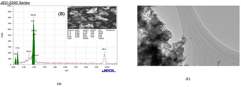
Characterization of gPbNPs (A) EDX spectrum and (B) image of gPbNPs by scanning electron microscope (C) Image of gPbNPs by scanning electron microscope (JEM 1011).
3.2 Viability of cells
By measuring the dehydrogenase conversion of the tetrazolium compound to water-insoluble formazan crystals—a reduction reaction that occurs in the mitochondria of living cells—the assay was utilized to evaluate the cytotoxic activity of g-PbNPs. The colon cancer (Caco2) cell line was cultivated in a 96-well plate. Subsequently, for a whole day, the cells were subjected to varying concentrations of g-PbNPs (1, 5, 10, 25, 50, 100, and 150 μg/mL). The impact of g-PbNPs on the Caco2 cell line's viability was demonstrated by the results. Furthermore, as seen in Fig. 2A, the viability of the Caco2 cell line was dramatically (p < 0.05) decreased in 24 h by g-PbNPs to 68, 61, 59, 55, 47, 41, and 35 %, respectively.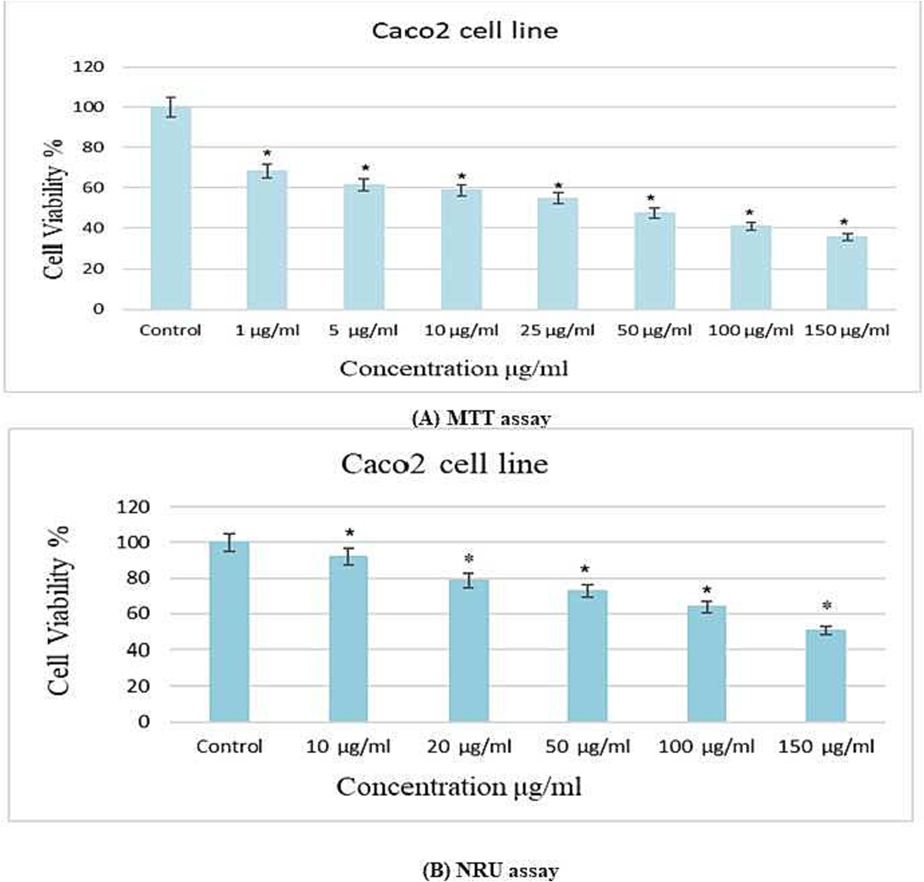
Different concentractions gPbNPs induced cytotoxicity in Caco2 cell line after 24 h exposure (A) MTT assay (B) NRU assay. *p ≤ 0.05 vs control.
The outcomes were compared to control cells, which show an inhibition that is dosage dependent. As indicated in Fig. 3, the inhibitory concentration (IC50) value for g-PbNPs treated Caco-2 cells at 24 h was determined to be 21.75 μg/mL. Table 1 displays the sublethal dosages of g-PbNPs that were determined for additional tests based on the IC50.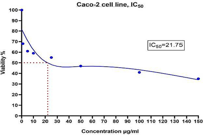
The half maximal inhibitory concentration (IC50) value 24 h calculated for g-PbNPs in treated cells of Caco2 cell line based on MTT results.
Zi-PbNP treatment of the Caco2 cell line demonstrated the cytotoxicity of the NRU assay. Following Zi-PbNP treatment, the lysosomal cytotoxic rose dramatically (p < 0.05). As a result, as seen in Fig. 2B, the cell viability was reduced to 91, 78, 72, 63, and 50 %, respectively. Both the MTT and NRU results demonstrated a similar decrease in cell viability (Fig. 2A–B).
3.3 Generation of reactive oxygen species
After being exposed to g-PbNPs for 24 h, the Caco2 cell line developed reactive oxygen species. As demonstrated in Fig. 4B, the Caco2 cell line exhibited a slightly elevated level of DCF as green fluorescence when treated to 6, 11, and 16 μg/mL, respectively, of g-PbNPs. Fig. 4A illustrates the dose-dependent production of ROS within cells. After being exposed to a Caco2 cell line for 24 h, 16 µg/mL of g-PbNPs produced higher ROS than the control group, as seen in Fig. 4A and B.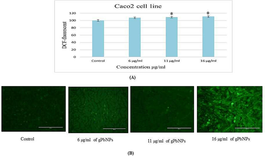
(A) Percentage change in intracellular in the Caco2 cell line (B) Generation of green fluorescence in Caco2 cell line after exposure to various concentration of gPbNPs for 24 h. The magnification power was 20x. *p ≤ 0.05 versus control.
3.4 Apoptosis
3.4.1 Enzyme activity of caspase −3/7
We assessed caspase-3/7 activity in Cacoa-2 cells in this experiment. As the exposure dose was raised, caspase-3/7 activities increased in a dose-dependent manner, and its level rose in Cacoa-2 cells (Fig. 5).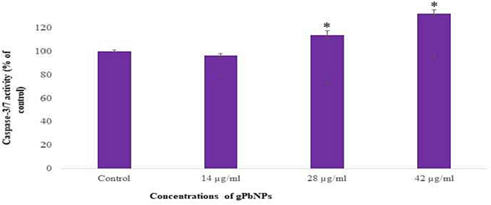
Caspase-3/7 activity in the Caco2 cell line after exposure of gPbNPs. Each value represents the mean ±SE of three experiments * p ≤ 0.05 versus control.
3.4.2 Proteomic profile of apoptosis
Using the Human Apoptosis protein array, we examined the primary proteins implicated in cell death and apoptosis in this setting following a 24-hour exposure of the Caco-2 cells to the produced g-PbNPs. as depicted in Fig. 6A,B. We discovered changes in certain proteins. Several proteins exhibited either up- or down-regulation in accordance with their function within the apoptotic cascade. Furthermore, bcl-2 was downregulated and several of these proteins, such as Bax, Bad, p27, p53, and caspse-3, were increased erratically (Fig. 6A, B).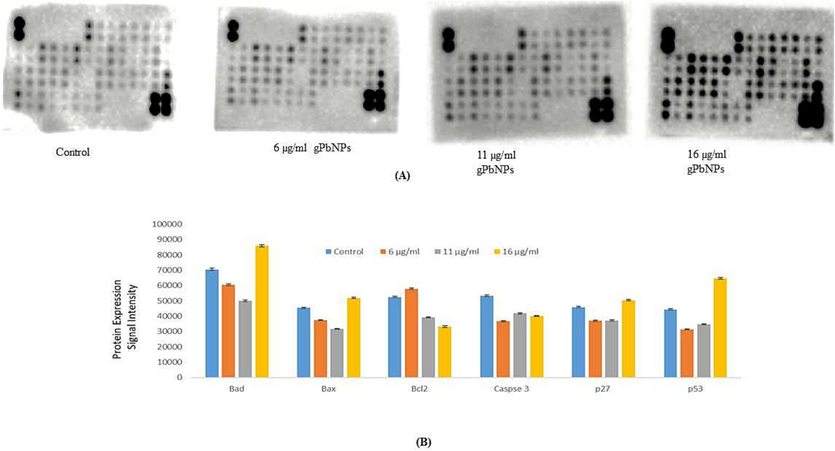
The levels of apoptosis – related proteins inCaco2 cell line with gPbNPs exposure was measured using the RayBio human apoptosis antibody array. (A) Equal total lysate protein was loaded on each RayBio Human Apoptosis Array membrane and selected pro-apoptosis an anti-apoptosis protein signal intensity are indicated in ref/blue boxes. (B) Representative bar graph of the apoptotic up/down regulate proteins. Error bars, ±S.E.
3.4.3 Polymerase chain reaction in quantitative real-time
In Caco2 cells, the messenger RNA (mRNA) levels of the apoptotic genes Bax and Bcl-2 were assessed using qRT-PCR following a 24-hour exposure to gPbNPs at concentrations of 6, 11, and 16 μg/mL, respectively. The gPbNPs treatment on the Caco2 cell line elevated Bax and downregulated Bcl-2, according to the qRT-PCR results, at 16 μg/mL of exposure to g-PbNPs (Fig. 7).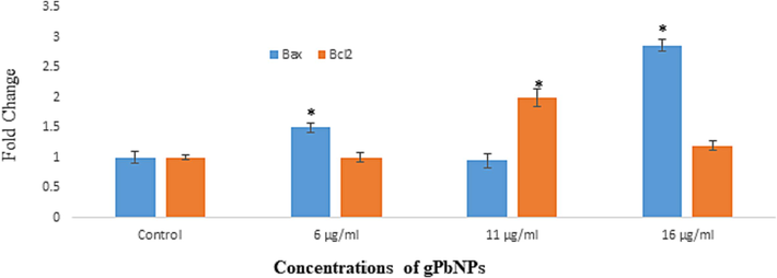
Mrna levels of Bax and Bcl2 in Caco2 cell line after exposure to different concentration of gPbNPs for 24 h. * p ≤ 0.05 versus control.
4 Discussion
The world's cancer death rate is high at the moment, and treatment guidelines are ineffective. There are currently numerous studies being carried out to create environmentally friendly production techniques for nanoparticles and their possible application in the treatment of cancer (Thawini and Al-Shawi, 2021). Plant-derived biomolecules and bioreducing substances, including proteins, flavonoids, terpenoids, and cofactors, provide a versatile, cost-effective, environmentally friendly option for synthesizing nanoparticles and serving as a capping agent (Khani et al., 2018; Saman et al., 2021).
Additionally, nanoparticles may become more stable and cytotoxic, which would increase their bioavailability. According to Huang et al. (2019), the natural substance effectively treats cancer by enhancing the immune system, decreasing the negative effects of cancer therapy, and inhibiting tumor development and spread. Numerous investigations have demonstrated that Z. spina-christi inhibits colon cancer cell line proliferation and induces apoptosis (Guo et al., 2011; Kaenda, 2012). Using the MTT assay, this study assessed the gPbNPs' potential for toxicity in the Caco2 cell line. The outcome demonstrated a decline in cell viability as gPbNP concentration increased, suggesting that more gPbNPs may collect inside cells, increasing stress and ultimately resulting in cell death. In 24 h, gPbNPs reduced the development of the Caco2 cell line with an IC50 value of 21.75 μg/mL.
Additionally, there was a correlation between the MTT assay findings and the NRU assay results. The lysosomal integrity of viable cells was assessed using the NRU test, and the results demonstrated that exposure of the Caco2 cell line reduced cell viability at all concentrations in a dose-dependent manner. Meanwhile, the NRU test revealed somewhat reduced cytotoxicity as compared to the MTT result assay. Furthermore, our outcomes aligned with those of Rajendrasozhan et al. (2021), who found that the colon cancer cell line treated with the diethyl ether fraction of Z. spina-christi bark extract agents showed anti-cancer activity, with an IC50 of 11.6 μg/mL, according to the MTT assay. It is noteworthy that g-PbNPs enhanced the DCF fluorescence in the Caco2 cell line in a dose-dependent manner (6, 11 and 16 μg/mL, respectively). This suggests that NPs generate ROS and may cause oxidative stress in the cells. On the other hand, since this can happen in populations that are severely exposed, it is imperative to emphasize that even a small number of in vitro effects may be connected to substantial biological impacts in vivo, particularly in the event of chronic exposure.
According to Reed (1999), there was a downregulation of apoptosis in correlation with an unbalanced pattern of gene expression that influences both cell death and growth. Using qRT-PCR, the expression of the genes Bax and Bcl-2 after exposure to g-PbNPs was investigated. The induction of apoptosis is determined by the ratio of Bax/Bcl-2 expression. The effects of g-PbNPs resulted in a dose-dependent increase in Bax expression and an increase in Bcl-2 expression at higher concentrations of g-PbNPs. Furthermore, in the Caco2 cell line, resistance to g-PbNP-induced apoptosis is linked to elevated levels of Bcl-2 expression. Bax genes were upregulated and bcl2 was downregulated in response to caspase-3 activation. Research conducted in vitro on specific tumor cell lines revealed that overexpression of Bcl-2 inhibits cell death (Findley et al., 1997). Bcl-2 inhibits apoptosis via directly interacting with mitochondria and by preventing membranes from becoming more permeable, according to Yang and Korsmeyer (1996). According to Pillai (2020), human colorectal cancer (HCT116) cell line exposure to an aqueous extract of Ziziphus jujube fruits raised Bax expression levels, with the exception of Bcl-2, in a time- and dose-dependent manner. These findings support the hypothesis that g-PbNPs selectively limit tumor growth by inducing apoptosis and acting as an inhibitor. It might be a useful strategy to improve a successful cancer therapy. All things considered, our results clearly imply that g-PbNPs should be thoroughly studied for in vivo testing to validate these characteristics.
Acknowledgement
This research was funded by Researchers Supporting Project number (RSP2023R27), King Saud University, Riyadh, Saudi Arabia.
Declaration of competing interest
The authors declare that they have no known competing financial interests or personal relationships that could have appeared to influence the work reported in this paper.
References
- Nanoalumina induces apoptosis by impairing antioxidant enzyme systems in human hepatocarcinoma cells. Int. J. Nanomed.. 2015;10(1):3751-3760.
- [Google Scholar]
- Oxidative Stress-Induced DNA Damage by Manganese Dioxide Nanoparticles in Human Neuronal Cells. Biomed Res Int.. 2017;2017:1-10.
- [Google Scholar]
- Genus Ziziphus for the treatment of chronic inflammatory diseases. Saudi J. Biol. Sci.. 2021 Dec;28(12):6897-6914.
- [Google Scholar]
- Apoptotic and DNA-damaging effects of yttria-stabilized zirconia nanoparticles on human skin epithelial cells. Int. J. Nanomed.. 2019;14:7003.
- [Google Scholar]
- A rapid and sensitive method for the quantitation of microgram quantities of protein utilizing the principle of protein dye binding. Anal. Biochem.. 1976;72:248-254.
- [Google Scholar]
- Airway oxidative stress and inflammation El-Sayed IH, Huang X, El-Sayed MA. Surface plasmon resonance scattering and absorption of anti-EGFR antibody conjugated gold nanoparticles in cancerdiagnostics: applications in oral cancer. Nano Lett.. 2005;5:829-834.
- [Google Scholar]
- Expression and regulation of Bcl-2, Bcl-xl, and Bax correlate With p53 status and sensitivity to apoptosis in childhood acute lymphoblastic leukemia. Blood. 1997;89(8):2986-2993.
- [Google Scholar]
- Cellular targets and mechanisms in the cytotoxic action of non-biodegradable engineered nanoparticles. Curr Drug Metab.. 2013;14(9):976-988.
- [Google Scholar]
- Sulforaphene inhibits esophageal cancer progression via suppressing SCD and CDH3 expression, and activating the GADD45B-MAP2K3-p38-p53 feedback loop. Cell Death Dis.. 2020;11(8):713.
- [Google Scholar]
- Natural products for treating colorectal cancer: A mechanistic review. Biomed. Pharmacother.. 2019;117:109142.
- [Google Scholar]
- Anticancer, antioxidant and antimicrobial screening of extracts from Ziziphus Mucronata, heliotropium ciliatum and Gnidia polycephala from the Oshikoto region of Namibia. Asian J Tropical Biotechnol. 2012
- [Google Scholar]
- Green synthesis of copper nanoparticles by fruit extract of Ziziphus spina-christi (L.) Willd.: application for adsorption of triphenylmethane dye and antibacterial assay. J. Mol. Liq.. 2018;255:541-549.
- [Google Scholar]
- Three novel dietary phenolic compounds from pickled Raphanus Sativus L. inhibit lipid accumulation in obese mice by modulating the gut microbiota composition. Mol. Nutr. Food Res.. 2021;65
- [Google Scholar]
- Nanotechnology advances in the detection and treatment of cancer: an overview. Nanotheranostics. 2022;6(4):400-423.
- [Google Scholar]
- Antiproliferative actions of Ziziphus jujube fruit extract is mediated through alterations in Bcl2-Bax ratio and through the activation of caspases. Chem. Biol. Lett.. 2020;7(1):41-46.
- [Google Scholar]
- In vitro cytotoxicity analysis of Zizyphus spina-christi stem bark extract on human cancer cell lines. Bioinformation. 2021;17(5):583.
- [Google Scholar]
- Green synthesis of MgO nanocatalyst by using Ziziphus mauritiana leaves and seeds for biodiesel production. Appl. Organomet. Chem.. 2021;35(5):e6199.
- [Google Scholar]
- MTT assay for cell viability: intracellular localization of the formazan product is in lipid droplets. Acta Histochem.. 2012;114(8):785-796.
- [Google Scholar]
- A polysaccharide of Ziziphus spina-Christi L., and it’s Silver nanoparticles induced reactive oxygen species and late apoptosis of Liver cancer cells. Nanomed. Res. J.. 2021;6(3):237-247.
- [Google Scholar]
- Effect of silver nanoparticles on gene transcription of land snail Helix aspersa. Sci Rep. 2022;12(1):1-12.
- [Google Scholar]
- Molecular Thanatopsis: a discourse on the BCL2 family and cell death. Blood. 1996;88(2):386-401.
- [Google Scholar]
Appendix A
Supplementary material
Supplementary data to this article can be found online at https://doi.org/10.1016/j.jksus.2023.103041.
Appendix A
Supplementary material
The following are the Supplementary data to this article:


