

Translate this page into:
Mathematical model based on fractional trace operator for COVID-19 image enhancement
⁎Corresponding author. hamidjalab@um.edu.my (Hamid A. Jalab),
-
Received: ,
Accepted: ,
This article was originally published by Elsevier and was migrated to Scientific Scholar after the change of Publisher.
Peer review under responsibility of King Saud University.
Abstract
The medical image enhancement is major class in the image processing which aims for improving the medical diagnosis results. The improving of the quality of the captured medical images is considered as a challenging task in medical image. In this study, a trace operator in fractional calculus linked with the derivative of fractional Rényi entropy is proposed to enhance the low contrast COVID-19 images. The pixel probability values of the input image are obtained first in the proposed image enhancement model. Then the covariance matrix between the input image and the probability of a pixel intensity of the input image to be calculated. Finally, the image enhancement is performed by using the convolution of covariance matrix result with the input image. The proposed enhanced image algorithm is tested against three medical image datasets with different qualities. The experimental results show that the proposed medical image enhancement algorithm achieves the good image quality assessments using both the BRISQUE, and PIQE quality measures. Moreover, the experimental results indicated that the final enhancement of medical images using the proposed algorithm has outperformed other methods. Overall, the proposed algorithm has significantly improved the image which can be useful for medical diagnosis process.
Keywords
Fractional calculus
Trace operator
Fractional Rényi entropy
COVI-19 images
Image enhancement

1 Introduction
Image enhancement is a method of improving the content of image for specific application. Medical image enhancement is one of categories of image processing which has a big effect on the diagnosis decisions (Zhou et al., 2020). One of important topic in medical imaging is the “image contrast” which is related to the brightest and the darkest pixels inside the image. The research on detecting and treating COVID-19 infection has sparked people's interest across the world. Furthermore, the widespread availability of X-ray and CT imaging modalities makes these two imaging modalities ideal for detecting COVID-19 infection early. To have a better understanding of the state of the COVID-19 infection, chest X-rays and CT scans are used to view the disease spreading across the lungs. Most of captured medical images suffering from low contrast of organs and tissues, which makes it difficult to distinguish between the normal and abnormal tissues. Therefore, the process of medical image enhancement is an important stage for correctly diagnosing task. Different image processing algorithms for image enhancement are developed for image contrast enhancement. The applications of fractional calculus in science, and engineering, have been significantly increased due to the ability of fractional calculus in obtaining accurate solutions which makes the fractional-order-based methods more suitable approaches for many image processing methods like image enhancement (Ibrahim Rabha et al., 2021; Jalab et al., 2021a; Jalab et al., 2017).
In the literature, several spatial domain algorithms-based images enhancing methods have been reported. Fractional calculus is the most extensively utilized concept in image enhancement methods (Ibrahim & Baleanu, 2021a, 2021b; Jalab et al., 2022; Veeman et al., 2021). Due to the unpredictable change in the quality of the acquired images, enhancing medical imagery is a difficult undertaking. Based on this, we present an automated approach for improving low contrast COVID-19 images utilizing the trace operator in fractional calculus and the derivative of fractional Rényi entropy (FToRE). We aim to provide a new medical image enhancement algorithm which improves the perception information in an image for other image processing algorithms. The following are the study's major contributions:
-
A new dynamic image enhancement algorithm to achieve better contrast enhancement.
-
A new FToRE image enhancement based on trace operator in fractional calculus which is associated with the derivative of fractional Rényi entropy.
-
A new convolution window developed by using the covariance between the input image and the probability image.
2 Related work
Medical image processing has received significant interest recently due to the spread of COVID-19 infection. Most of the medical imaging systems generate images with noise or with low contrast quality, therefore, the diagnosis process accuracy may be affected. In the medical imaging, quality of captured images is affected by the organ structure, tissue complexity, as well as the imaging system noise, whereas the quality of these images could be enhancement to obtain a clearer image. Image enhancement algorithms are used to improve the visual look of images in such a way without modifying the image information. In this context, numerous challenges of medical image processing have been proposed researchers, and most of them focusing on image enhancement. In the literature, several image enhancement methods with different mathematical models have been proposed. Several studies concentrating on increasing image quality have recently suggested the concept of fractional-order-based image enhancement algorithms. Roy et al. 2016 (Roy et al., 2016) applied the “Fractional Poisson” model to enhance video frames as a pre-processing step for text detection and recognition. The main aim was to remove the generated Laplacian noise from video frames to enhance the detection accuracy. Since this approach was developed as a pre-processing for video frames enhancement, so, it may not work well for medical image enhancement. Similarly, “local fractional entropy” based image enhancement method was developed by Al-Shamasneh et al. 2018 (Al-Shamasneh et al., 2018) for MRI kidney images enhancement. This model achieved good results for MRI kidney image enhancement only. In the same research direction the “Riesz fractional operator” model was developed by Raghunandan et al. 2017 (Raghunandan et al., 2017) to enhance the low contrast license plate. This model worked well for license plate images only. A dual illumination estimation for improving the image illumination is presented in Zhang Q et al 2019, (Zhang et al., 2019). This approach was developed for only low illuminated images but not for images with low contrast. Zhang et al. 2019 (Zhang & Yan, 2019) also proposed a fractional mask-based image enhancing method. This approach relied on the output of the image segmentation for enhancing the input images which was unable to enhance the image contrast efficiently.
Recently, (Ibrahim Rabha et al., 2021) presented a fractional partial differential equations(FPDEs) based mathematical model for medical image enhancement. The FPDE's key benefit is its ability to improve low-contrast photos. Similarly, (Jalab et al., 2021b,c) proposed a new fractional calculus-based medical image enhancing method. This enhancement method improves image contrast while preserving image features, but it is ineffective for improving non-uniform illumination areas within input images. Furthermore, (Jalab et al., 2021b,c) developed a novel fractional integral entropy (FITE) model for image enhancement. This model well enhanced the input images using the image contents linked with the fractional power to enhance the illumination pixel value of the image. While, in Ibrahim et al (Ibrahim Rabha et al., 2021), a new class of fractional partial differential equations (FPDEs) was presented for medical image enhancement. The achieved results showed the benefit of using FPDEs in improving the medical image quality. In Qin et al, 2020 (Qin et al., 2020), a medical low contrast image enhancement method was proposed based on multi-scale Retinex method. This method significantly improved the information of the medical image in dark area. However, it has high complexity. In Rahman et, al., 2021 (Rahman et al., 2021), the effect of five standard image enhancement algorithms for detection of COVID-19 from the X-ray images using six different deep learning CNN models was explored. This work proved the benefit of using image enhancement as a pre-processing step for achieving good accuracy for COVID-19 image classification. Likewise, in Qiu T 2020 (Qiu et al., 2019) an effective medical image enhancement approach using CNN has been proposed. The experimental results indicated that the edge details of the input image are much enhanced. However, the limitation of CNN based image enhancement was with the training images.
More recently the, the fractional calculus operators have proven to be effective as new approach in image enhancement (Aldawish & Ibrahim, 2022; Aldawish & Jalab, 2022). There is still room for improvement, regardless of the results of the fractional operators as an image enhancement. This study presents a new mathematical model (FToRE) based on fractional trace operator associated with the derivative of fractional Rényi entropy to enhance the low contrast image intensities, which is the study's key contribution. The main advantage of proposed (FToRE) is its ability to enhance the low contrast image intensities by the derivative of fractional Rényi entropy.
3 Methods
This section gives a quick overview of the background of the deep connection between the trace operator and the fractional concept to develop the image enhancement model using the drivitive of Rényi entropy which is considered to be a fractional entropy of poewer α.
3.1 The trace operator
The trace operator covers the idea of the constraint of a function to the boundary of its domain to extend this function into a Sobolev space W(Ω), where Ω is the bounded domain with Lipschitz boundary L( Ω). This is mainly significant for the invetigation of methematical formila containing the derivative. The trace operator can be formulated for functions in W(Ω). Let be a bounded domain with Lipschitz boundary. Then, the bounded linear trace operator be given as follows (Ekeland & Temam, 1999):
Such that
Since the trase operator is defined on the boundary ; therefore, the drivitive of the trase operator can be considered. This study aims to generalize the trase operator in view of fractional calculus. For this purpose we sugest to use the drivitive of Rényi entropy which is considered to be a fractional entropy of poewer α to enhance the low contrast image intensities.
The recommended fractional order trace operator was used to convert the formula to the appropriate kinds. It also has the potential to provide useful information about the images.
3.2 Rényi entropy
The image enhancement is a function used for improving the image quality (Jalab et al., 2017). This concept motivated us to develop a new medical image enhancement model using based Rényi fractional entropy (RFE) which is presented as follows (Jalab et al., 2021a):
is obviously a non-increasing concave function in
for all discrete random variables. In addition, when
, we obtain the Shannon Entropy. Moreover, when α → 0, RFE shows the logarithm of the dimension of the space that is
The temporal derivative of RFE can be used to connect RFE with the trace operator (DeMars et al., 2013).
We next go on to look into the characteristics of (3).
Theorem 1
Consider Eq. (2) for a set of pixel probabilities. Then the upper bound of Eq.(3) is given by
Proof.
By substituting the trace operator theorem Eq. (1) in Eq.(3), we have
In view of [3]-Relation (9), we conclude that there are two positive constants satisfy the inequalities respectively.
And
Substituting the last two inequalities in inequality (5), we conclude that
The following is the proposed image enhancement model (FToRE):
-
Consider the input image (I).
-
Initialize the fractal power (α = 0.036) as tune image enhancement value.
-
Find the pixel probability value (ρ).
-
Calculate the covariance matrix ℵ.
-
Calculate the proposed window W mask using Eq. (9).
-
Calculate the enhanced image using Eq. (10).
The fractional power α is the parameter for fine detail enhancement in the proposed FToRE model. The value of α is obtained empirically by calculating the average BRISQUE score for all images in the dataset, as shown in Fig. 1. It should be noted that the best BRISQUE score is reached when the value of is equal to 0.036 (lower is better).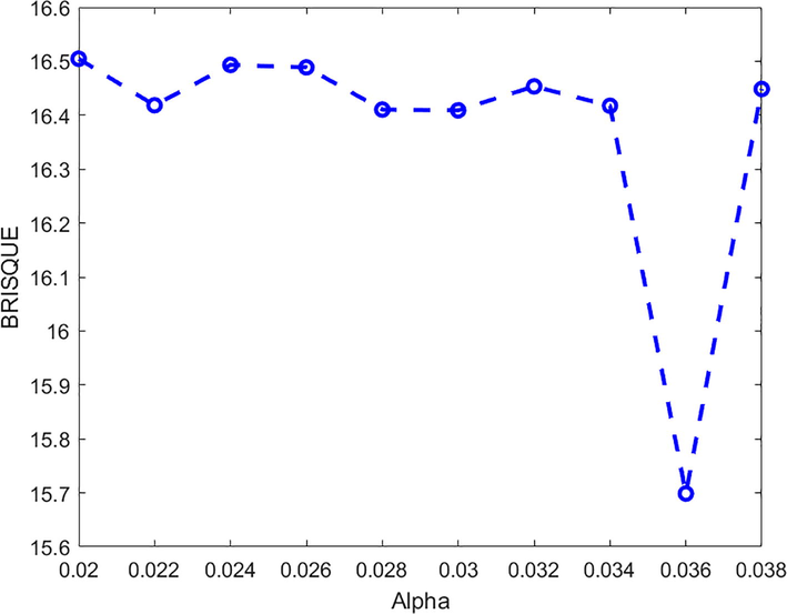
The average scour of BRISQUE for various values of α.
4 The results
This section focuses on the proposed medical image enhancement model's performance analysis. All experiments are executed using MATLAB 2021a on Windows 10, Intel(R), i7 with 8 GB RAM and GeForce GTX 950M GPU(The Mathworks (2021). ‘‘Matlab”. In. Massachusetts, USA).
4.1 Datasets
In this study, two datasets are obtained from:
-
The dataset contains 137 of COVID-19 and 317 in total containing Pneumonia and Normal Chest X-ray. (Covid-19 Chest X-rays Dataset.: COVID-19, Viral Pneumonia, Normal).
-
The CT scan dataset named as COVID-19 DATABASE is created by “Italian Society of Medical and Interventional Radiology” (Radiology, 2020).
4.2 Evaluation metrics
The following quality assessment metrics are used to assess the improved image's quality (The Mathworks (2021). ‘‘Matlab”. In. Massachusetts, USA):
1- “The blind referenceless image spatial quality evaluator (BRISQUE)”, which calculates the image perceived quality (Mittal et al., 2011).
2- “Perception based image quality evaluator (PIQE)”, which measures the image with arbitrary distortion (Venkatanath et al., 2015).
4.3 Results of proposed FToRE model
The qualitative achieved results of the proposed image enhancement model (FToRE) including the input images; the enhanced images, as well as the histogram plot are shown in Fig. 2. It is clear from Fig. 2 (b) that the input image pixel probability looks dense, while the enhanced image pixels probability in Fig. 2 (d) looks distributed. Some of image details of input images are unclearly shown as illustrated in Fig. 2a. The achieved image contrast is well enhanced by proposed FToRE model as shown in Fig. 2c. This indicates that the image’s contrast has been improved by proposed image enhancement model (FToRE), which proved the efficacy of the proposed FToRE as a medical image enhancement technique.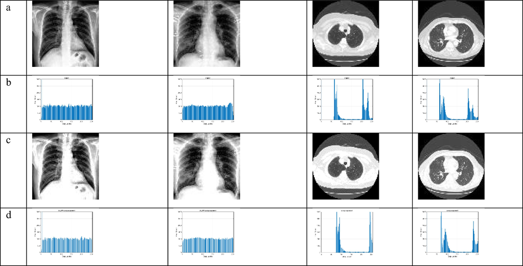
The output of the proposed FToRE enhancement algorithm. (a) Input images, (b) Histogram of input images, (c) Enhanced images, (d) Histogram of enhanced images.
4.4 Comparative analysis
For comparative analysis, we considered the following image enhancement methods, which are proposed to improve images with low contrast.
Shamasneh et al.(LFE) (Al-Shamasneh et al., 2018), proposed local fractional entropy for enhancing the MRI Kidney images with low contract, while, Raghunandan et al. (RF) (Raghunandan et al., 2017), proposed Riesz fractional method for license plate images enhancement.
Zhang Q et al. (DIE) (Zhang et al., 2019), presented a dual illumination estimation method to improve the illumination of poor illuminated. Jalab et al (FITE) (Jalab et al., 2021b,c) introduced a new fractional integral entropy (FITE) for medical image improvement. Ibrahim et al (FPDE) (Ibrahim Rabha et al., 2021), proposed a new class of fractional partial differential equations for medical image improvement using a similar approach.
The qualitative enhancement results using the three standard datasets are shown in Fig. 3. The input images are shown in Fig. 3(a) while the corresponding enhanced images by proposed method which are shown in Fig. 3(g). The brightness of fine details in enhanced images such as edge pixels’ increases compared to the edge pixels in the input images. Therefore, the proposed FToRE algorithm enhancement images effectively independent of image contents.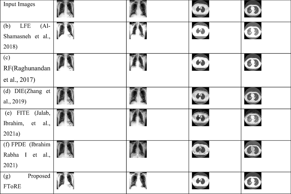
The comparative analysis of enhancement results. (a) Input Image, (b) LFE (Al-Shamasneh et al., 2018), (c) RF (Raghunandan et al., 2017), (d) DIE (Zhang et al., 2019), (e)FITE (Jalab et al., 2021b,c), (f) FPDE (Ibrahim Rabha et al., 2021), (g) Proposed FToRE.
The quantitative enhancement results of proposed FToRE and the existing methods are illustrated in Table 1. The proposed FToRE algorithm has achieved the most accepted scores for BRISQUE, and PIQE measures with respect to the existing enhancement methods. In which the lower scores of BRISQUE, and PIQE represent the better image quality. This supports the robustness of proposed FToRE in improving the fine details of low contrast images whatever image content.
Input Image
Enhanced Image
Methods
BRISQUE
PIQE
BRISQUE
PIQE
X-ray
LFE (Al-Shamasneh et al., 2018)
19.3789
22.0338
41.7218
39.7814
RF (Raghunandan et al., 2017)
25.4803
24.5502
DIE (Zhang et al., 2019)
14.7144
15.3783
FITE (Jalab et al., 2021b,c)
19.2169
23.2188
FPDE (Ibrahim Rabha et al., 2021)
16.9769
29.5673
Proposed FToRE
16.4486
21.0140
CT
LFE(Al-Shamasneh et al., 2018)
43.0733
45.3261
48.2256
49.6971
RF(Raghunandan et al., 2017)
41.1033
42.0481
DIE (Zhang et al., 2019)
41.7663
39.1558
FITE (Jalab et al., 2021b,c)
37.2590
40.4642
FPDE (Ibrahim Rabha et al., 2021)
43.2146
44.3182
Proposed FToRE
36.7163
41.4708
Overall, the brightening enhancement of the proposed FToRE algorithm makes becomes the image boundaries clear and well-seen. This is due to the model's ability to produce fair visual results for images with low contrast. This is the contribution of the fractional trace operator with the fractional Rényi entropy in this study.
5 Conclusions
In this study, we developed a novel image enhancing method based on a trace operator in fractional calculus which is associated with the derivative of fractional Rényi entropy to enhance the low contrast images. The proposed method involved dynamic enhancement using the convolution operation between the window mask with the input image. The proposed image enhancement model started with finding the pixel probability values of the input image, then the covariance between the input image and the calculated probability image be calculated as the enhancement window. Finally, the proposed image enhancement is performed by using the convolution window with the input image. The proposed FToRE algorithm enhances images more successfully than existing image enhancement algorithms, according to the findings of the experiments. In comparison to previous approaches, the proposed FToRE algorithm has the benefit of being better suitable for low contrast image enhancement. The proposed enhancement model's limitation is that it may tend to over-emphasize smooth regions and retain or amplify noise as well. We will investigate how to customize the proposed FToRE algorithm for specific applications in future work to attain the best possible image contrast enhancement outcomes.
Acknowledgement
To Princess Nourah bint Abdulrahman University Researchers Supporting Project number (PNURSP2022R300), Princess Nourah bint Abdulrahman University, Riyadh, Saudi Arabia.
Declaration of Competing Interest
The authors declare that they have no known competing financial interests or personal relationships that could have appeared to influence the work reported in this paper.
References
- A new mathematical model of multi-faced COVID-19 formulated by fractional derivative chains. Adv. Contin. Discr. Models. 2022;2022(1):1-10.
- [Google Scholar]
- A mathematical model for COVID-19 image enhancement based on Mittag-Leffler-Chebyshev shift. Comput. Mater. Continua. 2022;73(1):1307-1316.
- [Google Scholar]
- A new local fractional entropy-based model for kidney MRI image enhancement. Entropy. 2018;20(5):344.
- [Google Scholar]
- Covid-19 Chest X-rays Dataset. : COVID-19, Viral Pneumonia, Normal. https://www.kaggle.com/pranavraikokte/covid19-image-dataset.
- Entropy-based approach for uncertainty propagation of nonlinear dynamical systems. J. Guidance Control Dyn.. 2013;36(4):1047-1057.
- [Google Scholar]
- Geometric behavior of a class of algebraic differential equations in a complex domain using a majorization concept. AIMS Math.. 2021;6(1):806-820.
- [Google Scholar]
- On quantum hybrid fractional conformable differential and integral operators in a complex domain. Revista de la Real Academia de Ciencias Exactas, Físicas y Naturales. Serie A. Matemáticas. 2021;115(1):1-13.
- [Google Scholar]
- A medical image enhancement based on generalized class of fractional partial differential equations. Quant. Imag. Medi. Surg.. 2021;20:1-26.
- [Google Scholar]
- Fractional Renyi entropy image enhancement for deep segmentation of kidney MRI. CMC-Comput. Mater. Continua. 2021;67(2):2061-2075.
- [Google Scholar]
- A novel Pixel’s fractional mean-based image enhancement algorithm for better image splicing detection. J. King Saud Univ.-Sci.. 2022;34(2):101805.
- [Google Scholar]
- Jalab, H. A., Ibrahim, R. W., Hasan, A. M., Karim, F. K., Al-Shamasneh, A. a. R., & Baleanu, D. (2021b). A new medical image enhancement algorithm based on fractional calculus.
- Image denoising algorithm based on the convolution of fractional Tsallis entropy with the Riesz fractional derivative. Neural Comput. Appl.. 2017;28(1):217-223.
- [Google Scholar]
- A new medical image enhancement algorithm based on fractional calculus. CMC-Comput. Mater. Continua. 2021;68(2):1467-1483.
- [Google Scholar]
- The Mathworks, (2021). ‘‘Matlab”. In. Massachusetts, USA. In.
- Blind/referenceless image spatial quality evaluator. In: 2011 Conference Record of The Forty Fifth Asilomar Conference On Signals, Systems And Computers (ASILOMAR). 2011.
- [Google Scholar]
- A medical image enhancement method based on improved multi-scale retinex algorithm. J. Med. Imag. Health Inform.. 2020;10(1):152-157.
- [Google Scholar]
- Efficient medical image enhancement based on CNN-FBB model. IET Image Proc.. 2019;13(10):1736-1744.
- [Google Scholar]
- Radiology, I. S. o. M. a. I. (2020). COVID-19 CT Scans. https://www.sirm.org/category/senza-categoria/covid-19/ , (accessed on 10 June 2021).
- Riesz fractional based model for enhancing license plate detection and recognition. IEEE Trans. Circuits Syst. Video Technol.. 2017;28(9):2276-2288.
- [Google Scholar]
- Exploring the effect of image enhancement techniques on COVID-19 detection using chest X-ray images. Comput. Biol. Med.. 2021;132
- [Google Scholar]
- Fractional poisson enhancement model for text detection and recognition in video frames. Pattern Recogn.. 2016;52:433-447.
- [Google Scholar]
- A new megastable chaotic oscillator with blinking oscillation terms. Complexity. 2021;2021:1-12.
- [Google Scholar]
- Blind image quality evaluation using perception based features. In: 2015 Twenty First National Conference on Communications (NCC). 2015.
- [Google Scholar]
- Dual illumination estimation for robust exposure correction. Comput. Graphics Forum 2019
- [Google Scholar]
- Image enhancement algorithm using adaptive fractional differential mask technique. Math. Found. Comput.. 2019;2(4):347.
- [CrossRef] [Google Scholar]
- New improved optimized method for medical image enhancement based on modified shark smell optimization algorithm. Sensing Imaging. 2020;21(1):1-22.
- [Google Scholar]


