

Translate this page into:
Isolation and identification of pathogens causing dragon stripe disease on Kadsura coccinea
⁎Corresponding author. yhua7710@hunau.edu.cn (Yang Hua)
-
Received: ,
Accepted: ,
This article was originally published by Elsevier and was migrated to Scientific Scholar after the change of Publisher.
Peer review under responsibility of King Saud University.
Abstract
The Kadsura coccinea (commonly known as black tiger plant) is an edible plant with wide applications in traditional Chinese medicine and is an important source for various bioactive metabolites. The incidence of dragon stripe disease, a serious infection caused by various fungal pathogens, is high in K. coccinea. The disease result in reduced productivity of the plant and thereby cause economic loss to the farmers. It is therefore necessary to have clear information on the pathogenic strains of fungi that causes dragon stripe disease on K. coccinea for its effective diagnosis and management. The study isolated and purified different pathogenic microorganisms, which were further, identified using sequence analysis and phylogenetic tree construction of rDNA-ITS genes. The pathogenicity of the identified organisms was verified according to Kochʼs rule. A total of 26 fungi were isolated from 100 pieces of fungal infected leaf tissues. The major ones were Apiospora jiangxiensis, Apiospora chromolaenae, Neopestalotiopsis formicarum, Diaporthe kochmanii, Diaporthe longicolla, Botryosphaeria fabicerciana, and Alternaria alstroemeriae. The present study first report that the K. coccinea as a host for Neopestalotiopsis formicarum. Another six species of fungi identified may exist in the plant tissues of K. coccinea as endophytic or saprophytic fungi, the role of which need to be further evaluated. Overall, the results of the study may be useful for the understanding of the pathogenic nature of dragon stripe disease on K. coccinea, as well as to provide a basis for field diagnosis and comprehensive management of the disease.
Keywords
Kadsura coccinea
Dragon Stripe Disease
Pathology
Fungal identification
Neopestalotiopsis formicarum
rDNA-ITS

1 Introduction
Kadsura coccinea (Lem.) is a woody vine belonging to Schisandraceae family and distributed in Hunan, Jiangxi and Guangxi provinces (Li et al., 2021). It is a common herbal medicine utilized by the Miao and the Dong folk communities of China. The plant had been used in folk medicines for the management of menalgia during menstrual period. In addition, the fruit pulp of the plant is a rich source of vitamin C (Yang et al., 2020), and therefore it has cosmetic applications including skin whitening and increasing the glow. The presence of bioactive stress volatile myrcene has result in a pleasant and unique aroma for the flower of K. coccinea and hence it is widely utilized for the preparation of perfumes and to provide fragrance of beverages. In addition, the leaves of K. coccinea are attractive with emerald green color and have graceful and charming branches. The flowers are yellow or red and the fruit shape is the unique and composite with long fruiting period. All these together enhance the value of K. coccinea as an ornamental plant (Zhong-hou et al., 2017). The higher pharmacological and nutritional values of K. coccinea resulted in an increase on its market demand.
To meet the increasing demand, the K. coccinea has been cultivated in large in China. However, the cultivation is not devoid of issues; the predominant concerns associated with the cultivation include the prevalence of bacterial and fungal diseases. It has been observed that the incidence of root rot, leaf blight, Dragon Stripe Disease and leaf spot are predominant diseases that affect K. coccinea during 2015–2017. Further, the Dragon Stripe Disease and leaf blight are more deleterious and result in significant reduction in the productivity of K. coccinea in Hengyang and Tongdao province (Zhong-hou and Jing-Na, 2018). In 2019, Zhong et al., (2019) have observed that the leaf spot of K. coccinea in the nursery of K. coccinea in Changsha is caused by Corynespora cassiicola. Later, Shi et al., (2019) have found that the pathogenic bacterium that causes blanch blight of K. coccinea is Neofusicoccum parvum. A study by Xie, et al., (2018) reported that Neopestalotiopsis clavispora causes the leaf spot of K. coccinea in the plantation in Guangxi province. However, the cause and pathogenicity of Dragon Stripe Disease in K. coccinea is not been clear, despite its higher prevalence. Hence, the present study isolated the microorganisms from the infected leaves of K. coccinea which was then purified and identified using theoretical basis and molecular phylogenetic techniques.
2 Material and method
2.1 Experimental material
The leaves of K. coccinea that are infected and developed Dragon Stripe Disease was collected from Hunan. The collected leaves were having typical symptom and they were put in clean sealed collecting bag and marked.
2.2 Test method
2.2.1 Isolation and purification of the pathogenic bacteria
The collected leaves of K. coccinea with symptoms of Dragon Stripe Disease were brought to the lab and cleaned. The bacterial strains were collected to PDA culture medium using single spore method. A portion of the infected leaves were kept as reserve under 4 °C and −8 °C (Wang et al., 2017).
2.2.2 Molecular determination of the pathogenic bacteria
Shanghai Shenggong Bioengineering Corporation is entrusted to carry out DNA extraction, PCR amplification and sequencing. The primer used for PCR amplification was ITS1/ITS4. The BLAST matching was carried out in GenBank on NCBI network based on the sequences obtained and matching homologous fungoid ITS sequence were determined. The process was run was repeated at 1000 replication of bootstraps with neighbor-joining method in MEGA 7.0 (Neighbor-Joining, NJ) and the Maximum Composite Likelihood phylogenetic tree and analyzing its kinship was prepared.
2.2.3 Morphological identification of the pathogenic bacteria
Clump of isolated and purified strains of pathogenic bacteria were inoculated to new PDA culture medium. The diameter of the colony was measured under 28 °C after 5d and recorded the morphological characteristics by selecting hypha under the optical microscope. The morphological characteristics and sizes of the conidiophores were also recorded. The determination of classification and the position of the pathogenic bacteria were done in accordance with the Manual of Identification of fungoid (Wei, 1979).
2.3 Pathogenicity test
The process was carried out according to the wound method (Kwon et al., 2022). The tender leaves of health K. coccinea was selected and disinfected with 75% of alcohol for 10 s and fully washed for 3 times with sterile water. A small scratch on the part to be inoculated was made with sterilized scalpel along the symmetrical point of the leaf vein on the center of each leaf. Then several fungoid cakes were made with cultured strains using aseptic puncher with the pore diameter of 5 mm. The hyphae from inoculated fungoid cakes go downward to the wound of leaves; for each strain a replicate of 3 leaves were carried out and selected abacterial PDA piece as control. Treated leaves were put in to a sterile plastic petri dish of 150 mm in diameter and 15 mm in height, the filter paper with certain amount of sterile water is paved on the bottom of the petri dish, the base of petiole is packed with wet cotton and then put into the intelligent climate incubator to cultivate under 25 °C. The morbidity of leaves were recorded regularly. Isolation of pathogenic bacteria was carried out after the disease symptoms become evident and confirmed that the isolated strains are the same as that of the originals.
3 Result and analysis
3.1 Field symptoms of the disease of dragon stripe disease of K. coccinea
The common onset seasons of Dragon Stripe Disease of K. coccinea are from May to September through observation, which mainly harm leaves (Fig. 1D). At initial stage, brown elliptic small spots develop on the diseased part (Fig. 1A), which mainly concentrated at the edges of leaves, then gradually expanded inwards as dark brown spots in irregular shapes. It showed a greyish white colour at the center of spots and scattered small black points were also visible in an ordered arrangement on center, which are acervuli of the pathogenic bacteria with brown halation around, the spots have obvious crackles of the concentric circles (Fig. 1B), the spots would expand to the whole leaf gradually with time lapse. The leaves would perish and drop in serious with extreme infection (Fig. 1C).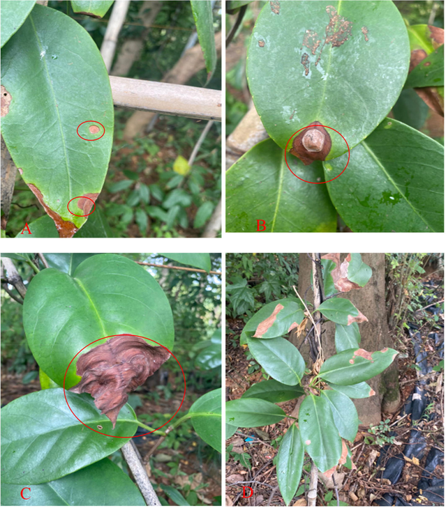
Symptoms of disease of Dragon Stripe Disease of K. coccinea on farm A: Leaves of invasion at initial stage; B: Leaves of invasion at medium term; C: Leaves of invasion at later stage; D: Diseased plant in morbidity.
3.2 Isolation and identification of the pathogenic bacteria
A total of 26 strains in total are isolated and obtained from 100 leaf tissues at the junction of diseased leaves and healthy ones. The isolated strains were initially divided into 11 categories according to the pattern and color of bacterial colony and one strain was selected from each category to deliver to Shenggong Biology Corporation to carry out molecular determination. The result shows that 11 strains are divided into 7 kinds of pathogenic bacteria, which are Apiospora jiangxiensis, Apiospora chromolaenae, Neopestalotiopsis formicarum, Diaporthe kochmanii, Diaporthe longicolla, Botryosphaeria fabicerciana and Alternaria alstroemeriae respectively. The strains HLH (12) B and HLH (10) A obtain amplified fragment in the size of 541 bp through PCR amplification and then test for electrophoresis of 1% of Sepharose, the similarities with A. jiangxiensis in GenBank (Accession No: NR_157464.1) are 100% and 99.63% respectively. The phylogenetic tree constructed using MEGA 7.0 (Fig. 2) shows that strains HLH(12)B and HLH(10)A come together with A. jiangxiensis, which shows that HLH(12)B and HLH(10)A are A. jiangxiensis; the amplified fragment of the strain HLH(7)A is 574 bp, the similarity with A. chromolaenae (Accession No: NR_168859.1) in GenBank is 95.11%, the phylogenetic tree comes together with A. chromolaenae, which verifies that the strain HLH(7)A is A. chromolaenae; the amplified fragments of the strains HLH7A and HLH18A are 537 bp and 526 bp respectively, the similarities with N. formicarum in GenBank (Accession No: NR_145242.1) are 98.7% and 99.62% respectively, HLH7A and HLH18A on the phylogenetic tree come together with N. formicarum, which verifies that the strain HLH1A is the strain D. longicolla; the sequence length of the strain HLH9A is 550 bp, the similarity with the D. kochmanii in Genbank (Accession No: NR_144924.1) is 98.36%, the phylogenetic tree comes together with D. longicolla, which verifies that the strain HLH1A is D. longicolla; the sequence length of the strain HLH9A is 562 bp, similar with B. fabicerciana in Genbank (Accession No: NR_137763.1) is 99.11%, the phylogenetic tree comes together with B. fabicerciana, which verifies that the strain HLH19A is B. fabicerciana; the amplified fragments of the strains HLH20A, HLH(17)A and HLH2A are 556 bp, 549 bp and 563 bp respectively the similarity with A. alstroemeriae in Genbank (Accession No: NR_163686.1) are 98.92%,99.27% and 98.58% respectively, HLH20A, HLH(17)A and HLH2A on the phylogenetic tree come together with A. alstroemeriae, which verifies that the strains HLH20A, HLH(17)A and HLH2A are A. alstroemeriae.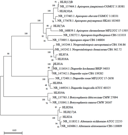
Phylogenetic tree constructed based on the similarity of rDNA-ITS sequence.
3.3 Determination of pathogenicity of the pathogenic bacteria
A total of 7 strains were inoculated to the healthy leaves of K. coccinea and then to leaves with the strain HLH7 after 3 days to start invasion, brown halation could be seen on leaves. The symptoms of subsequent invasion are basically the same with natural invasion on farm small black spots could be seen in the control group and at stabbing points of leaves that inoculate other strains, however, without any expansion (Fig. 3). According to Kochʼs theory, tissue isolation was again carried out from the diseased leaves to obtain the strain with the same character with the original (Fig. 4), which shows that the strain HLH7 is the disease germ to the Dragon Stripe Disease of K. coccinea.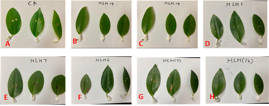
Mobilities of isolated strains on the leaves of K. coccinea after inoculation B-H: Disease symptoms of inoculations of HLH19, HLH9, HLH1, HLH7, HLH2, HLH(7)and HLH12 respectively; A: control.
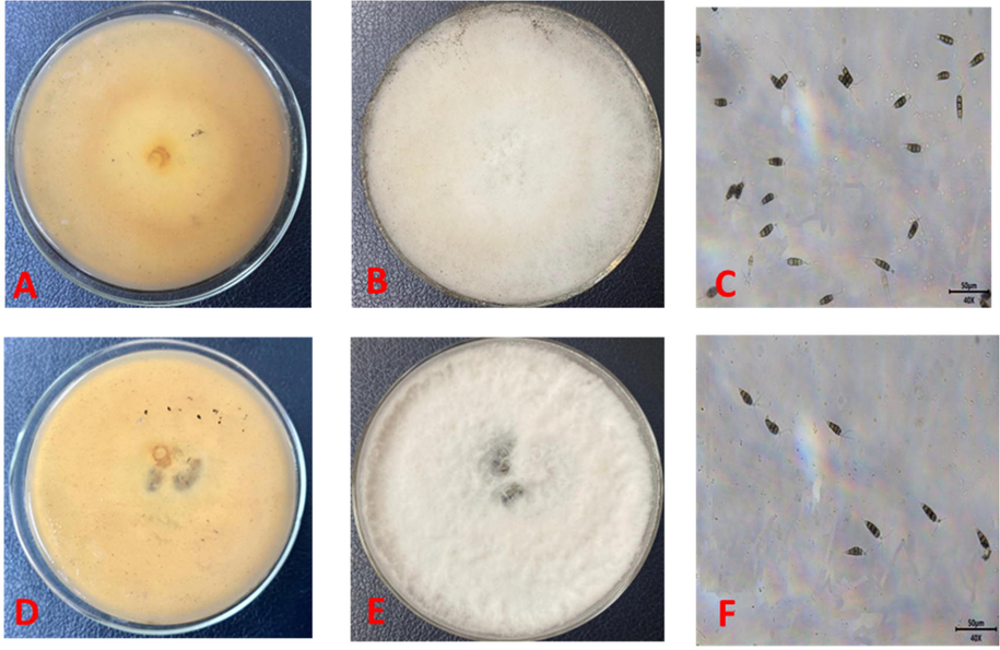
Forms of colony and thallus of the pathogenic bacteria of Dragon Stripe Disease of K. coccinea.
3.4 Morphological characteristics of the pathogenic bacteria
The pathogenic bacterium N. formicarum grows vigorously on PDA culture medium. At the end of 5d of culture, the bacterial colony grows in the petri dish to a diameter of 65 mm. The front of colony was white with the regular edge and tiled. Aerial hyphae were flourishing and dense, which show flocculence (Fig. 4E). It started to produce irregular sores in black spots (Fig. 4B) and showed fawn color at back of the colony without obvious increments (Fig. 4A, D). Conidiophore are fusiform to oval shapes, upright or slightly curved, 4 partitions and 5 cells with the sizes of 19.8–30.3 × 7.2–9.5 μm (n = 100; base cells are achromatic in conical type, the base is truncate with folds, the wall is thin with the length of 4.2–5.6 μm, three-color cells in the middle are brown in columelliform shape in length of 14.7–16.5 μm, the partition strangulates, the colors of diaphragm and perine are deeper than that of other parts with thick wall and verrucae, the second cell up the base is light brown with the length of 3.9–6.5 μm, the third cell is dark brown with the length of 4.1–6 μm; the fourth cell is brown with the length of 3.9–6.1 μm the top cell is in subcylindrical type and transparent, the wall is thin and smooth with the length of 3.7–5.2 μm, 2–3 colorless appendages grow on the top cell in filarious type, bending without virgation in length of 20.9–34.6 μm; 1 base appendage grows on the cercospora in medium style and tubulose type with the length of 3.1–7.6 μm (Fig. 4C, F).
The back of the colony of the strain HLH7; B front of the colony of the strain HLH7; C conidiophores of the strain HLH7; D back of the colony of the strain for the diseased leaf that was isolated again after inoculation; E front of the colony of the strain for the diseased leaf that was isolated again after inoculation; F conidiophores of the diseased leaf that is isolated again after inoculation.
4 Discussion
Kadsura coccinea (Lem.) is a traditionally used medicinal plant all over the China; the plant belongs to Schisandraceae family and widely used in folk medicines as analgesic agent, especially by the Miao and the Dong folk communities. The plant is highly cultivated over China for various nutritional products and bioactive constituents. However, the increased incidence of diseases that affect the plant significantly brought down the productivity of these plantations. Among the various diseases, the dragon stripe disease is increasingly occurring in different areas, however, the actual pathogenic species is yet to be discovered.
The Neopestalotiopsis is a genus of pathogenic fungi, which parasitize various plant species. These pathogenic species is distributed throughout the tropical and subtropical area (Maharachchikumbura et al., 2013). Studies have reported that the fungus harm various plants belonging to more than 50 families; therefore, the infection causes significant damage to the cash crops as well as ornamental plants. It has been reported that the fungi invade the plants through simple dermal wounds and then causing symptoms such as leaf spot, leaf blight, blanch blight, root rot and anabrosis (Song, 2015). The Neopestalotiopsis is a new category of fungoid in Pestalotiopsis and was confirmed based on the data from ITS, TUB and TEF genes (Mahadevakumar and Janardhana, 2014). Since the infection of the fungi causes numerous variety of diseases with multiple names and also the symptoms are shared among these diseases, it is necessary to utilize the morphology and molecular systematics for proper identification of the disease (Wei and Xu, 2004).
The present study identified that N. formicarum is the pathogenic organism of the disease using rDNA-ITS sequence analysis, pathogenicity test, morphological identification and inspection of Kochʼs Rule. N. formicarum is a kind of saprophyte reported from the dead ant specimen from Ghana in western Africa in 2014 and also from the plant fragments in Cuba. The close species Neopestalotiopsis clavispora differs from the N. formicarum in the large conidiophores and long top appendages (Maharachchikumbura et al., 2014). Living habits of N. formicarum are diverse including saprophyte, endophyte and disease germ. The species has been reported to be an important pathogen of plants and anamorphic fungus with certain economic value. Hidayat has previously isolated endophytic fungus N. formicarum from the bark and leaves of Styrax sumatrana (Hidayat et al., 2021). Likewise, Slamet also isolated the fungus from the bark and caudex of Styrax benzoin (Slamet et al., 2021).
It has been reported that N. formicarum could cause leaf spot in Pestalotiopsis to Hevea brasiliensis (Pornsuriya et al., 2020). Also, it causes leaf spot to Euterpe oleraceae and E. precatoria, Elaeis guineenses and Musa paradisiaca (Gualberto et al., 2021) and leaf blight and brown spots to Plinia cauliflora (Lin et al., 2022). The present study reports the new host of N. formicarum, K. coccinea from home and abroad. Field symptoms of the Dragon Stripe Disease of K. coccinea are similar with anthracnose, obvious or in apparent increase could be seen on diseased part and then expand to fusiform or irregular lesions on leaves at later stage. In plantation of K. coccinea, farmers and technicians call this disease as anthracnose, through isolation and identification of the pathogenic bacterium, this study clarifies that this disease is not the anthracnose; we call it the Dragon Stripe Disease based on the characteristics of disease. The field symptoms of this disease and morphological characteristics of the pathogenic bacterium are so similar with ringspot disease of K. coccinea while the pathogenic bacterium of ringspot disease of K. coccinea is N. clavispora. This study has finally determined that N. formicarum is the pathogenic bacterium of Dragon Stripe Disease in K. coccinea through rDNA-ITS sequence analysis and construction of phylogenetic tree. Due to superposition of morphological parameters among interspecies in Neopestalotiopsis, studies show that independent application of ITS could not distinguish species of Pestalotiopsis, (including species of Neopestalotiopsis), while constructing phylogenetic tree in application of combination of ITS gene and EF1-α and (or) β-tublin gene, the interspecies have obvious boundary (Song, 2016; Xie, 2020). Therefore, it shall expand area and number of samples and apply polygenic combination to identify the pathogenic bacterium of the Dragon Stripe Disease of K. coccinea correctly and comprehensively next.
Besides N. formicarum, the study has also isolated 6 kinds of pathogenic bacteria namely A.jiangxiensis, A. chromolaenae, D.kochmanii, D.longicolla, B. fabicerciana and A. alstroemeriae from infected leaves of K. coccinea. The test result of pathogenicity shows that N. formicarum is the only pathogenic bacterium and that diversity of fungoid on leaves of K. coccinea. Further, the 6 fungi are isolated from the diseased leaves of K. coccinea could not make incursions into the excised leaves of K. coccinea, which shows that this kind of fungus may be easier to infest leaves under living condition. B. fabicerciana has been once reported as the disease germ of leaf spot to Malania oleifera (Pan et al., 2022), therefore, to further verify the pathogenicity of this kind of fungus to K. coccinea, successive studies are required to carry out wound inoculation under living condition to improve overall accuracy of the test. Further explorations are required to see if this kind of fungus may exist in the plant tissues of K. coccinea in forms of endophytes or saprophytic bacteria.
5 Conclusion
The edible plant K. coccinea is of high value in traditional, folk and modern medicines as well as a rich source of several nutrients. The Dragon Stripe Disease caused by various fungal pathogens often reduces the productivity of the plant. The result of the study for the first time identified the role of N. formicarum based on the field symptoms as well as based on the analysis with Koch’s Rule. Molecule characterization and phylogenetic analysis confirmed the presence of N. formicarum in the affected plants. Overall, our finding will be helpful for the farmers and scientists for early detection, identification and management of the Dragon Stripe Disease in K. coccinea.
Acknowledgement
The authors are thankful to the higher authorities for the facilities provided.
Authors’ contribution
This study was done by the authors named in this article, and the authors accept all liabilities resulting from claims which relate to this article and its contents.
Availability of data and materials
The data used to support the findings of this study are available from the corresponding author upon request.
Funding
Natural Science Foundation of Hunan Province (2022JJ60049); Innovation Capability Demonstration Project of Hunan Provincial Development and Reform Commission (No.319, Xiang Development Reform Investment [2021]); Hunan Forestry Science and Technology Project (No.42 [2020], Xiangcaizihuan; No.9 [2021] of Xiangcaizi Ring Index; XLK201959); Applied Berry-like Liana Germplasm Bank in Wuling Mountain Area (2020TP3004); Research Project of Hunan Education Department (19C0651).
Declaration of Competing Interest
The authors declare that they have no known competing financial interests or personal relationships that could have appeared to influence the work reported in this paper.
References
- Pseudopestalotiopsis gilvanii sp. nov. and Neopestalotiopsis formicarum leaves spot pathogens from guarana plant: a new threat to global tropical hosts. Phytotexa. 2021;489(2):121-139.
- [CrossRef] [Google Scholar]
- Diversity of endophytic fungi isolated from benzoin-producing tree styrax sumatrana. IOP Conf. Ser.: Earth Environ. Sci.. 2021;762(1):012002
- [CrossRef] [Google Scholar]
- Identification and Characterization of Bacillus tequilensis GYUN-300: An Antagonistic Bacterium Against Red Pepper Anthracnose Caused by Colletotrichum acutatum in Korea. Front. Microbiol.. 2022;13:826827
- [CrossRef] [Google Scholar]
- Optimization of ultrasound-assisted extraction process and antioxidant activity of total flavonoids from panax nigra. Sci. Technol. Food Ind.. 2021;42(13):179-183.
- [Google Scholar]
- First report of leaf brown blight caused by Neopestalotiopsis formicarum on jabuticaba in Taiwan. Plant Dis. 2022
- [CrossRef] [Google Scholar]
- First report on the association of Pestalotiopsis mangiferae with leaf blight disease of Canthium dicoccum in India. For. Pathol.. 2014;44(5):424-426.
- [CrossRef] [Google Scholar]
- A destructive new disease of Syzygium samarangense in Thailand caused by the new species Pestalotiopsis samarangensis. Trop. Plant Pathol.. 2013;38(3):227-235.
- [CrossRef] [Google Scholar]
- First Report of Leaf Spot on Malania oleifera Caused by Botryosphaeria fabicerciana in China. Plant Dis. 2022 PDIS09212038PDN
- [CrossRef] [Google Scholar]
- Identification and characterization of Neopestalotiopsis fungi associated with a novel leaf fall disease of rubber trees (Hevea brasiliensis) in Thailand. J.Phytopathol.. 2020;168(7–8):416-427.
- [CrossRef] [Google Scholar]
- Pathogen identification and biological characteristics of black tiger blight. Plant Pathol.. 2019;49(06):866-870.
- [Google Scholar]
- Diversity of endophytic fungal species from styrax benzoin found in benzoin-producing locations in North Sumatra. IOP Conf. Ser.: Earth Environ. Sci.. 2021;914(1):012041
- [CrossRef] [Google Scholar]
- Fungal systematics of Pestalotiopsis and evaluation of barcoding genes. Guiyang: Guizhou University; 2015.
- Taxonomy, Molecular systematics and Molecular barcoding of Pestalotiopsis. Guangxi: Guangxi University; 2016.
- Identification of anthracnose of Camellia oleifera in Hunan province. Scientia Silvae Sinicae. 2017;53(08):43-53.
- [Google Scholar]
- Handbook of Fungal Identification. Shanghai: Shanghai Science and Technology Press; 1979.
- Pestalotiopsis kunmingensis sp. nov., an endophyte from Podocarpus macrophyllus. Fungal Divers. 2004;15:247-254.
- [Google Scholar]
- Pathogen identification, toxin component analysis and disease control of black tiger wheel spot. Guangxi: Guangxi University; 2020.
- First report of ring spot on Kadsura coccinea caused by Neopestalotiopsis clavispora in China. Plant Dis.. 2018;102(10):2032.
- [CrossRef] [Google Scholar]
- The complete chloroplast genome of ‘black tiger 2’ (Kadsura coccinea (lem.) A.C. Smith) in southeast of China and phylogenetic relationships AQ1. Mitochondrial DNA Part B. 2020;5(1):296-297.
- [CrossRef] [Google Scholar]
- First report of Corynespora cassiicola causing leaf spot on Kadsura coccinea in China. Plant Dis.. 2019;103(2):366.
- [CrossRef] [Google Scholar]
- Investigation on main diseases and pests of medicinal plant black tiger. J. Anhui Agric. Univ.. 2018;46(34):138-140.
- [Google Scholar]


