

Translate this page into:
Investigation of phytochemical profile and in vivo anti-proliferative effect of Laetiporus versisporus (Lloyd) Imazeki mushroom against diethylnitrosamine-induced hepatocellular carcinoma
⁎Corresponding authors. thiyagaramesh@gmail.com (Thiyagarajan Ramesh), biotechurn@gmail.com (Usha Raja Nanthini Ayyakannu)
-
Received: ,
Accepted: ,
This article was originally published by Elsevier and was migrated to Scientific Scholar after the change of Publisher.
Peer review under responsibility of King Saud University.
Abstract
The purpose of the study is to explore the bioactive compounds present in Laetiporus versisporus (LVEE) Lloyd Imazeki ethanolic extract and its anticancer activity against hepatocellular carcinoma (HCC) induced by DEN in rats. The bioactive compounds present in the LVEE was analyzed by GC–MS. The anticancer activity of LVEE was analyzed with the DEN induced rats were orally treated with LVEE (250 mg/kg), cyclophosphamide (50 mg/kg) to the respective treatment groups for 45 days. The levels of tissue markers, phase-I metabolizing enzymes was measured calorimetrically. ELISA was used to determine the level of AFP and CEA. The expression of apoptotic genes Bcl-2, p53, caspase 3 and caspase 9 was assessed by real time RT-PCR. Results of the phytochemical screening analysis showed that 12 bioactive molecules were existing in LVEE. Results of pharmacodynamics analysis showed that the 45 days of treatment with LVEE and cyclophosphamide (CPA) therapy significantly (p < 0.05) decreased the levels of MDA, AFP, CEA, cytochrome P450 and cytochrome b5, and increased levels of tissue markers and bilirubin in serum. The findings of RT-PCR analysis showed that the LVEE and CPA were significantly (p < 0.05) downregulated the pro-apoptotic genes Bcl-2 and upregulated the p53, caspase 3 and caspase 9 mRNA expression through mechanisms involved in promoting the cell apoptosis, and preserving the functional integrity of the liver membrane. L. versisporus (LV) can be considered as new anticancer agent that offers new opportunities for anticancer therapy.
Keywords
Laetiporus versisporus
Bioactive compounds
DEN
HCC
Histopathology

1 Introduction
According to the World Health Organization report in 2018, liver cancer is the fourth most important cause of morbidity and mortality worldwide. Among 9.6 million cancer deaths in 2018, liver cancer accounts for 782,000 deaths. Hepatocellular carcinoma (HCC) represents approximately 90% cases of principal liver cancer and leads to chronic liver disease and cirrhosis, which predominantly occurs in patients (Llovet et al., 2016). Most of the essential biological features called metastasis are the key cause of treatment failure and death due to liver cancer. Metastasis and recurrence rate can reach up to 50% even in the case of small HCC. Liver cancer is a disease which is chronic and associated with a defect in the regulation of a various number of signaling pathways which results from several metabolic processes (Khan et al., 2011). Alcohol consumption, viral infection with hepatitis B or hepatitis C, chronic inflammation of the liver due to metabolic dysfunction that causes either cirrhosis or fibrosis or both, and ultimately contributes to the growth of HCC. Severe inflammatory damage (necrosis), liver cirrhosis and disease have been related to liver immune functions that lead to liver defense (Llovet et al., 2016).
Apoptosis is strictly regulated by the mechanism of cell death and is necessary for cytotoxicity induced by anticancer compounds (Cotter, 2009). The Beta cell lymphoma-2 (Bcl-2) protein family is an important metabolic regulator of apoptosis; it is expressed in some tumor cells that are recognized as a key factor in regulating apoptosis. Bcl-2 protein family regulates intracellular signals both pro- apoptotic and anti-apoptotic and mediates the mitochondrial membrane potential. Bcl-2 protein overexpression facilitates survival of cells (Charo et al., 2005). Tumor-suppressor protein p53 acts as the genome’s most essential shield. Most human cancers, including HCC, are linked to p53 mutations and demonstrate their promising role in the process of HCC. Caspase-3 belongs to the cysteine-aspartic acid protease family of proteins and had a significant role in the process of execution of cell apoptosis. Inhibition of caspase-3 can cause hepatocyte death due to DNA damage, causing liver cancer (Shang et al., 2018). Caspase-9 belongs to protease family that initiates mitochondrial apoptotic pathways by activating multimeric-protein complexes and is a primary therapeutic drug target for various pathogenesis induces apoptosis.
Fifty percent of the approved anticancer drugs may be either original natural products, semi-synthetic products, natural product-based synthesized biomolecules, and pharmacophores discovered by the anticancer drugs investigation. Natural products include a wide range of bioactive or chemical compounds that are biologically active, including anticancer activities (Begnini et al., 2014). Edible mushrooms are commonly used as a health supplements for medicinal purposes and have therapeutically active compounds that have pharmacological activities such as anticancer, chronic bronchitis and immune enhancement (Wasser and Weis, 1999). Laetiporus Murrill is a world-wide edible mushroom; it belongs to the family Fomitopsidaceae and the order Polyporales is a cosmopolitan genus and is generally referred to as “Chicken of the woods” or “sulphur shelf”. This fungus is found in trees, boreal areas, tropical zones, on the stumps, stems and logs of several tree species (Klaus et al., 2013). However, the morphological characteristics of Laetiporus versisporus (Lloyd) Imazeki are entirely different. Aging fruit bodies are semiglobose and have hymenophore in the form of more chlamydospores. Initially, the color of the fruit bodies is lemon yellow, and then when matured, turns to white and brown. Edible mushrooms have been studied for their potent bioactivities, but no scientific assessment of the anticancer activity of Laetiporus versisporus (Lloyd) Imazeki has been published to date. Therefore, in this present study, our objective is to examine the underlying potential anticancer defense mechanism by treating with Laetiporus versiporus ethanolic extract (LVEE) against diethylnitrosamine (DEN)-induced hepatocarcinoma in male wistar albino rats.
2 Materials and methods
2.1 Preparation of the mushroom extract
Laetiporus versisporus mushrooms were collected from Kodaikanal (Latitude: 10.2278 and Longitude: 77.4595), India during the rainy season and was taxonomically identified and stored at Biotechnology Department, Mother Teresa Women’s University, Kodaikanal, India under voucher specimen number MTWU P26. They were cleaned and washed with distilled water. The fruiting bodies of mushrooms were cut into small pieces and dried at room temperature and finely ground. The fruiting bodies powder were extracted with 70% ethanol using the Soxhlet apparatus. The dried content was obtained after the removal of solvent by the rotary evaporator. Finally, the collected ethanolic extract of fruiting bodies of mushroom was stored at −20 °C for further studies.
2.2 Bioactive compounds analysis of LVEE using GC–MS
Laetiporus versisporus mushroom was subjected to Gas Chromatography-Mass Spectrometry (GC–MS) analysis to determine bioactive constituents using Clarus 500 GC/MS – PerkinElmer. The sample data was acquired on a Elite-5MS capillary column. Helium is utilized as the carrier gas with the flow rate of 1 ml/min. 1 µl of sample (LVEE) was infused in to the column with the injector temperature at 270 °C. The spectrum of bioactive constituents present in extracts can be achieved with 70 eV electron ionization and detector scan mode was set to 40–450 amu. A 0.5 sec scan interval and fragments range 40–450 Da was maintained with the total running duration of 30 min.
2.3 Animals and diets
In this experimental study, healthy Male wistar albino rats and its weight range 120–150 g were procured from Tamilnadu Veterinary & Animal Sciences University (TANUVAS), Tamil Nadu, India and was maintained at 25 ± 5 °C in an animal house cage that is well ventilated with 50 ± 15% humidity with a normal photoperiod of 12 h light and dark cycles for 7 days period. The commercial pellet diet for rats with ad libitum water was provided for rats. The animal care and handling of animals was done as per regulations of Committee for the Purpose of Control and Supervision of Experiments on Animals (CPCSEA) and experimental model reviewed and approved by Institutional Animal care Ethics Committee with Institutional Animal Ethics Committee (IAEC) number 11/2016/IAEC/MTWU, Mother Teresa Women’s University, Kodaikanal, Tamil Nadu, India. All the experimental studies were carried out as per the guidelines suggested by IAEC.
2.4 Experimental model
Wistar albino rats male weight ranges between 120 and 150 g was selected for experiment and were consigned into six rats in each of the four groups. The DEN (0.01%) mixed with drinking water and provided for the duration of 16 weeks to induce hepatocellular carcinoma as the method described by Ramakrishnan et al. (2006). Control group rats (Group 1) were administered orally with normal saline. Group 2 (HCC control) rats with overnight fasting were received 0.01% DEN. Group 3 HCC rats were provided with LVEE (250 mg/kg) for the duration of 45 days. HCC rats treated (Group 4) with 50 mg/kg of cyclophosphamide (CPA; positive control) for the period of 45 days.
2.5 Experimental procedures
All the rats were sacrificed by cervical decapitation under mild anesthesia after the completion of experimental regimens. From the rats blood samples were collected into the centrifuge tubes by cardiac puncturing. Hepatic marker enzymes were analyzed using separated serum. From the rats, liver was collected then washed with saline (ice cold) and stored in formalin (10%). Harvested tissues were used for histopathological examination.
2.6 Blood chemistry analysis for liver function and cancer markers
Aspartate transaminase (AST) and alanine transaminase (ALT) in serum were examined for hepatic marker enzymes by the method of Reitman and Frankel (1957) alkaline phosphatase (ALP) activity was predicted by the method of Bessey et al. (1946); Acid phosphatase (ACP) and lactic acid dehydrogenase (LDH) was estimated by the method of King (1965). γ-Glutamyl transferase (γ-GT) and 5′-Nucleotidase (5′NT) activity was predicted by the method of Persijn and Silk (1976); Bilirubin was measured by Malloy and Evelyn (1937).
2.7 Determination of AFP and CEA by ELISA
The solid phase type of enzyme linked immunosorbent assay (ELISA) was performed by utilizing UBI MAGIWELL (USA) enzyme immunoassay kit to estimate the markers enzymes namely Alpha-feto protein (AFP) and carcino embryonic antigen (CEA).
2.8 Analyses of phase I metabolizing enzymes in serum
Cytochrome P450 reductase (cyto P450) is a phase I enzymes was examined by the method of Omura and Takesue (1970) and cytochrome b5 reductase (cyto b5) activity was estimated by the method of Mihara and Sato (1972).
2.9 Analysis of gene expression by quantitative RT-PCR
The RNA in the cell was extracted from the liver tissues at the end of the experiment, based on the manual protocol using an RNeasy lipid tissue kit (Qiagen, Valencia, CA). Extracted RNA was quantified with UV/Vis spectrometer-ACT gene. cDNA using oligo (dT) primers was synthesized with cellular RNA (500 ng) and for RT-PCR (Invitrogen) Superscript III first strand synthesis system will provide reverse transcriptase. Using the Applied Biosystem 7500 PCR system (Applied Biosystem, Foster City, CA) fast-real time PCR were carried out. The gene expression levels of BCL-2 (Forward -AAACTGAACCAGAAAGGGAGAC and Reverse-CTGCTGCCATGGGGATTACC), p53 (Forward-ATGAGCGTTGCTCTGATGGT and Reverse-GGTGGCTCATACGGTACCAC), caspase-3 (Forward-GGACAACAACGAAACCTCCG and Reverse-ACACAAGCCCATTTCAGGGT) and caspase-9 (Forward-TTGAGGTGGCCTTCACTTCC and Reverse-GTGGGCAAACTTGACACTGC) were normalized against the β-actin (Forward- CCACCATGTACCCAGGCATT and Reverse-GAGCCACCAATCCACACAGA) gene (housekeeping gene).
2.10 Tissue collection and histopathological examination
The liver tissues were sliced from normal, DEN, DEN + LVEE, and DEN + CPA groups were subjected to histopathological examination to depict the cellular architecture. After fixation of liver, kidney and spleen tissue to the neutral formalin solution (10%) for 48 h, followed by 6 h fixation with bovine solution. Tissues embedded in paraffin after the process of fixation, sections were subjected cut using microtome and placed on the slides and stained with H & E stain visualized in the light microscope, finally the images were acquired by the method of Dunn (1974).
2.11 Statistical analysis
All the experimental values were reported as mean ± standard error of the mean for six animals in each group. All the data’s were statistically evaluated and compared by Statistical Package of Social Science (SPSS-16.0, SPSS, Inc., Chicago, IL). Differences among means were analyzed by LSD and significant level is represented as p < 0.05.
3 Results
3.1 Bioactive constituent’s analysis
GC–MS chromatogram of LVEE shows many bioactive constituents with the subsequent peaks at different retention time (Fig. 1). Thirteen peaks of LVEE were identified on GC–MS. The identified bioactive constituent’s retention time, molecular formula and their biological activities are shown in Table 1.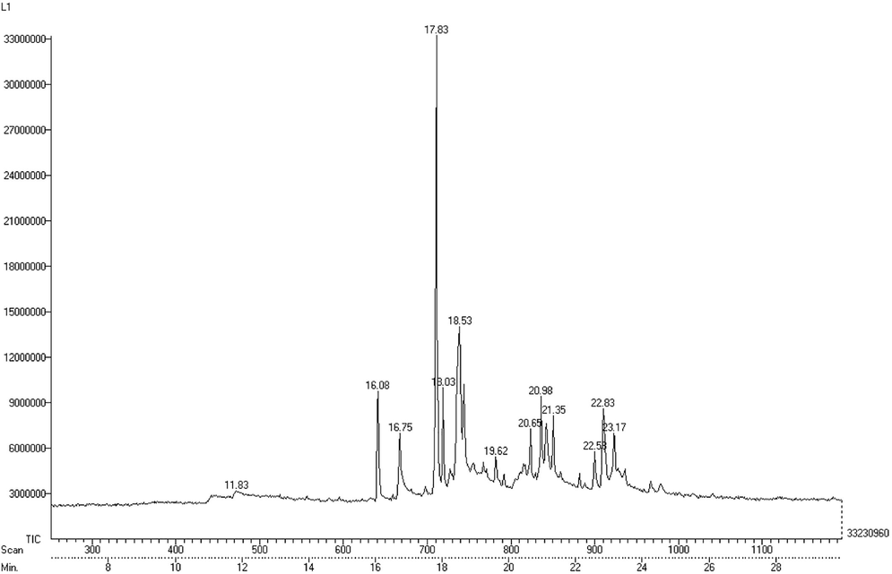
The Gas chromatography-mass spectroscopy (GC–MS) chromatogram of LVEE with 13 peaks by comparing retention time with standard.
Number
Retention time (min)
Molecular formula
Compounds
Pubchem Id
Biological activities
1
11.83
C10H18O
α-terpineol
442,501
Anticancer activity (Hassan et al., 2010), Antioxidant activity (Bicas et al., 2011), Cardioprotective activity (Sabino et al., 2013), Antiulcer activity (Matsunaga et al., 2000).
2
16.08
C15H10O2
Flavone
10,680
Anti-inflammatory and Antimicrobial (Cushnie and Lamb, 2005), Anti-allergic and Antioxidant (Havsteen, 1983).
3
16.75
C16H32O2
n-Hexadecanoic acid
985
Antitumor activity (Harada et al., 2002).
4
17.83
C19H36O2
11-Octadecenoic acid, methyl ester, (Z)
5,364,505
Antibacterial, Antifungal and Antioxidant (Asghar et al., 2011)
5
18.03
C19H38O2
Octadecanoic acid, methyl ester
8201
Antimicrobial (Gehan et al., 2009)
6
19.62
C13H16N2O5
8-Carbethoxy-1-methyl-1,4,5,6,7,8-hexahydropyrrolo[2,3-b] azepin-4-one-3-carboxylic acid
624,386
Not found
7
20.65
C17H20O5
2,6-Naphthalenedicarboxylic acid, 3,4-dihydro-3,7-dimethyl-5-methoxy-, dimethyl ester
627,617
Not found
8
20.98
C15H8N2O6
Coumarine, 3-(2,4-dinitrophenyl)-
596,144
Not found
9
21.35
C21H38O2
6,11-Eicosadienoic acid, methyl ester
5,364,621
Not found
10
22.58
C20H20N2O
Hexanediamide, 2,5-bis(Methylene)-N,N’-diphenyl
615,074
Not found
11
22.83
C22H42O2
E-13-Docosenoic acid(Brassidic Acid)
5,282,772
Not found
12
23.17
C21H42O3
Methoxyacetic acid, octadecyl ester
542,201
Not found
13
18.53
C20H40
1-Eicosene
18,936
Not found
3.2 Effect of LVEE on body weight and relative liver weight
To evaluate anticancer activity of LVEE in DEN-induced rats, LVEE treatment was started on 10th week since induction of DEN and lasting for 45 days. Body weight and liver weight of the rats were analyzed. The results of the effects of DEN, LVEE and CPA on body and liver weights are shown in Fig. 2a-b. Compared with DEN-induced HCC rats, LVEE (250 mg/kg) and CPA (50 mg/kg) showing significant increase in the body weight (p < 0.05). While, decreased in liver weight than that in DEN induced group (p < 0.05).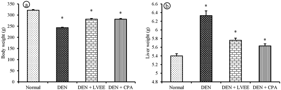
Effect of LVEE on (a) body weight and (b) liver weight of DEN group rats. Results are shown as mean ± S.D (n = 6). * p < 0.05 compared with normal group.
3.3 Effects of LVEE on liver function related biomarker in serum
The hepatic marker enzymes activities namely AST, ALP, ACP, ALT, LDH, γ-GT, 5′NT, and bilirubin (mg/dl) in serum were determined to evaluate the hepatic cellular damage. The current result shows that animals treated with DEN significantly increased the activities of serum AST, ALT, ALP, ACP, LDH, γ-GT, and 5′NT compared to control group (Fig. 3a-d) but when treated they decreased in the dose dependent manner. The DEN treated rats showed significant enhancement in the activity of serum bilirubin compared to control rats (Fig. 4a). This result confirmed that the LVEE has hepatoprotective activity.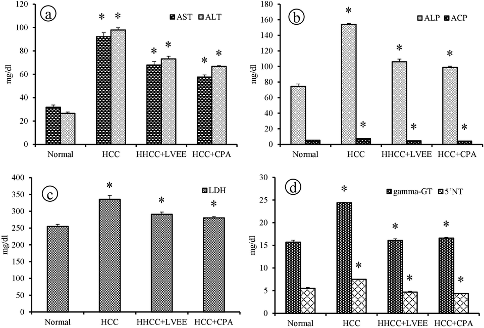
Effects of LVEE on liver function markers of (a) AST and ALT, (b) ALP and ACP, (c) LDH and (d) γ-GT and 5′NT activity in DEN group rats. Results are shown as mean ± S.D (n = 6). *p < 0.05 compared with normal group.
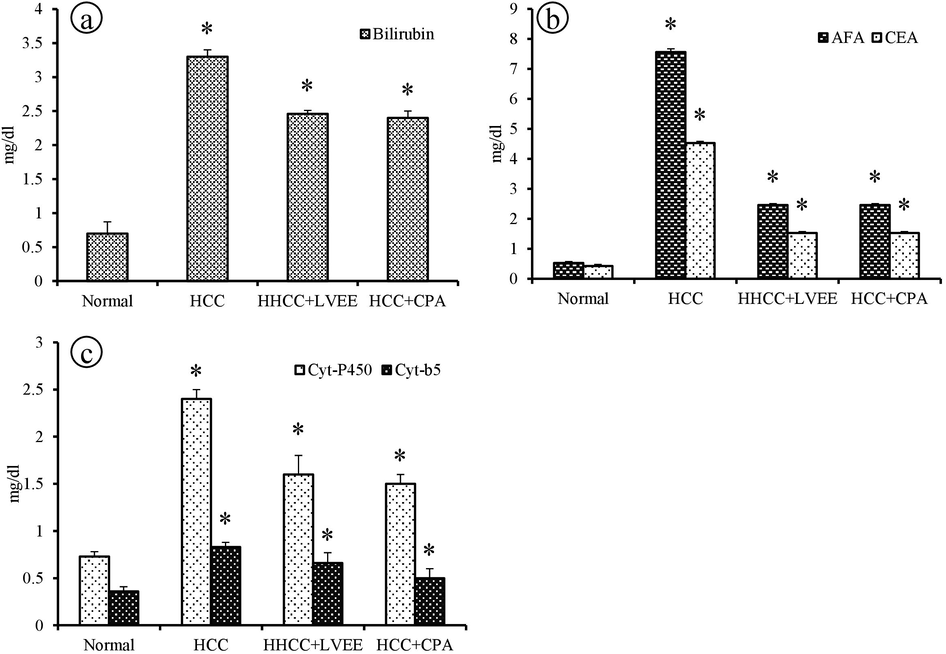
Effects of LVEE on cancer markers of (a) bilirubin, (b) AFP and CEA, (c) cyto-P450 and cyt-b5, in DEN group rats. Results are shown as mean ± S.D; (n = 6). *p < 0.05 compared with normal group.
3.4 Effect of LVEE on AFP and CEA in serum
The most important indicators of serum AFP and CEA for hepatocellular carcinoma was determined by utilizing ELISA assay. From the results, LVEE and CPA could decrease the serum AFP and CEA levels when compared with DEN-induced HCC rats. Meanwhile, LVEE treatment shows the reduction of the serum AFP and CEA level compared with CPA treated rats (Fig. 4b).
3.5 Effect of LVEE on phase I metabolizing enzymes in serum
DEN-induced HCC rats exhibited significantly increased the level of cyt P450 and cyt b5 reductase activity with respect to normal rats (Fig. 4c). When DEN-induced rats treated with LVEE shows significant reduction in the level of cyt P450 and cyt b5 reductase activity (p < 0.05). Moreover, no significant difference was observed in LVEE treated rats with respect to CPA treated rats.
3.6 Effect of LVEE on the apoptotic related mRNA expression
To demonstrate the apoptotic activity of LVEE, Bcl-2, p53, caspase-3, and caspase-9 were measured. The DEN induced HCC rats showing the increase in the level of anti-apoptotic protein Bcl-2 when compared with normal group rats, but it was significantly reduced in LVEE (250 mg/kg) and CPA (50 mg/kg) group rats. Compared to DEN induced group rats, increase in the level of caspase-3 and caspase-9 in LVEE treated group rats, suggesting the activation of caspases. Furthermore, we investigated significant negative regulator p53, mRNA expression was decreased in DEN-induced HCC rats, but it was increased in LVEE and CPA group rats (Fig. 5).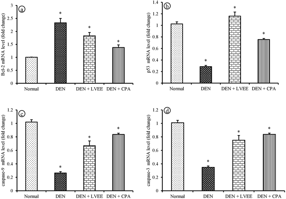
Gene expression analysis of LVEE effect at the mRNA quantification using qRT-PCR. Gene expression of (a) Bcl-2, (b) p53, (c) Caspase 9 and (d) Caspase 3 at mRNA level in the liver tissue of DEN group rats. Values are shown mean ± S.D (n = 6). *p < 0.05 compared with normal group.
3.7 Effect of LVEE on liver histopathology
The therapeutic effects exhibited by LVEE in DEN-induced hepatocarcinoma were studied by the histopathological assessment (Fig. 6). Microscopically, the control rats hepatic section of liver tissue showed central vein with usual cell architecture, and sinusoids with mild dilatation (Fig. 6a). DEN-induced rats liver tissue showed the caused observable histological injury in the hepatocytes, abnormal cells with focal area of fatty change, inflammation, necrosis, congestion and some loss of liver parenchymal cells (Fig. 6b). DEN-induced rats treated with LVEE depicted mild injury with normal cellular architecture and hepatocyte showing central vein with slight inflammation and reduces the fatty change (Fig. 6c). CPA treated group showed normal lobular architecture with slight inflammation (Fig. 6d).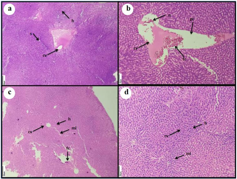
Histopathology study on the effect of LVEE + DEN group rats liver. After 45 days treatment, liver tissue was collected from six albino rats in each groups and viewed under light microscope after H & E staining (40 × magnification) (a) Group −1 (control) rats depicting normal hepatocytes (h) with central vein (cv), and sinusoid (s) (b) HCC control group rats depicting massive fatty accumulation (fc) in central vein (cv), congestion, some reduction in parenchymal cells (pc) and necrosis injury (n), (c) HCC rats groups treated with LVEE (250 mg/kg b.wt/day) showing decreased fatty accumulation, distinct progress of sinusoid and central vein with mild inflammation (mi) in hepatocytes (h); (d) HCC group treated with CPA (50 mg/kg b.wt/day) showing a normal sinusoids and central veins in the hepatocytes.
4 Discussion
DEN is a heptocarcinogen and produces promutagenic products that are responsible for carcinogenic activity namely, O6-ethyldeoxyguanosine, O4 and O6-ethyldeoxythymidine by its metabolic biotransformation process (Hussain et al., 2012). In the current study, we observed the decreased body weight in DEN induced rats that are in concurrence with an earlier report on the weight loss in rats when exposed to DEN which is a cytotoxic agent could damage DNA caused by electrophilic ethyl diazonium ion production by reacting with nucleophiles and subsequent cellular damage of the affected rats body organs due to more oxidative stress (Heindryckx et al., 2009). In DEN-induced rat, liver weight increases significantly when compared to control group and that could be due to the effect of DEN on the liver. The liver weight increase might be due to progression of the tumor growth in the liver which shows liver enlargement markedly in DEN treated rats. After treatment with LVEE, the liver enlargement caused by DEN was reduced. This study showing that liver weight of treated rats has decreased as compared with DEN induced liver cancer, so in this manner these results are consistent with that C. carandas showed positive effect against hepatic carcinoma (Singh et al., 2018), the liver weight increased in DEN treated rats as compared with that of normal group.
Current clinical settings, liver cancer markers in tissues such as ALT, ALP, AST, and γ-GT are commonly used indicators to detect the liver diseases. This study results revealed that the level of AST, ALT, ALP, ACP, LDH, and γ-GT in serum were elevated extensively in rats groups induced with DEN than that of normal group rats. Meanwhile, LVEE and CPA treated rats group showed the lowered level of these enzyme activities, which put forth LVEE protective effect by stopping membrane damage and loss of integrity against DEN-induced HCC rats. This study results are in agreement with Krishnan et al. (2017) who shows a similar observation on Tetilla dactyloidea crude methanolic extract against DEN-induced HCC rats shows the liver marker enzymes activities levels decreases significantly after the treatment with CPA.
The reversible oxidation of L-lactate to pyruvate was catalysed by the enzyme LDH which is present in the cytosol. The enhancement of hepatocyte membrane permeability and cellular leakage was due to increase of LDH activity in serum. The increase in LDH activity during cancer condition increases rate of glycolysis in the tumor cells which lead to the energy producing pathway for the malignant cells (Nandakumar and Balasubramanian, 2012). γ-GT is early enzyme marker of hepatocarcinogenesis and it is located in the hepatic cells outer membrane, their elevation leads to bile duct necrosis and cholestasis (Reynaert et al., 2001). 5′NT is an another marker enzyme and serves as a diagnostic tool for liver damage. In this study, LVEE treatment shows significant reduction in the levels of 5-NT activity in the serum that is because of the secondary metabolites in LVEE to effectively restrain expansion. Elevation of bilirubin level was indicator with many liver diseases. The slight increase in the serum bilirubin associated with liver injury in rats or leads to bile duct obstruction (Rasekh et al., 2008). The release of the unconjugated bilirubin and mass obstruction of conjugated response from injured hepatocytes was associated with the increase level in the serum bilirubin in the DEN administered mice. After the treatment of LVEE, decrease in the serum bilirubin demonstrates the viability of the L. versisporus mushroom which enhances the liver health.
To estimate the protective function of LVEE in DEN-induced HCC rats, the tumor markers AFP and CEA activity has been studied. AFP is a onco-fetal protein present in serum that is gradually lost in the development process and completely absent in the healthy individuals (adult) and increase in circulating AFP level will found in DEN-induced rats. AFP is a tumor marker enzyme and its increased level is found in HCC because more than 70% of HCC patients have increase AFP level in the serum because of tumor secretion. CEA is a heavily glycosylated protein with molecular weight of 180–200 kDa, belongs to immunoglobulin family and elevated levels of CEA used as tumor marker enzyme which widely utilized to detect presence of different types of cancers. Increased CEA levels associated with an increase tumor size and metastasis. The study results showed after the treatment of LVEE and CPA could decrease the level of serum AFP and CEA in DEN induced HCC rats, which suggests there was rate of decrease in tumor production. This study results similar with Vishnu Priya et al. (2018) have revealed the decrease in tumor markers after administration of T. chebula aqueous extract that reduce tumors production rate.
Cytochrome P450 is a membrane bound heme proteins that is found predominantly in the hepatocyte cells endoplasmic reticulum which involved in metabolism of endogenous xenobiotics and compounds (Moon et al., 2006). Along with cyt P450 reductase, cyt b5 involves in the molecular oxygen activation by cyt P450, which requires electrons produced by donor NADH cyt b5 reductase. Therefore, we determined these enzymes serum levels of DEN-induced HCC rats. Compared with DEN induced HCC rats, LVEE and CPA decreased cyt-P450 activity and cyt-b5 activity. The present study results are agreed with Mangalamani Daisy and Devaki (2011) who reported that chrysin (5, 7 dihydroxy flavone) is an isoflavone naturally occurring biological compound from flowering plants, effectively modulates the phase I and phase II metabolizing enzymes during DEN induced liver carcinoma.
The principal mechanism played by chemotherapeutic agents in the initiation of cell apoptosis in tumor formation and this may be appropriate for HCC development. Bcl-2 family proteins have a vital regulatory function in the mitochondrial pathway that involves the pro and anti-apoptotic member of proteins. The DEN induced HCC rats showing the increased level of anti-apoptotic Bcl-2 protein while up on treatment with LVEE and CPA, Bcl-2 was significantly (p < 0.05) decreased. Caspase-3 is an important mediator of apoptosis which is triggered by caspase-9 an activator. In this experimental study, compared to DEN induced group rats, the increased level of caspase-3 and caspase-9 expression after the treatment with LVEE which activates the caspase-3 expression. Caspase-3 and caspase-9 are an activator proteins that play an important function in the execution period of cell death (apoptosis) (Yin and Ding, 2003).
It was shown that p53 tumor suppressor gene act as an important regulator of apoptosis mechanism and arrests cell cycle. Furthermore, this regulation subsequently inhibits the activity of cell cycle-regulating enzymes and the Bcl-2 protein (anti-apoptotic) expression (Santoro et al., 2014). Reduction in the expression of Bcl-2 signaling molecules has been reported as anticancer mechanisms in diverse types of cancers. Our data also revealed that LVEE activated the p53 gene, causing a series of events which leads to the activation of caspase-3 and finally cell apoptosis. Our study results supported with Moreira et al. (2015) who stated that p53 levels were decreased in DEN-induced HCC rats and causes downregulation of p53 gene. Taken together, the data suggest that LVEE had anti-cancer activity against DEN-induced HCC rats by activating p53 induced apoptotic signaling pathway. Meantime, LVEE could control the biochemical markers. Hence, LVEE can be a good therapeutic source for HCC.
5 Conclusion
In conclusion, LVEE treatment exerted chemoprotective effect on DEN- induced hepatocellular carcinoma rats as this mushroom extract can revive serum liver marker enzymes, cancer marker enzymes – AFP and CEA, and phase-I enzymes – cyt P450 and cyt b5 activity. LVEE preserves the cellular architecture of liver tissue towards DEN induced HCC rats. Furthermore, downregulation of anti-apoptoptic Bcl-2 activity and upregulation of p53, caspase-3 and caspase-9 activities upon LVEE treatment suggests its hepatoprotective activity against DEN-induced HCC rats. Thus, the results of this investigation have established the efficacy of LVEE as an effectual chemotherapeutic agent.
Acknowledgements
This research was supported by Department of Science & Technology (Project No. DST/SSTP/TN/104/2017-2018), India. Also, we appreciate the contributions of all those who participated in this research and the comments of the reviewers of this manuscript.
Declaration of Competing Interest
The authors declare that they have no known competing financial interests or personal relationships that could have appeared to influence the work reported in this paper.
References
- Gas chromatography-mass spectrometry(GC-MS) analysis of petroleum ether extract (oil) and bio-assays of crude extract of Iris germanica. Int. J. Gen. Mol. Biol.. 2011;3:95-100.
- [Google Scholar]
- Brazilian red propolis induces apoptosis-like cell death and decreases migration potential in bladder cancer cells. Evid. Based Complement Alternat. Med. 2014:1-13. 639856
- [Google Scholar]
- A method for the rapid determination of alkaline phosphatase with five cubic millimeters of serum. J. Biol. Chem.. 1946;164(1):321-329.
- [Google Scholar]
- Evaluation of the antioxidant and antiproliferative potential of bioflavors. Food Chem. Toxicol.. 2011;49(7):1610-1615.
- [Google Scholar]
- Bcl-2 over expression enhances tumor-specific T-cell survival. Cancer Res.. 2005;65(5):2001-2008.
- [Google Scholar]
- Apoptosis and cancer: the genesis of a research field. Nat. Rev. Cancer. 2009;9(7):501-507.
- [Google Scholar]
- Hand Book of Histopathological and Histochemical Techniques, (third ed.). Trowbridge and Esher, Butterworths, London: Redwood, Burn, Ltd; 1974.
- Marine natural products and their potential applications as antiinfective agents. World Sci. J.. 2009;7:872-880.
- [Google Scholar]
- Antitumor activity of palmitic acid found as a selective cytotoxic substance in a marine red alga. Anticancer Res.. 2002;22:2587-2590.
- [Google Scholar]
- Alpha terpineol: a potential anticancer agent which acts through suppressing NF-kappaB signalling. Anticancer Res.. 2010;450(30):1911-1919.
- [Google Scholar]
- Flavonoids, a class of natural products of high pharmacological potency. Biochem. Pharmacol.. 1983;32(7):1141-1148.
- [Google Scholar]
- Experimental mouse models for hepatocellular carcinoma research. Int. J. Exp. Pathol.. 2009;90:367-386.
- [Google Scholar]
- Evaluation of chemopreventive effect of Fumaria indica against N-nitrosodiethylamine and CCl4-induced hepatocellular carcinoma in Wistar rats. Asian Pac. J. Trop. Med.. 2012;5(8):623-629.
- [Google Scholar]
- Chrysin abrogates early hepatocarcinogenesis and induces apoptosis in N-nitrosodiethylamine-induced preneoplastic nodules in rats. Toxicol. Appl. Pharmacol.. 2011;251(1):85-94.
- [Google Scholar]
- The hydrolases-acid and alkaline phosphatases. In: Van D., ed. Practical Clinical Enzymology. London, UK: Van Nostrand; 1965. p. :191-208.
- [Google Scholar]
- The edible mushroom Laetiporus sulphureus as potential source of natural antioxidants. Int. J. Food Sci. Nutr.. 2013;64(5):599-610.
- [Google Scholar]
- In vitro, In silico and In vivo Antitumor Activity of Crude Methanolic Extract of Tetilla dactyloidea (Carter, 1869) on DEN Induced HCC in a Rat Model. Biomed. Pharmacother.. 2017;95:795-807.
- [Google Scholar]
- The determination of bilirubin with the photoelectric colorimeter. J. Biol. Chem.. 1937;119(2):481-490.
- [Google Scholar]
- Chrysin attenuates the instability of xenobiotic metabolizing and mitochondrial enzymes during Diethyl nitrosamine induced liver carcinoma. J. Pharm. Res.. 2011;4:1839-1845.
- [Google Scholar]
- Isolation of the antiulcer compound in essential oil from the leaves of Cryptomeria japonica. Biol. Pharm. Bull.. 2000;23(5):595-598.
- [Google Scholar]
- Partial purification of NADH-cytochrome b 5 reductase from rabbit liver microsomes with detergents and its properties. J. Biochem.. 1972;71:725-735.
- [Google Scholar]
- Dietary flavonoids: effects on xenobiotic and carcinogen metabolism. Toxicol. in Vitro. 2006;20:187-210.
- [Google Scholar]
- Massively parallel sequencing identifies recurrent mutations in TP53 in thymic carcinoma associated with poor prognosis. J. Thorac. Oncol.. 2015;10(2):373-380.
- [Google Scholar]
- Hesperidin a citrus bioflavonoid modulates hepatic biotransformation enzymes and enhances intrinsic antioxidants in experimental breast cancer rats challenged with 7, 12-dimethylbenz (a) anthracene. J. Exp. Ther. Oncol.. 2012;9:321-335.
- [Google Scholar]
- A new method for simultaneous purification of cytochrome b5 and NADPH-cytochrome c reductase from rat liver microsomes. J. Biochem.. 1970;67:249-257.
- [Google Scholar]
- A New Method for the Determination of γ-Glutamyltransferase in Serum. J. Clin. Chem. Clin. Biochem.. 1976;14:421-428.
- [Google Scholar]
- Suppression of N-nitrosodiethylamine induced hepatocarcinogenesis by silymarin in rats. Chem.-Biol. Interact.. 2006;161(2):104-114.
- [Google Scholar]
- Acute and subchronic oral toxicity of Galega officinalis in rats. J. Ethnopharmacol.. 2008;116(1):21-26.
- [Google Scholar]
- A colorimetric method for the determination of serum glutamic oxalacetic and glutamic pyruvic transaminases. Am. J. Clin. Pathol.. 1957;28(1):56-63.
- [Google Scholar]
- Somatostatin suppresses endothelin-1-induced rat hepatic stellate cell contraction via somatostatin receptor subtype 1. Gastroenterol.. 2001;121(4):915-930.
- [Google Scholar]
- Cardiovascular effects induced by α- 540 terpineol in hypertensive rats. Flav. Frag. J.. 2013;28:333-339.
- [Google Scholar]
- Beta-Catenin and epithelial tumors: a study based on 374 oropharyngeal cancers. Biomed Res. Int.. 2014;2014:1-13.
- [Google Scholar]
- Caspase-3 suppresses diethylnitrosamine-induced hepatocyte death, compensatory proliferation and hepatocarcinogenesis through inhibiting p38 activation. Cell Death Dis.. 2018;9:558.
- [Google Scholar]
- Attenuation of diethylnitrosamine (DEN) - Induced hepatic cancer in experimental model of Wistar rats by Carissa carandas embedded silver nanoparticles. Biomed. Pharmacother.. 2018;108:757-765.
- [Google Scholar]
- Biochemical Evidence for the Antitumor Potential of Garcinia mangostana Linn On Diethylnitrosamine-Induced Hepatic Carcinoma. Pharmacogn. Mag.. 2018;14(54):186-190.
- [Google Scholar]
- Medicinal properties of substances occurring in higher basidiomycetes mushrooms: Current perspectives. Int. J. Med. Mush.. 1999;1:47-50.
- [Google Scholar]
- Death receptor activation-induced hepatocyte apoptosis and liver injury. Curr. Mol. Med.. 2003;3(6):491-508.
- [Google Scholar]


