Translate this page into:
Inhibition of inducible nitric oxide synthase gene expression (iNOS) and cytotoxic activity of Salvia sclarea L. essential oil
⁎Corresponding author at: Kongunadu Arts and Science College, Coimbatore 641029, Tamil Nadu, India. Mobile: +91 9894793074. rtsampandan@yahoo.com (Ramaraj Thirugnanasampandan)
-
Received: ,
Accepted: ,
This article was originally published by Elsevier and was migrated to Scientific Scholar after the change of Publisher.
Peer review under responsibility of King Saud University.
Abstract
Essential oil obtained from Salvia sclarea L. leaves was tested for hepatoprotective, inducible nitric oxide synthase (iNOS) gene down regulation and cytotoxic activities. Histopathology of liver tissue showed severe inflammation and necrosis after treatment with 1.5 μg lipopolysaccharide (LPS)/30 g body weight of BALB/c mice. A decrease in inflammation was observed with the treatment of different concentrations of essential oil. Necrosis and infiltration of hepatocytes were barely seen at 100 μg of oil. Reverse transcriptase polymerase chain reaction (RT-PCR) analysis showed down regulation of iNOS at transcriptional level. Growth of HeLa cells was inhibited by the oil with an IC50 of 80.69 ± 0.01 μg/mL. Propidium iodide (PI) staining revealed the presence of apoptosis in oil treated cells. The findings indicate that S. sclarea essential oil is a promising natural source which may be useful for herbal medicine preparation.
Keywords
Essential oil
Lipopolysaccharide
Antiinflammation
Cytotoxicity
Blebbing
1 Introduction
Liver diseases are one of the most serious health problems nowadays. Studies on pathogenesis of hepatic diseases, oxidative stress and inflammation are well established. Though there are numerous advances in modern medicine still prevention and treatment of liver diseases are limited (Malhi and Gores, 2008; Tacke et al., 2009). Cervical cancer is the second most common cancer among the women worldwide. Data suggest that infection with human papilloma virus plays a major role in cervical cancer. Current treatment available for liver diseases and cervical cancer are mainly synthetic drugs. Serious side effects limit the use of such drugs. Therefore, using plant products may be the right choice for prevention and treatment with fewer side effects.
The genus Salvia belonging to the family Lamiaceae has been used in herbal medicine for thousands of years in the treatment of many different ailments and exhibit various pharmacological properties such as antioxidant, antiinflammatory, antimicrobial and antiplasmodial and is used in treating cold, bronchitis, tuberculosis, hemorrhage and menstrual disorders (Tepe et al., 2004; Topcu, 2006). The study species, Salvia sclarea L. (Clary sage), an aromatic perennial herb is considered economically and one of the most cultivated species for its essential oil. Traditionally the species has been used to promote blood circulation, remove stagnation, tranquilize the mind, clear heat from blood, resolve swelling and used in the treatment of coronary heart disease for the alleviation of angina pectoris, coronary artery spasm and myocardial infarction (Zhou et al., 2005). Based on the reports available on the medicinal uses of S. sclarea, the present study was aimed to carry out the in vivo, in vitro antiinflammatory effects and cytotoxic activity of essential oil.
2 Materials and methods
2.1 Collection of plant material and hydrodistillation of leaves
The leaves of S. sclarea L. was collected from Cinchona, Nilgiri hills, Tamil Nadu, India. Freshly collected leaves of S. sclarea (1000 g) were hydrodistilled for 4 h using Clevenger apparatus for essential oil extraction. Extracted oil was treated with anhydrous sodium sulfate to remove water and filled in small vials, tightly sealed and stored in a refrigerator (4 °C) for further studies.
2.2 Animals and experimental method
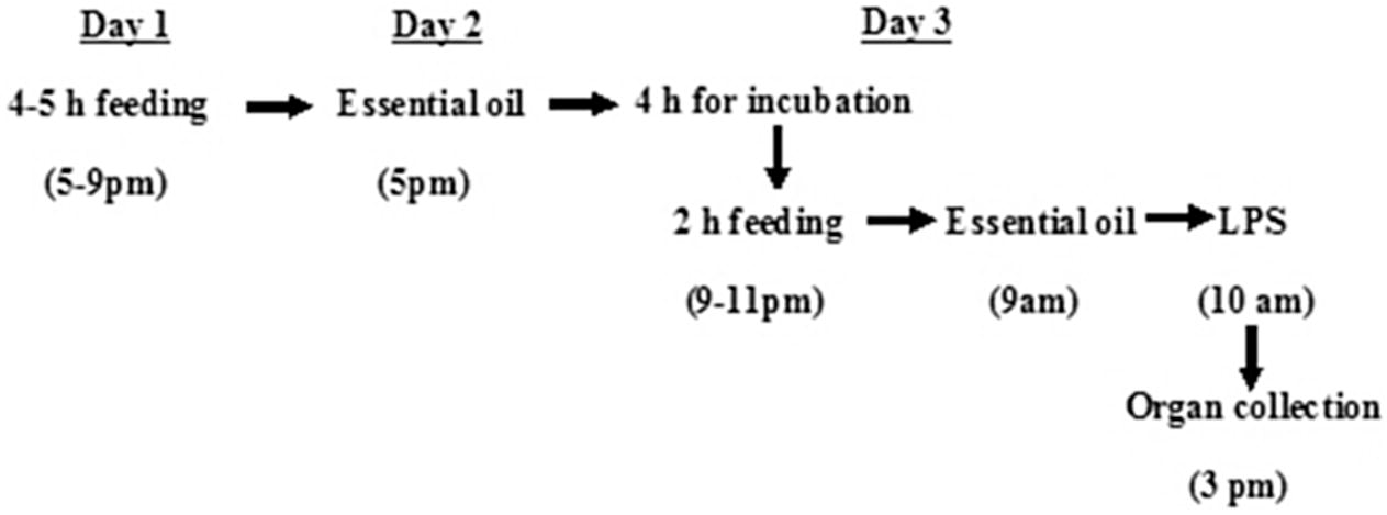
The animal care and handling were done according to the regulations of Council Directive CPCSEA no: 659/02/2 about Good Laboratory Practice on animal experimentation. Adult BALB/c mice weighing 30 g was used. Animal was purchased from KMCH Hospital, Coimbatore, Tamil Nadu, India and maintained in micro isolators with autoclaved bedding and cages, fed with autoclaved food pellets and deionized water. The animals were maintained under standard conditions of humidity, temperature (25 ± 2 °C) and light (12 h light/dark). Although the mice were not germfree, sterile procedures were used in handling these animals so as to prevent unintentional introduction of microbes that could activate iNOS production. Adult BALB/c mice were randomly assigned in to three groups, normal (n = 4), LPS treated (n = 8) and experimental (n = 8) (essential oil + LPS). Mice were placed on restricted, once a day diet and given different concentrations of essential oil (25, 50, 75 and 100 μg/30 g body weight) and LPS (1.5 μg/30 g body weight). After 5–6 h of LPS treatment, the animals were sacrificed and liver was collected. The following experimental design was as described by Chan et al. (1998) (Fig. 1).
Schedule for feeding and S. sclarea essential oil treatment.
2.3 Histopathology
The tissues were impregnated with histology grade paraffin wax (melting point 58–60 °C) at 60 °C for two changes of 1 h each. The wax impregnated tissues were embedded in paraffin blocks, mounted and cut with rotary microtome at 3 μM thickness. The sections were stained in Ehrlich’s hematoxylin (0.75%) for 8 min. Further they were counter stained in 1% aqueous eosin (1 g in 100 mL water) for 1 min and the excess stain was washed in tap water and allowed to dry. When the section was cooled, they were mounted in DPX (Distrene, Plasticiser and Xylene) mount having the optical index of glass (the sections were wetted in xylene and inverted on to the mount placed on cover slip). The architecture was observed at low power objective lens with 10× magnification. The liver cell injury and other aspects were observed under high power dry objectives.
2.4 RNA isolation and reverse transcriptase polymerase (RT-PCR) analysis
The liver tissue sample for RNA isolation was transferred to RNA later solution. The tissue sample was homogenized with 1 mL of trizol and transferred to fresh 1.5 mL centrifuge tubes and incubated at room temperature for 10 min at 4 °C to lyse the cells. About 200 μL of chloroform was added and mixed by pipetting for 30 s and centrifuged at 14,000 rpm for 15 min at 4 °C. The aqueous phase was carefully transferred to a fresh microfuge tube and 500 μL of isopropyl alcohol was added and incubated at room temperature for 10 min. The tubes were then centrifuged at 1400 rpm for 10 min at 4 °C and the supernatant was discarded. RNA pellet was washed with 75% ethanol by spinning at 1400 rpm for 5 min at 4 °C. The supernatant was discarded, the pellet was air dried and suspended in 9 μL of deionized autoclaved diethyl pyro carbonate (DEPC) treated water. Reverse transcription was carried out as follows. To the above sample, 2 μL of dNTP, 5 μL of cDNA synthesis buffer, 1 μL of oligo d (T), 1 μL of reverse transcriptase enzyme mix (Thermo Fischer Scientific, India) and 9 μL of nuclease free water were added and reverse transcription was carried out in a thermocycler (Eppendorf) to synthesize cDNA. The synthesized cDNA was further used for PCR. Primers and PCR conditions for murine iNOS and β-actin were in accordance with those of Yamasaki et al. (1998) and Kim et al. (2010), respectively. The PCR products were separated on 1.0% agarose gel and photographed using Biorad photo documentation system.
2.5 Cell culture
HeLa cell line was obtained from NCCS (National Center for Cell Science), Pune, India and routinely maintained in Dulbecco’s modified eagle medium (DMEM) supplemented with 10% fetal bovine serum, l-glutamine (1%), streptomycin and penicillin (1%) at 37 °C in a humidified incubator containing 5% CO2.
2.6 Cytotoxicity
The cytotoxic activity of S. sclarea essential oil on HeLa cell was determined by MTT assay (Yu et al., 2011). Cells were seeded at a density of 5 × 10³ cells/well in a 96 well plate. The cells were allowed to adhere for 24 h and then treated with essential oil at various concentrations (50–200 μg/mL) for 24 h. The culture medium was removed and 20 μL of MTT (5 mg/mL in DMEM) was added to each well, followed by incubation for 2 h. The formation of formazan crystals were visualized under a light microscope. The formazan crystals were dissolved by adding 100 μL isopropanol to each well. The absorbance was measured using a microplate reader at a wavelength of 570 nm. The effect of essential oil on HeLa cell proliferation was assessed as percentage cell viability over that of control, where vehicle treated control cells (0.1% DMSO) were taken as 100% viable.
2.7 PI staining
Apoptosis in HeLa cells was assessed using uptake of fluorescent dye PI (propidium iodide) as described by Brana et al. (2002). HeLa cells 1 × 10⁴ cells/well were seeded in a 24 well plate and grown until confluent. The cells were treated with essential oil at various concentrations (25–100 μg/mL) for 24 h. The cells were washed with ice cold PBS and fixed with 70% ethanol for 30 min. After fixation, the plates were rinsed again with ice cold PBS followed by staining with 200 μL of PI (500 μM) for 1 h, further the plates were washed twice with ice cold PBS and the nuclear staining of the apoptotic cells were observed under a fluorescence microscope.
2.8 Statistical analysis
The data obtained for cytotoxicity assay were processed using SPSS (16.00) for IC50 calculation.
3 Results
3.1 Extraction of essential oil
Hydrodistillation of 1 kg fresh leaves of S. sclarea yielded 500 μL of pale yellow colored oil with strong aromatic smell. The isolated essential oil was further used to evaluate in vivo and in vitro antiinflammatory and cytotoxic activities.
3.2 Histopathological analysis
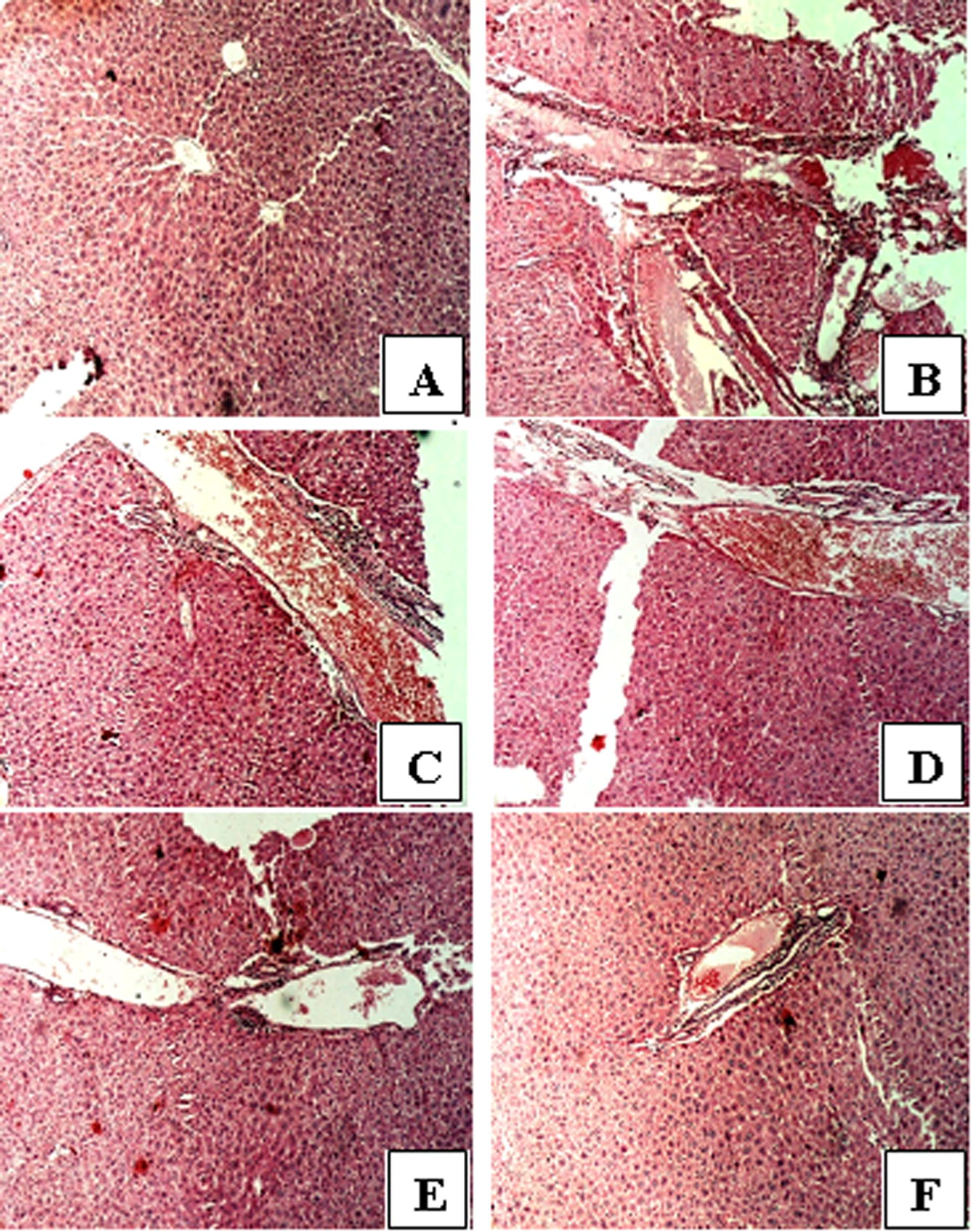
Histopathological study of the hepatocytes of BALB/c mice control group showed no obvious abnormality. The hepatocytes were seen intact with central vein presenting normal architecture (Fig. 2A). 1.5 μg LPS/30 g body weight induced hepatic tissue damage. Severe hemorrhage with cell necrosis was seen. The section revealed hepatic central vein distended and the capillary wall disturbed. Severe inflammation with lymphocytic cellular infiltration was also observed (Fig. 2B). 25 μg oil did not show any positive effect on hepato protection in LPS treated animals. Severe necrosis with inflammation, sinusoidal dilatation, congestion and hepatic lesions were observed (Fig. 2C). The histology of the hepatocytes was observed to be slightly altered with mild inflammation, cellular necrosis and sparse lymphocytic infiltration at 50 μg oil (Fig. 2D). At 75 μg of oil, the hepatocytes appeared with normal architecture and no visible inflammatory cell infiltration in tissue section (Fig. 2E). Essential oil at 100 μg/30 g body weight treated liver section showed normal architecture with the absence of inflammation or any other cell necrosis (Fig. 2F). Histopathology data thus showed that oil is a potent hepatoprotective agent.
Histopathological analysis of BALB/c mice liver treated with S. sclarea essential oil. (A) Normal control representing no obvious abnormality of inflammation. (B) Positive control (1.5 μg/mL LPS) representing abnormality of inflammation. (C) The tissue exhibited severe necrosis with inflammation at 25 μg/mL of oil. (D) The histology of the hepatocytes observed to be slightly altered with mild inflammation and cellular necrosis at 50 μg/mL of oil. (E) The hepatocytes appear normal with very mild necrosis observed at 75 μg/mL oil. (F) No obvious abnormality at 100 μg/mL of oil.
3.3 Down regulation of iNOS
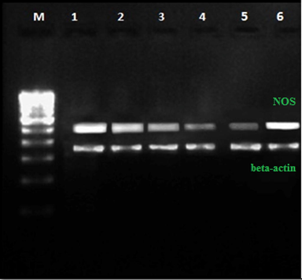
The total RNA of hepatocytes was isolated and RT-PCR was performed to test the down regulation activity of oil against iNOS at transcriptional level. The results of RT-PCR clearly indicated that LPS treated liver cells showed over expression of iNOS. However oil treated liver cells showed significant down regulation of iNOS. Down regulation activity of oil was time and concentration dependent. β-actin served as an internal marker (Fig. 3).
Efficacy of S. sclarea essential oil on down regulation of iNOS. (M) 100 bp DNA ladder, (1) positive control (LPS at 1.5 μg/mL), (2) (3) (4) and (5) iNOS down regulation activity of oil at 25, 50, 75 and 100 μg/mL respectively, (6) normal control (NOS gene).
3.4 Cytotoxicity and apoptosis induction
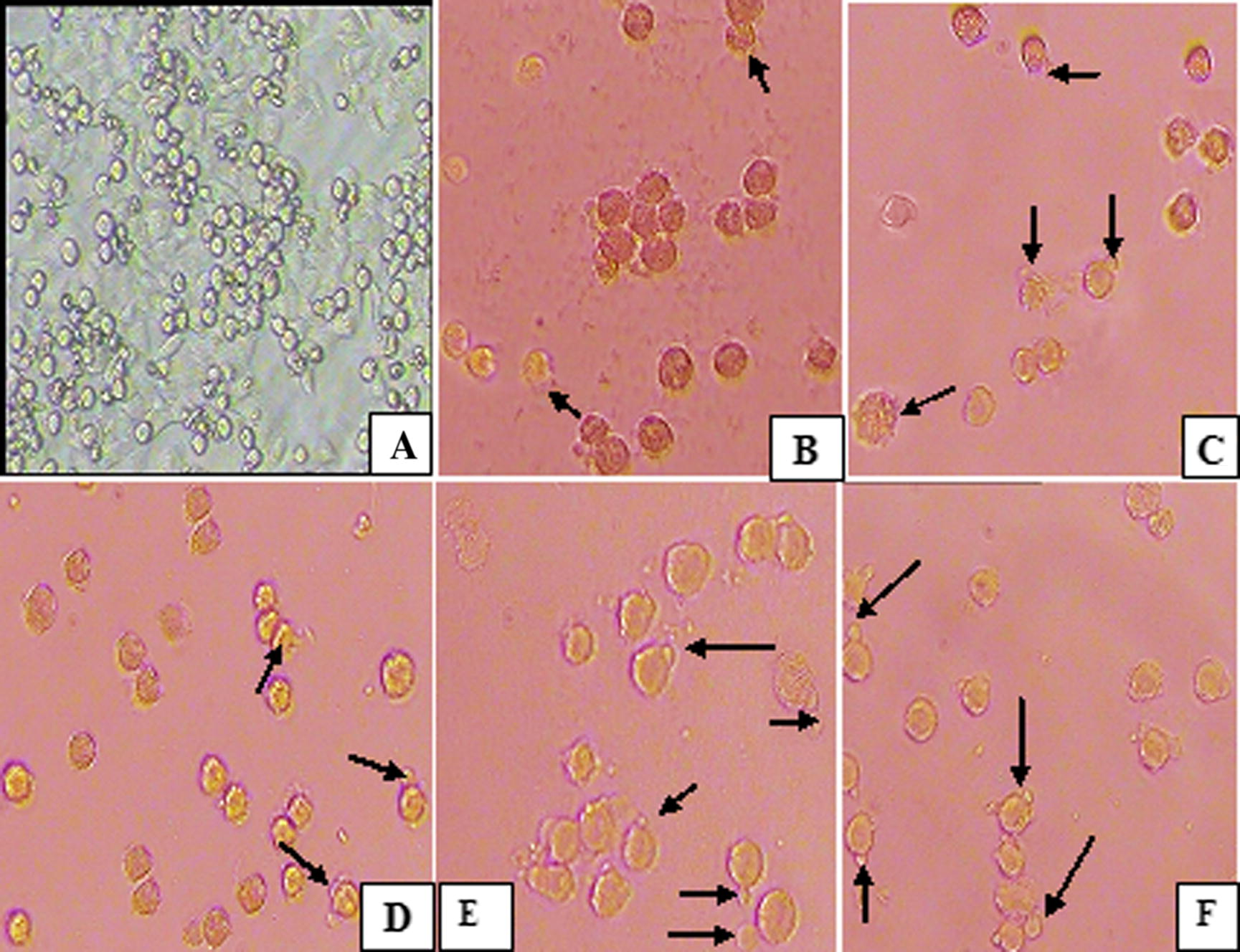
S. sclarea essential oil inhibited the growth of HeLa cells in a dose dependent manner after 24 h of treatment. Cytotoxic activity was also performed at 48 and 72 h and that no differences with result obtained at 24 h were observed. Out of five different concentrations (25, 50, 75, 100 and 125 μg/mL) tested, oil showed cytotoxicity with IC50 value of 80.69 ± 0.01 μg/mL. The PI staining of essential oil treated HeLa cells showed characteristics of apoptotic bodies including blebbing, cell breakage and chromatin condensation. The high level of apoptosis was observed at 100 μg/mL oil (Fig. 4).
Apoptotic activity of S. sclarea essential oil on HeLa cells. (A) Control, (B) (C) (D) (E) and (F) apoptotic induction of oil at a concentration of 25, 50, 75, 100 and 125 μg/mL, respectively.
4 Discussion
Herbs, spices and their essential oils have been used as pharmaceuticals in alternative medicine since long ago. Essential oil possess antioxidant, antibacterial, antifungal, cytotoxic, antiinflammatory and immune modulatory activities. The main objective of this study was to evaluate antiinflammatory and cytotoxic activities of S. sclarea essential oil. Essential oil of S. sclarea has been found to demonstrate antibacterial, antifungal, antioxidant, antiviral, antimalarial and anticholinesterase (Jirovetz et al., 2006; Kuzma et al., 2009; Ogutcu et al., 2008; Ozek et al., 2010 and Orhan et al., 2008) properties and has been used in perfume industries and liquor production. The major compounds present in the essential oil are linalyl acetate and linalool (Sharopov and Setzer, 2012).
LPS is the major constituent of outer cell wall of gram negative bacteria and widely used to examine the mechanism of inflammation that produce hepatic necrosis followed by fulminant hepatic failure (Vincent et al., 2002). In the present study 1.5 μg LPS induced severe hepatic cell damage and subsequent treatment of different concentrations of S. sclarea essential oil showed hepatoprotective activity. The optimum concentration required for hepatoprotection is around 75–100 μg oil. The observed activity of oil may be related to the suppression of LPS induced nuclear factor kappa B (NF-κB) mediated mitogen activated protein kinase (MAPKs) and subsequent regulation of cyclooxygenase (COX-2) and iNOS expression (Mestre et al., 2001). The result of this study suggests that hepatoprotection of oil might be associated with inhibition of iNOS and the activity may be due to the presence of active constituents like linalyl acetate, linalool etc. Similar result was reported by Peana et al. (1999) where plants producing considerable amount of these monoterpenes are reported as potential antiinflammatory agents.
Apoptosis induction is currently recognized as a key strategy to arrest proliferation of cancer cells. Several classes of anticancer agents have been developed but there is a problem in the use of these agents in cancer treatment because of undesirable side effects. Therefore plant based medicines have more attention toward cancer treatment (Chauthe et al., 2012). The present study demonstrated that S. sclarea essential oil induced cytotoxicity and apoptosis in HeLa cells. Mechanisms accounting for the reported cytotoxic effect may include increasing membrane fluidity, leakage of ions and cytoplasmic content and reduced ATP production, alteration of pH gradient and loss of mitochondrial potential (Azmi et al., 2006; Wei and Shibamoto, 2010 and Tuttolomondo et al., 2013). However the detailed mechanism of apoptosis has not been identified. The anticancer activity of each compound present in the oil was not evaluated so it is not possible to say which compound is responsible. So it is assumed that anticancer effect of oil may be related to synergistic effect of its constituents. Essential oil of many salvia species are reported to have cytotoxic and apoptotic activities against a number of human cancer cells (Kamatou et al., 2008; Cardile et al., 2009; Russo et al., 2013; Parsaee et al., 2013 and Nikolic et al., 2014).
5 Conclusion
In conclusion, we found that treatment with S. sclarea essential oil had a strong hepatoprotective activity and significantly inhibited the mRNA expression of iNOS which could help to develop essential oil based therapeutics for liver diseases. Cytotoxic and apoptotic activity of oil may create awareness among public to use plant based medicines for treatment of cervical cancer in developing countries.
Acknowledgement
We are grateful to our college management for financial support.
References
- Plant polyphenols mobilize endogenous copper in human peripheral lymphocytes leading to oxidative DNA breakage: a putative mechanism for anticancer properties. FEBS Lett.. 2006;580:533-538.
- [Google Scholar]
- A method for characterising cell death in vitro by combining propidium iodide staining with immune histochemistry. Brain Res. Protoc.. 2002;10:109114.
- [Google Scholar]
- Essential oils of Salvia bracteata and Salvia rubifolia from Lebanon. Chemical composition, antimicrobial activity and inhibitory effect on human melanoma cells. J. Ethnopharmacol.. 2009;126:265-272.
- [Google Scholar]
- In vivo inhibition of nitric oxide synthase gene expression by curcumin, a cancer preventive natural product with anti-inflammatory properties. Biochem. Pharmacol.. 1998;55:1955-1962.
- [Google Scholar]
- One pot synthesis and anticancer activity of dimeric phloroglucinols. Bioorg. Med. Chem. Lett.. 2012;22:2251-2256.
- [Google Scholar]
- Chemical composition, antimicrobial activities and odor descriptions of various Salvia sp. and Thuja sp. essential oils. Nutrition. 2006;90:152-159.
- [Google Scholar]
- Antimalarial and anticancer activities of selected South African Salvia species and isolated compounds from S. radula. S. Afr. J. Bot.. 2008;74:238-243.
- [Google Scholar]
- Hepatoprotective effect of pinoresinol on carbon tetrachloride induced hepatic damage in mice. J. Pharmacol. Sci.. 2010;112:105-112.
- [Google Scholar]
- Chemical composition and biological activities of essential oil from Salvia sclarea plants regenerated in vitro. Molecules. 2009;14:1438-1447.
- [Google Scholar]
- Cellular and molecular mechanisms of liver injury. Gastroenterology. 2008;134:1641-1654.
- [Google Scholar]
- Redundancy in the signalling pathways and promoter elements regulating cyclooxygenase-2 gene expression in endotoxin treated macrophage/monocytic cells. J. Biol. Chem.. 2001;276:3977-3982.
- [Google Scholar]
- Chemical composition, antimicrobial, and cytotoxic properties of five Lamiaceae essential oils. Ind. Crops Prod.. 2014;61:225-232.
- [Google Scholar]
- Bioactivities of the various extracts and essential oils of Salvia limbata C.A. Mey. and Salvia sclarea L. Turk. J. Biol.. 2008;32:181-192.
- [Google Scholar]
- Activity of essential oils and individual components against acetyl and butyrylcholinesterase. Z. Naturforsch.. 2008;63:547-553.
- [Google Scholar]
- Enantiomeric distribution of some linalool containing essential oils and their biological activities. Rec. Nat. Prod.. 2010;4:180-192.
- [Google Scholar]
- Apoptosis induction of Salvia chorassanica root extract on human cervical cancer cell line. Iran. J. Pharm. Res.. 2013;12:75-83.
- [Google Scholar]
- Chemical composition and antimicrobial action of the essential oils of Salvia desoleana and S. sclarea. Planta Med.. 1999;65:752-754.
- [Google Scholar]
- Chemical composition and anticancer activity of essential oils of mediterranean sage (Salvia officinalis L.) grown in different environmental conditions. Food Chem. Toxicol.. 2013;55:42-47.
- [Google Scholar]
- The essential oil of Salvia sclarea L. from Tajikistan. Rec. Nat. Prod.. 2012;6:75-79.
- [Google Scholar]
- Inflammatory pathways in liver homeostasis and liver injury. Clin. Rev. Allergy Immunol.. 2009;36:4-12.
- [Google Scholar]
- Antimicrobial and antioxidative activities of the essential oils and methanol extracts of Salvia cryptantha Benth. and Salvia multicaulis Vahl. Food Chem.. 2004;84:519-525.
- [Google Scholar]
- Biomolecular characterization of wild sicilian oregano: phytochemical screening of essential oils and extracts, and evaluation of their antioxidant activities. Chem. Biodivers.. 2013;10:411-433.
- [Google Scholar]
- Clinical trials of immunomodulatory therapies in severe sepsis and septic shock. Clin. Infect. Dis.. 2002;34:1084-1093.
- [Google Scholar]
- Antioxidant/lipoxygenase inhibitory activities and chemical compositions of selected essential oils. J. Agric. Food Chem.. 2010;58:7218-7225.
- [Google Scholar]
- Reversal of impaired wound repair in iNOS-deficient mice by topical adenoviral mediated iNOS gene transfer. J. Clin. Invest.. 1998;101:967-971.
- [Google Scholar]
- Anticancer, antioxidant and antimicrobial activities of the essential oil of Lycopus lucidus Turcz. var. hirtus Regel. Food Chem.. 2011;126:1593-1598.
- [Google Scholar]
- Danshen: an overview of its chemistry, pharmacology, pharmacokinetics and clinical use. J. Clin. Pharmacol.. 2005;45:1345-1359.
- [Google Scholar]


