

Translate this page into:
Influence of heat treated files on conserving the remaining dentin thickness during endodontic retreatment – An invitro CBCT study
⁎Corresponding author. sbhandi@roseman.edu (Shilpa Bhandi)
-
Received: ,
Accepted: ,
This article was originally published by Elsevier and was migrated to Scientific Scholar after the change of Publisher.
Peer review under responsibility of King Saud University.
Abstract
Objectives
Non-surgical endodontic retreatment is to be a conservative option over endodontic surgery in case of recurrent infections. Root canal retreatment procedures needs to be initiated faster and proficiently with proper root canal retreatment files. The main aim of this study is to evaluate the remaining root dentin thickness post Gutta Percha (GP) retrieval from the root canal after using two different rotary instruments.
Materials and methods
A total of sixty extracted single rooted teeth were chosen. Shaping and Cleaning was done Step Back (Telescopic) technique with apical preparation of size 40 k File. Irrigation was carried out using NaOCl (3 %) and Ethyelenediamine tetraacetic acid (EDTA) to clear the smear layer. The canals were dried using paper points and obturation was completed using lateral compaction technique with AH plus resin sealer. Upon completing the obturation, it was categorized into two groups: Group I- ProTaper retreatment files (PTR) and Group II- Solite RS3 (S-RS3) Retreatment files. Cone Beam Computed Tomography (CBCT) was used to assess the remaining dentin thickness post gutta-percha retrieval at 3,5 and 7 mm respectively. To test the significance between groups independent t test was used.
Results
It can be noted from the results that more amount of remaining dentin thickness was seen after using Solite RS3 files at 3 mm, 5 mm and 7 mm on the mesial side compared to ProTaper retreatment files (p < 0.05). There is no statistically significant difference seen between the files at three levels on the distal side (p > 0.05).
Conclusion
Within the limitations of the study, it can be concluded that Solite RS3 files have the potential to preserve the remaining dentin thickness. However, more studies including various other parameters should be performed to arrive at a definitive conclusion.
Keywords
Gutta percha
ProTaper Retreatment files
Remaining dentin thickness
Retreatment
Retrieval
Solite RS3

1 Introduction
The success of endodontic treatment relies on key factors such as proper instrumentation and complete disinfection of entire root canal system. Instrumentation directly influences the remaining dentin thickness which in turn impacts the fracture resistance of the root (Torabinejad et al., 2009). Previous studies have found out that a minimum of 0.3–0.5 mm of remaining dentin thickness is required to ensure the longevity of endodontically treated teeth (Subramanian et al., 2021). Preservation of remaining dentin thickness is pivotal for fracture resistance and strength of the teeth.
The concept of preserving remaining root dentin thickness becomes highly important in orthograde retreatment cases (Kumar et al., 2022).Moreover, it is important to know that, not only the thickness is important, but also how the presence of root microcracks due to the high processing torque of the instruments inside the canal can significantly influence the resistance of the tooth over time, and therefore from here the importance of careful instrumentation and low operating torque limits, as indicated (Gambarini et al.,2020).This is because of the procedural errors that occur during retreatment procedures such as perforation and excessive dentin removal (Yang et al., 2015). Orthograde retreatment procedure involves complete removal of obturating material, further cleaning and shaping and re obturating the canal (Karaoğlan et al., 2022). The success of orthograde root canal retreatment compared to other modes of treatment is 74 to 98 % (Kang et al., 2015). Although the success of retreatment is mostly favorable, the outcome is also influenced by the procedural pattern.
Majority of the dentin is removed during instrumentation. Previously, hand files were used to retrieve the obturating material (Bier et al., 2009). Initially, carbon files were used, the disadvantage of these files are prone for corrosion. Later, stainless-steel files were developed (Shahriari et al., 2009). However, the disadvantages of stainless-steel files are less flexible (Moore et al., 2009) (Pilo et al., 1998). In order to overcome the above limitations, rotary instruments made of Nickel titanium were introduced. Nevertheless, it was difficult to achieve a good canal taper and simultaneously preserve the remaining dentin (Zandbiglari et al., 2006). Preserving the remaining dentin becomes imperative especially in retreatment cases. Hence it is important to choose appropriate files especially during retreatment (Wong, 2004).
Rotary instruments such as the ProTaper retreatment system and R Endo systems are widely used in orthograde retreatment procedures. ProTaper retreatment (Dentsply Sirona,Tulsa, Swiss) system comprises three files D1, D2 and D3. They have progressive tapers and lengths (Giuliani et al., 2008). Solite RS3 retreatment file (Kedo Dental, Chennai, India) system is a novel file system that comprises three files RS1, RS2 and RS3 which come in three different tapers, lengths and cutting tips. RS2 and RS3 files are heat-treated files which make them more flexible. Conventional retreatment files tend to remove more dentin during retrieving of guttapercha (Kulkarni NR, Kamat SB, Hugar SI, Nanjannawar GS, 2019) and often leads to more iatrogenic errors. The advantage of heat-treated retreatment files is flexible, ability to retrieve the gutta percha by preserving the dentin during retreatment.(Krishnan et al., 2019) The present study aimed at comparing the remaining dentin thickness during retreatment at 3 mm,5mm and 7 mm using two different retreatment files namely ProTaper retreatment files and Solite RS3 heat treated retreatment files respectively.
2 Materials and methods
2.1 Specimen preparation
Sixty freshly extracted single-rooted mandibular premolar teeth which were extracted due to periodontal reasons with completely formed apices were selected for the study. All the teeth were stored in 5 % Glutaraldehyde solution - Korsolex Rapid (Raman & Well, Hamburg, Germany). Institutional Ethical clearance (SRB/SDC/ENDO-2080/20/092) was obtained from the Institutional Human Ethical Committee. The inclusion criteria as follows: presence of one canal with single root, no signs of crazes lines or cracks, without any kind of resorption and a curvature of canals of less than 15° were included for the study. To establish a standardized length of 18 mm diamond disk was used to decoronate under copious coolant.
Endodontic access cavity preparation was made and a glidepath was created using #10 k-file. Shaping and cleaning of the canals was done using Step back technique till 40 K file as the master apical file, with copious irrigation using NaOCl (3 %) and EDTA (17 %) to remove organic and inorganic tissues from the tooth. Paper points were used to dry the canals followed by lateral compaction technique with AH plus resin sealer for obturation. A CBCT was taken after completing the obturation in order to assess the remaining dentin thickness before and after gutta percha retrieval. Subsequently, a week later, the obturated teeth were divided randomly into two groups using block randomisation (https://www.randomization.org), for endodontic retreatment using either ProTaper retreatment (PTR) files (Dentsply Sirona,Tulsa, Swiss) or Solite RS3 (S-RS3) retreatment files (Kedo dental, Chennai, India), respectively, under the groups assigned consisting of 30 teeth per group, solvents were not used. The canals irrigated with NaOCl (3 %) and EDTA (17 %) during gutta-percha retrieval and saline was used as a final rinse to remove the debris and remanent. A second CBCT was taken post gutta-percha retrieval to assess for remaining dentin thickness. Three investigators were participated in this study for the sample preparation, assessment of remaining dentin thickness in CBCT and for doing the statistical analysis.
2.2 Evaluation of remaining dentin thickness
The remaining dentin thickness was assessed after obturation in axial sections at 3 mm, 5 mm and 7 mm of CBCT respectively for both the groups. After the retrieval of gutta percha using PTR files or S-RS3 files, same axial planes the remaining dentin thickness was measured in the second CBCT and was compared with the post obturation CBCT. (Fig. 1 and Fig. 2).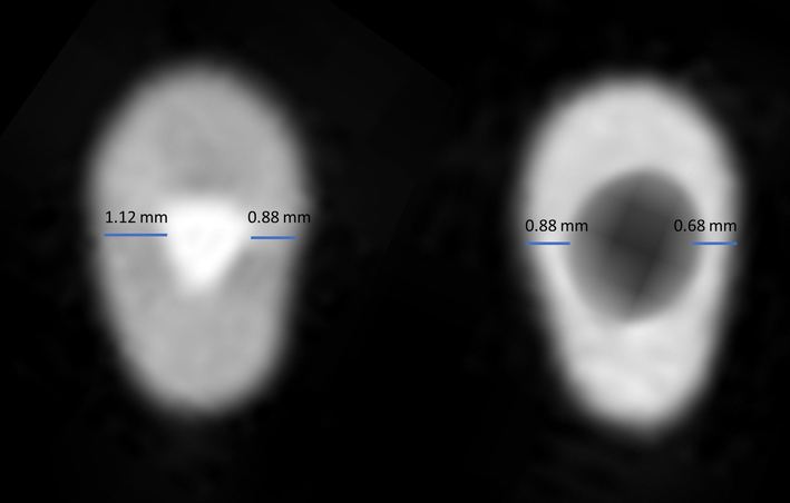
Obturation and post guttapercha retrieval CBCT images in the axial section under the ProTaper universal retreatment files group at 5 mm.
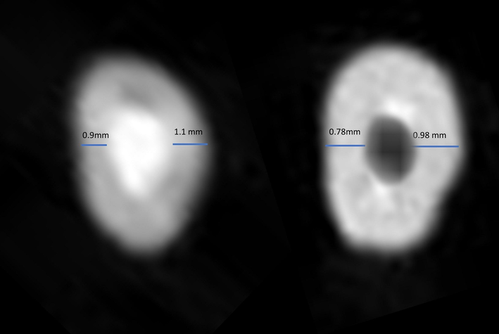
Obturation and post guttapercha retrieval CBCT images in the axial section under the Solite RS3 files group at 5 mm.
2.3 Statistical analysis
The obtained values were analysed statistically using SPSS version 23.0 software with Independent t test at 3 mm,5mm and 7 mm, both mesially and distally, keeping p value < 0.05 was statistically significant.
3 Results
The obtained result showed the mean and standard deviation for the remaining dentin thickness at the mesial side from CEJ 3 mm,5mm,7mm for PTR group (0.781 ± 0.270,0.611 ± 0.061, 0.446 ± 0.64), for S-RS3 (0.840 ± 0.180, 0.726 ± 0.031,0.484 ± 0.022) (Table 1). Independent t test resulted that there was a statistically significant difference between PTR and S-RS3 groups at all levels in mesial side (p < 0.05), (Table 2), (Fig. 3).
GROUPS
Mean
Std. Deviation
At 3 mm Mesial
PTR
0.781
0.270
S-RS3
0.840
0.180
At 3 mm Distal
PTR
0.750
0.071
S-RS3
0.886
0.119
At 5 mm Mesial
PTR
0.611
0.061
S-RS3
0.726
0.031
At 5 mm Distal
PTR
0.645
0.049
S-RS3
0.710
0.076
At 7 mm Mesial
PTR
0.446
0.064
S-RS3
0.484
0.022
At 7 mm Distal
PTR
0.452
0.067
S-RS3
0.474
0.033
GROUPS
p value
At 3 mm Mesial
PTR
0.023*
S-RS3
At 3 mm Distal
PTR
0.555
S-RS3
At 5 mm Mesial
PTR
0.047*
S-RS3
At 5 mm Distal
PTR
0.596
S-RS3
At 7 mm Mesial
PTR
0.040*
S-RS3
At 7 mm Distal
PTR
0.079
S-RS3
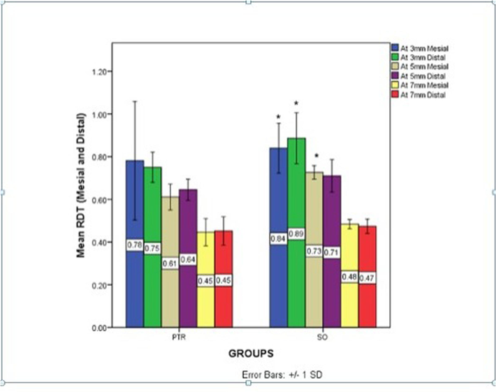
The bar graph depicts the mean and standard deviation remaining dentin thickness of ProTaper Retreatment files and Solite RS3 Retreatment files at 3 mm, 5 mm and 7 mm on mesial and distal sides. * Indicates there is significant difference at 3 mm, 5 mm and 7 mm on the mesial side (p < 0.05).
The obtained result showed the mean and standard deviation values for the remaining dentin thickness at the distal side from CEJ 3 mm,5mm,7mm for PTR group (0.750 ± 0.071, 0.645 ± 0.049, 0.452 ± 0.067), for S-RS3 (0.886 ± 0.119, 0.710 ± 0.076, 0.474 ± 0.033) (Table 1). Independent t test showed that there was no significant difference between PTR and S-RS3 groups at all levels in distal side (p > 0.05), (Table 2), (Fig. 3).
4 Discussion
Successful root canal therapy depends on the triad of thorough irrigation, disinfection and complete sealing of the entire root canal system (Gorni and Gagliani, 2004). Cleaning and shaping are considered to be the most effective step during root canal procedure that determines the remaining dentin thickness, duly noted that it should be prepared adequately to permit the flow of irrigants and also allow for good obturation (Celikten et al., 2015). During shaping and cleaning, the canals should not be over enlarged that comprises the remaining dentin thickness (Venino et al., 2017), the fracture resistance of endodontically treated teeth is determined by the remaining dentin thickness, and thus directly influences the longevity of the tooth (Celikten et al., 2015)(Venino et al., 2017). Previous research has found that it is during mechanical instrumentation that more dentin was removed especially in the mesial and distal direction during retreatment (Shemesh et al., 2011).
It is important to have an adequate canal taper for the irrigants to flow into the intricacies (Kulkarni NR, Kamat SB, Hugar SI, Nanjannawar GS, 2019). It is important to be cautious while instrumentation during retreatment procedure as it can directly influence the thickness of root dentin as already the dentinal thickness would have been compromised during root canal procedure (Xu et al., 2017) (Malagnino VA, Grande NM, Plotino G, 2008). Hence it is important to choose the appropriate retreatment files to serve the purpose of retrieving the obturating material as well as preserving the remaining dentin thickness (Gorni and Gagliani, 2004) (Celikten et al., 2015) (Venino et al., 2017).
In the current study, two retreatment files were used namely: PTR and S-RS3 Retreatment files. ProTaper Retreatment files are nickel titanium files come in three different lengths, diameters and unique taper in a sequential manner to retrieve gutta percha, these are not heat treated (Ganesh et al., 2014). Solite RS3 retreatment files come in three different tapers, lengths and color coded to identify easily. The three files are RS1, RS2 and RS3 in blue, red and yellow respectively. RS2 and RS3 files are heat-treated which is established to retrieve the gutta percha even in curved canals. The Solite RS1 has cutting tip with tip diameter 0.30 mm, 8 % taper, length of the file is 15 mm. It has a modified convex triangle cross section having three -point contact to disengage the gutta percha from the coronal third of the root canal. Solite RS2 has cutting tip with tip diameter 0.25 mm with approximately 7 % taper, length of the file is 18 mm. Solite RS3 has non-cutting tip with tip diameter 0.20 mm with 6 % taper, length of the file is 23 mm. Both RS2 and RS3 are heat-treated file facilitating the gutta percha removal from the curved root canals in middle and apical third respectively.
In a study conducted by Raiden et al, it was concluded that conventional radiographs do not reveal the exact thickness of dentin and might show greater thickness of dentin than the original thickness. Conventional radiographs, Cone Beam Computed Tomography (CBCT), Micro Computed Tomography and Scanning Electron Microscope are the recommended methods for evaluation of residual filling material and remaining dentin thickness. In the present study we have used CBCT to assess the remaining dentin thickness as it allows to evaluate the prepared canal space in all dimensions (Subramanian et al., 2021). Three sections of the root canal system were evaluated: 3 mm, 5 mm, and 7 mm which represent apical, middle, and coronal thirds of the root canal, wherein there is a high chance of operational errors.
In the current study, the Solite RS3 retreatment file system reported less dentin was removed compared to ProTaper retreatment files. The reason being is due to the heat treatment of the files making it flexible enough to retrieve the obturating material and avoiding the effect on root dentin. The alternate cutting edges of the file enables it to penetrate into the obturating material and the unique flexibility prevents the excessive cutting of dentin. The three-point contact of ProTaper retreatment files enable the retrieval of obturating material effectively but concomitantly affects the root dentin as the files having 9 %, 8 % and 7 % taper for D1, D2 and D3 respectively.
Most of the single rooted teeth used in the present study have an oval cross section which is wider mesiodistally and narrow buccolingually (Ali et al., 2019). The rotary files that are round in cross section tend to cut excessive dentin in mesial and distal direction (da Silva Limoeiro et al., 2016)(Bürklein et al., 2015). ProTaper retreatment files have a triangular cross section with two alternative cutting edges and zero radial lands which will eliminate threading into the canal wall and remove excessive dentin (da Silva Limoeiro et al., 2016) (Bürklein et al., 2015)(Rubio et al., 2017). It can also be inferred that more dentin removal at the coronal third compared to apical third was seen, while using ProTaper retreatment files. Study done by Kulkarni et al 2019 showed that D race file system preserved more dentin when compared to ProTaper retreatment files (Kulkarni NR, Kamat SB, Hugar SI, Nanjannawar GS, 2019).
5 Limitations
The limitation of the study was one type of obturation technique was carried out, single rooted teeth were used and two different types of retreatment files were evaluated.
6 Conclusion
The result of the present research showed that Solite RS3 files have the potential to preserve the remaining dentin thickness, which is a main concern when it comes to retreatment. However, more studies including various other parameters such as obturation techniques, usage of solvents, other methods of gutta percha retrieval and various retreatment files should be performed in-order to arrive at a definitive conclusion. Future scope is to translate invitro study evaluation to clinical trials.
Funding
There is no external source of funding.
Ethical approval
The study was approved by the Institutional Review Board (or Ethics Committee) of Saveetha Dental College & Hospitals, Saveetha Institute of Medical and Technical Sciences, Saveetha University, Chennai. (SRB/SDC/ENDO-2080/20/092).
CRediT authorship contribution statement
Mulumoodi Rama Sowmya: Conceptualization, Resources. Pradeep Solete: Conceptualization, Data curation, Visualization, Project administration. Ganesh Jeevanandan: Methodology, Investigation, Funding acquisition. S. Surendar: Methodology, Formal analysis, Visualization, Project administration. Shashit Shetty Bavabeedu: Software, Investigation, Supervision. Amal shaiban: Software, Resources, Funding acquisition. Zeeshan Heera Ahmad: Validation, Formal analysis, Supervision. Thodur Madapusi Balaji: Formal analysis, Resources. Shilpa Bhandi: Validation, Data curation.
Acknowledgment
None.
Declaration of competing interest
The authors declare that they have no known competing financial interests or personal relationships that could have appeared to influence the work reported in this paper.
References
- Comparative assessment of manual, rotary and reciprocating instruments for removal of root fillings, using stereomicroscope: An in vitro study. Bangladesh Med. Res. Counc. Bull.. 2019;45:54-61.
- [CrossRef] [Google Scholar]
- The ability of different nickel-titanium rotary instruments to induce dentinal damage during canal preparation. Journal of Endodontia. 2009;35:236-238.
- [CrossRef] [Google Scholar]
- Shaping ability of ProTaper NEXT and BT-RaCe nickel-titanium instruments in severely curved root canals. Int. Endod. J.. 2015;48:774-781.
- [CrossRef] [Google Scholar]
- Comparative evaluation of shaping ability of two nickel-titanium rotary systems using cone beam computed tomography. BMC Oral Health. 2015;15:32.
- [CrossRef] [Google Scholar]
- Micro-Computed Tomographic Evaluation of 2 Nickel-Titanium Instrument Systems in Shaping Root Canals. Journal of Endodontia. 2016;42:496-499.
- [CrossRef] [Google Scholar]
- The relevance of operative torque and torsional resistance of nickel-titanium rotary instruments: A preliminary clinical investigation. Saudi Endod J. 2020;10:260-264.
- [Google Scholar]
- A comparative assessment of fracture resistance of endodontically treated and re-treated teeth: An in vitro study. J. Conserv. Dent.. 2014;17:61.
- [CrossRef] [Google Scholar]
- Efficacy of ProTaper universal retreatment files in removing filling materials during root canal retreatment. Journal of Endodontia. 2008;34:1381-1384.
- [CrossRef] [Google Scholar]
- The outcome of endodontic retreatment: a 2-yr follow-up. Journal of Endodontia. 2004;30:1-4.
- [CrossRef] [Google Scholar]
- Outcome of nonsurgical retreatment and endodontic microsurgery: a meta-analysis. Clin. Oral Invest.. 2015;19:569-582.
- [CrossRef] [Google Scholar]
- Outcome of single- versus two-visit root canal retreatment in teeth with periapical lesions: A randomized clinical trial. Int. Endod. J.. 2022;55:833-843.
- [CrossRef] [Google Scholar]
- An Overview of Thermomechanically Heat-treated Nickel–Titanium Alloy Used in Endodontics. Conserv. Dent. Endod. J.. 2019;4:34-38.
- [CrossRef] [Google Scholar]
- Kulkarni, N.R., Kamat, S.B., Hugar, S.I., Nanjannawar, G.S., P.P., 2019. Evaluation of remaining dentin thickness following use of three different rotary nickel–titanium retreatment files: A cone-beam computed tomography study. J Conserv Dent 22, 588–592.
- A comparative evaluation of remaining dentin thickness following biomechanical preparation of teeth using different rotary file systems: An in vitro study. J. Conserv. Dent.. 2022;25:32-36.
- [CrossRef] [Google Scholar]
- Malagnino, V.A., Grande, N.M., Plotino, G., S.F., 2008. The simultaneous technique for root canal preparation with the Mtwo NiTi rotary system. Endod. Prac. 11, 13.
- A micro-computed tomographic evaluation of apical root canal preparation using three instrumentation techniques. Int. Endod. J.. 2009;42:1057-1064.
- [CrossRef] [Google Scholar]
- Residual dentin thickness in mandibular premolars prepared with hand and rotatory instruments. Journal of Endodontia. 1998;24:401-404.
- [CrossRef] [Google Scholar]
- Comparison of Shaping Ability of 10 Rotary and Reciprocating Systems: an In Vitro Study with AutoCad. Acta Stomatologica Croatica. 2017;51(3):207-216.
- [Google Scholar]
- Comparison of removed dentin thickness with hand and rotary instruments. IranEndod. J.. 2009;4:69-73.
- [Google Scholar]
- Damage to root dentin during retreatment procedures. Journal of Endodontia. 2011;37:63-66.
- [CrossRef] [Google Scholar]
- Comparative Evaluation of the Remaining Dentin Thickness Using Different Root Canal Retreatment Techniques: A Cone-Beam Computed Tomography Study. J. Int. Dent. Med. Res.. 2021;14:901-909.
- [Google Scholar]
- Outcomes of nonsurgical retreatment and endodontic surgery: a systematic review. Journal of Endodontia. 2009;35:930-937.
- [CrossRef] [Google Scholar]
- A Micro-computed Tomography Evaluation of the Shaping Ability of Two Nickel-titanium Instruments, HyFlex EDM and ProTaper Next. Journal of Endodontia. 2017;43:628-632.
- [CrossRef] [Google Scholar]
- Conventional endodontic failure and retreatment. Dent. Clin. N. Am.. 2004;48:265-289.
- [CrossRef] [Google Scholar]
- Accuracy of Cone-beam Computed Tomography in Measuring Dentin Thickness and Its Potential of Predicting the Remaining Dentin Thickness after Removing Fractured Instruments. Journal of Endodontia. 2017;43:1522-1527.
- [CrossRef] [Google Scholar]
- The remaining dentin thickness investigation of the attempt to remove broken instrument from mesiobuccal canals of maxillary first molars with virtual simulation technique. BMC Oral Health. 2015;15:87.
- [CrossRef] [Google Scholar]
- Influence of instrument taper on the resistance to fracture of endodontically treated roots. Oral Surg. Oral Med. Oral Pathol. Oral Radiol. Endod.. 2006;101:126-131.
- [CrossRef] [Google Scholar]
Appendix A
Supplementary data
Supplementary data to this article can be found online at https://doi.org/10.1016/j.jksus.2023.102969.
Appendix A
Supplementary data
The following are the Supplementary data to this article:


