

Translate this page into:
Fungal metabolite isolated from Mycosphaerella nawae AM20 and its protective role in cerebral ischemia
⁎Corresponding author at: Medical Laboratory Sciences, College of Applied Medical Sciences, Majmaah University, Majmaah 11952, Saudi Arabia. a.abdelhadi@mu.edu.sa (Ahmed Abdel-Hadi)
-
Received: ,
Accepted: ,
This article was originally published by Elsevier and was migrated to Scientific Scholar after the change of Publisher.
Peer review under responsibility of King Saud University.
Abstract
Usnic acid is one of the important metabolites showed cardiovascular protective properties. The main aim of the study was to analyze antioxidant and cerebral ischemia preventive role in experimental Albino rats. The amount of usnic acid in the Mycosphaerella nawae AM20 culture extract was determined by High Performance Liquid Chromatography. Antioxidant property was evaluated by various methods and usnic acid has potential Fe2+ chelating activity. The influence of usnic acid on special learning and memory was studied in vivo using Albino rats. Morris water maze (MWM) analysis revealed that the experimental ischemic rats treated with usnic acid reached the platform at minimum time than ischemic rats without usnic acid treatment. At 0.1 mL usnic acid concentration, hydroxy-3-methyl-glutaryl-CoA reductase (HMGR) inhibition activity was 18.4 ± 1.1% and it increased as 80.3 ± 2.4% at 0.5 mL concentration in the reaction mixture. Likewise, acetylcholinesterase (ACE) inhibitor activity was observed and was dose dependent. At 0.2 mL concentration, 21.8 ± 1.7% enzyme activities inhibited and it increased as 48.3 ± 1.8% at 0.5 mL sample concentration. Fibrinolytic analysis revealed 13 ± 0 mm zone of clearance at 0.5 mL sample concentration and the standard plasmin showed 17 ± 2 mm zone of clearance. Biochemical analysis revealed that the amount of malondialdehyde (MDA) was higher in the hippocampal region of ischemia group animal (59.4 ± 3.5 μM). Moreover, usnic acid treated ischemia group showed reduced MDA content (29.8 ± 3.5 μM). Superoxide dismutase (SOD) activity was reduced in ischemic albino rat and improved SOD activity was observed in the treated animal.
Keywords
Fungi
Metabolites
Usnic acid
Cerebral ischemia
Stroke prevention

1 Introduction
Stroke in both mature and immature brains is a highly complex phenomenon that includes sequences of many pathological functions such as oxidative damage, inflammation, apoptosis and cell death (Abdullahi et al., 2018). Two types of stroke have distinct pathophysiological, however share some of the common phenomenon such as, decreased antioxidant abilities and increased oxidative stress, excitotoxicity, apoptosis and inflammatory response (Zhang et al., 2015). Ischemic stroke results from interrupted blood flow to the brain tissues, which affect glucose and oxygen deliver to brain tissues (Zamani et al., 2013) and causing functional and metabolic damage (Mestriner et al., 2013). One of the important pathophysiological characters of ischemic stroke is the disruption of brain-blood barrier and this pathophysiological character is common among stroke patients. The increased intracranial pressure is the results of primary brain injury and the pathophysiological responses to hematoma leads to the development of secondary brain injury (Chen et al., 2015). Effective treatment for stroke is very limited. Recombinant tissue plasminogen activator (rtPA) has been widely used to treat stroke, however, it was effective only within 4.5 h of incident (Fisher and Saver, 2015). Neurosurgical has been recommended to treat blood in patients or treat brain aneurysm in subarachnoid hemorrhage (Steiner et al., 2013).
During the state of ischemia, many pathogenic functions could contribute to damage including, excitotoxity, increase of intracellular calcium levels and failure of glucose, inflammation and the edema, the dysfunction of the blood–brain barrier and generation of free radicals (Sosa et al., 2015). Intracerebral hemorrhage (ICH) leads to the rupture of brain blood vessels and leakage of blood biochemical components into the parenchyma tissue of the brain. The role of hippocampus is memory and learning, was reported previously. The hippocampus region is highly susceptible to various oxidative stress causes by many kinds of injury in the brain region (Mergenthaler et al., 2013). Usnic acid is produced by various fungi and lichen species. It is one of the secondary metabolites and has various bioactive potential. It has several applications in pharmaceutical, arboricultural and agricultural fields. Usnic acid has antimicrobial properties and was effective against mycobacteria and Gram-negative bacteria. In addition to antimicrobial properties, it was effective against various plant pathogens. Usnic acid also exhibit antiprotozoal, anti-inflammatory, antiproliferative, antiviral and analgesic activity (Ingolfsdottir, 2002). The anticancer potential of usnic acid has been reported previously (Araujo et al., 2015). This compound also has wound healing properties (Pagano et al., 2019). Usnic acid is one of the useful secondary metabolite and metabolite is an attractive subject of research (Galanty et al., 2017). Mycosphaerella is one of the important genera of ascomycetous and comprising about 10,000 taxa. The genus Mycosphaerella produced various metabolites, including, (+)-oxymycousnine, (+)-isomycousnine, (−)-mycousnine, asteromine and cercosporin (Arnone et al., 1995; Moreno et al., 2011). This paper describes the use of usnic acid and its protective role in cerebral ischemia in experimental animal.
2 Materials and methods
2.1 Materials
Potato dextraose agar (PDA) and antibiotics were purchased from Hi-media Laboratories, Mumbai, India. Usnic acid and superoxide dismutase kit were purchased from Sigma Aldrich, USA. All other chemicals were analytical grade or American Chemical Society (ACS) grade.
2.2 Isolation and characterization of endophytic fungi
Tomato leaves were used for the isolation of endophytic fungal strain, Mycosphaerella nawae. The leaves were washed with tap water, followed by double distilled water. The leaves were sterilized for 60 s with 70% ethanol and further sterilized with 0.5% (w/v) sodium hypochlorite solution. It was cut into small pieces (2 × 2 cm) and the pieces were treated with culture medium containing 100 μg/mL of kanamycin and 100 μg/mL ampicillin, respectively for three days. Then the developed hyphae was placed on potato dextrose agar (PDA) medium and incubated at 28 ± 2 °C for 8–10 days. The fungal strains were sub-cultured periodically until pure colonies were appeared on PDA medium (Baazeem et al., 2021a).
2.3 Characterization of fungal strains
The fungal strain AM20 was cultured in potato dextrose broth medium and genomic DNA was extracted from the organism as suggested previously (Zhang et al., 2010). The internal transcribed spacer regions (ITS1 and ITS2) of the rDNA was amplified using the primers (5′-TCCTCCGCTTATTGATATGC-3′ and 5′-TCCTCCGCTTATTGATATGC-3′). It was amplified and sequenced using an ABI sequencer (Luo et al., 2015; El-Sheikh et al., 2020).
2.4 Submerged fermentation and extraction
The strain AM20 was grown in 500 mL Erlenmeyer flask containing PDA broth medium. The Erlenmeyer flask was incubated for 12 days in static condition at ambient temperature (28 ± 2 °C). The fermented broth was filtered and the filtrate was extracted with ethyl acetate. It was further dried using a rotary evaporator. In addition, the mycelium was ground using a mortar and further extracted using 80% acetone. Then it was condensed under reduced pressure and further extracted using ethyl acetate (Noël et al., 2021).
2.5 Fractionation of bioactive compounds
The ethyl acetate fraction of the crude extract was aseptically mixed with about 20 g silica gel (100 mesh). It was dried at 40 °C and loaded on a silica gel column (300 mesh). The secondary metabolites were eluted using petroleum ether (50%) and ethyl acetate (90%) with a linear gradient. A total of 50 fractions were further collected. It was further loaded on a thin-layer chromatography and the unique metabolites were pooled and further fractionated using Sephadex LH-20 column chromatography using MeOH/CHCl3 as the mobile phase. A total of 10 fractions (5 mL) were collected and combined and used for analysis (Yamamoto et al., 1985).
2.6 HPLC determination of usnic acid
The ethyl acetate extract was re-extracted with acetone and dried at ambient temperature. The extract was maintained in dark and stored at 4 °C. The standard usnic acid was prepared in reagent grade acetone. The stock concentration was prepared at 1 mg/mL concentration. A calibration curve was prepared at various concentrations using linear regression analysis (2–20 mg/L). The HPLC system equipped with LC pump, PDA detector, connected with auto sampler and data processing system. The flow rate was 0.5 mL/min and stainless steel column was used for analysis. Methanol and phosphate buffer were used at 70:30 ratio and usnic acid detection was performed at 245 nm. About 10 µL sample was injected and analysis was performed in triplicates (Ribeiro-Costa et al., 2004).
2.7 Antioxidant properties of usnic acid
2.7.1 Fe3+-reducing activity
Fe3+-reducing activity of the sample was performed according to the method of with little modifications. Usnic acid (0.5 mL, 20–100 µg/mL) was prepared and mixed with buffer (0.5 mL) and K3Fe(CN)6 (1.0 mL, 1.5%). The mixture was further incubated at 37 °C for 30 min. To this FeCl3 solution (0.05%, 1 mL) was added and the absorbance was read at 700 nm against reagent blank. α-Tocopherol was used as the standard (20–100 µg/mL) (Atif et al., 2020).
2.7.2 Fe2+ chelating activity
Fe2+ chelating activity of the sample was evaluated as described earlier with little modifications. Usnic acid was prepared at various concentrations (20–100 µg/mL), and an aliquot of FeCl2 solution (0.5 mM) was mixed with usnic acid. Finally, Fe2+ binding ability was estimated using a UV–Visible spectrophotometer at 522 nm against reagent blank. α-Tocopherol was used as the standard (20–100 µg/mL) (Atif et al., 2020).
2.7.3 Free radicals scavenging activities
The DPPH radical scavenging activity was performed as suggested previously. Briefly, DPPH (0.1 mM, 0.5 mL) solution was mixed with crude sample (20–100 µg/mL). DPPH solution was prepared previously with ethanol and incubated in dark for 30 min. The absorbance of the sample was read at 517 nm against reagent blank using a UV–visible spectrophotometer. Likewise, ABTS scavenging activity of the sample was estimated as described earlier. ABTS solution (2 mM) was prepared in double distilled water and oxidizing agent (K2S2O8) (2.3 mM). About 0.5 mL sample (20–100 µg/mL) was incubated with 1.0 mL of ABTS solution and incubated for 30 min. α-Tocopherol was used as the standard (20–100 µg/mL). The optical density of the sample was read at 734 nm against reagent blank.
2.8 Hydroxy-3-methyl-glutaryl-CoA reductase inhibitor property
Hydroxy-3-methyl-glutaryl-CoA reductase (HMGR) inhibitor activity was evaluated. This inhibitor assay is based on spectrophotomerty analysis of the decrease in absorbance of sample at 340 nm, which shows the oxidation of NADPH by the catalytic subunit of HMGR and the presence of HMG Co-A. The enzyme assay mixture consists of assay buffer (0.75 mL), NADPH (0.15 mL), and HMG Co-A (0.15 mL). The final reaction volume was 1.5 mL. The enzyme reaction was initiated by the supplementation of 10 µL HMGR and the reaction mixture was incubated at ambient temperature (28 ± 1 °C). The optical density of the sample was measured after 5 min incubation and HMGR activity was measured. Pravastatin was used as the standard.
2.9 ACE inhibitor effect
The ACE inhibitor property of usnic acid was evaluated. Captopril was used as the standard ACE inhibitor and considered as a positive control.
2.10 Fibrinolytic enzyme activity of usnic acid
Fibrinolytic enzyme activity of usnic activity was evaluated as described previously by Astrup and Müllertz (1952) with minor modification. Plasmin was used as the standard. Fibrinogen (1.25%) was prepared in sodium phosphate buffer (pH 6.0, 0.1 M), and 0.9% agarose and thrombin (50 U/mL) was added and incubated for 1 h. Plasmin was prepared at 10 U/mL and the zone of clearance was measured (mm) around the sample/standard well (Al Farraj et al., 2020).
2.11 Experimental animal
Twenty five male Albino rats (150–200 g) were procured and maintained in an animal house with adequate environmental conditions (humidity: 50–55%, 26–28 °C, 12/12 light and day cycle). All experimental and control animals were allowed for free access to food and water. All experiments were performed in the morning and daily handling time was 10 min/day before to perform the experiments. Usnic acid was prepared in DMS appropriately. This work was approved by institutional ethical committee.
2.12 In vivo experimental trials
The albino rats were divided into three groups (n = 7) and maintained in a separate animal cage. These groups included, sham group, ischemia/reperfusion group, usnic acid treated ischemia/reperfusion group. Experimental animals in ischemia/reperfusion were treated with usnic acid at 50 mg/kg doses at the starting point of reperfusion. To generate cerebral ischemia/reperfusion, occlusion was made in the common carotid arteries of experimental animal. In the case of sham group of experimental rats, surgical procedures were followed without any obstruction of their common carotid arteries.
2.13 Transient global cerebral ischemia/reperfusion animal model
Transient global cerebral ischemia/reperfusion was performed. The experimental animals were anesthetized using xylazine (20 mg/kg) and ketamine (75 mg/kg) (IP injection). Then the common bilateral carotid arteries were gently exposed and Yashargil aneurism clips were used for occlusion in the artery within 15 min of exposure. After the completion of occlusion time, the clips were gently removed from the animal and reperfusion period (48 h) was established. Restoration of blood flow was monitored continuously by visual observation. After the completion of surgery, experimental animals were maintained in cages and allowed for free water and food for 10 days.
2.14 Morris water maze experiment
This experiment is widely used for the determination of special learning and memory of the experimental animal. The experimental animal was maintained in the illuminated room and external clues were kept in various area of the room and were maintained in the constant position throughout the experimental period.
2.15 Spatial training experiment
This experiment was carried out for three days after exertion of the cerebral ischemia animal model. Adequate training was provided to the experimental animal for searching the hidden submerged platform for four days via five experimental trials. The selected platform was maintained in the northwest quadrant followed by allowing experimental animals in the water facing the wall of the cage from one random quadrant. In all experimental trials, the experimental animal could climb the platform or could swim. The experimental animals were placed near the platform for 30 s to find the platform. Finally, the escape latency was registered and the average of escape latency was observed after three experimental trials.
2.16 Samples for biochemical analysis
Animals from control and experimental groups were anesthetized using xylazine (20 mg/kg) and ketamine (75 mg/kg). The skull was opened and brain was removed for biochemical analysis. It was removed and the hippocampal region was homogenized with phosphate buffer with protease inhibitor, phenylmethylsulfonyl fluoride. It was homogenized using a glass homogenizer and centrifuged for 4500 rpm for 10 min and the clear supernatant was stored at −20 °C. Then the amount of MDA, glutathione and SOD were determined.
2.17 Malondialdehyde assay
Malondialdehyde (MDA) is considering as one of the indicators for the determination of lipid peroxidation products. In this study, hippocampus sample was used for the determination of MDA as the marker using MDA determination kit. For the determination of MDA, sample was prepared in ice-cold potassium chloride within 2 min. Ice cold trichloroacetic acid and thiobarbituric acid were mixed with samples and heated for 1 h at 100 °C. It was cooled and centrifuged at 10 rpm for 10 min. The precipitate was removed and the supernatant was used for analysis. The absorbance of the sample was read at 535 nm against the reagent blank.
2.18 Glutathione analysis
Glutathione analysis of the hippocampus sample was determined using a commercial kit. The brain tissues were divided and homogenized using phosphate buffer saline. The supernatant was collected and stored. It was used for the determination glutathione (mM). The sample was reacted with GSH chromogenic reagent and the OD was read at 412 nm using a UV–Visible spectrophotometer.
2.19 Superoxide dismutase (SOD) determination
Superoxide dismutase (SOD) assay was performed using the clear supernatant obtained using superoxide dismutase kit. About 1.0 mL of the sample was mixed with 1.0 mL SOD reagent according to the manufactures instruction. It was incubated with colour reagent, and the amount of SOD was calculated.
2.20 Statistical analysis
Analysis of variance (ANOVA) was performed and the p-value <0.05 was considered as statistically significant.
3 Results
3.1 Characterization of endophytic fungus
The endophytic fungus was isolated from the tomato leaves and the genomic DNA was used for the amplification of ITS1 and ITS2 region. It was identified as Mycosphaerella nawae AM20. After 8–10 days incubation, the fungal colonies were very dark green in colour. After three weeks incubation, dark brown colour colonies were observed. The diameter of the fungal strain was 15–20 mm in diameter at 28 ± 2 °C.
3.2 Determination of usnic acid from the culture
The bioactive usnic acid was determined from the extract of the selected fungal strain AM20. The amount of usnic acid was determined by using HPLC analysis. The identification of usnic acid peak in the chromatograph of fungal extract was performed by comparison of retention time of the peak with that of usnic acid (standard). Usnic acid yield was 2.5% of the dry weight of the fungal strain.
3.3 Antioxidant properties
Ferric ions (Fe3+) interacted with usnic acid and formed Fe4[Fe(CN–)6]3 complex and this complex has maximum absorbance at 700 nm. Fe3+ reductive potentials of the sample were carried out. The fungal extract has potential Fe3+ reducing activity. In our study, significant DPPH scavenging activity was observed. When an antioxidant sample reacts with DPPH radicals, they can donate hydrogen ions and further reduced DPPH. The final colour change was monitored spectrophotometrically (Fig. 1A–D).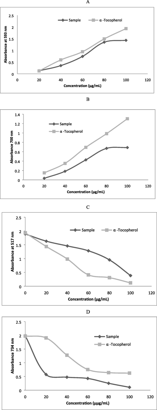
Antioxidant activity of usnic acid from the fungal strains. (A) Fe2+ reducing activity; (B) Fe2+ chelating activity; (C) DPPH scavenging activity; and (D) ABTS scavenging activity.
3.4 Cardioprotective properties of usnic acid
Cardiovascular-protective property of usnic acid was evaluated and the result was described in Table 1. Cardiovascular-protective activity was evaluated in terms of ACE inhibition and fibrinolytic enzyme activity of the sample. At 0.1 mL concentration, HMGR inhibition activity was 18.4 ± 1.1% and it increased as 80.3 ± 2.4% at 0.5 mL concentration in the reaction mixture. Likewise, ACE inhibitor activity was observed and was dose dependent. At 0.2 mL concentration, 21.8 ± 1.7% ACE inhibition was observed and it increased as 48.3 ± 1.8% at 0.5 mL sample concentration. The standard ACE inhibitor showed 45.1 ± 1.6% inhibition. The sample showed fibrinolytic property and was increased at higher concentrations. At 0.5 mL sample concentration, the zone of clearance was 13 ± 0 mm and the standard plasmin showed 17 ± 2 mm zone of clearance (Table 1). *Pravastatin (60 µg) was used as the standard for the determination of HMGR inhibitory effect. Captopril (20 µg) was used for the analysis of ACE inhibitor activity. Plasmin was prepared at 10 U/mL concentrations and the result was compared.
Sample (mL)
HMGR
inhibition (%)ACE
inhibition (%)Fibrinolytic
activity (mm)
0.1
18.4 ± 1.1
20.4 ± 1.9
6 ± 1
0.2
25.3 ± 0.9
21.8 ± 1.7
8 ± 1
0.3
76.4 ± 1.2
30.4 ± 2.2
10 ± 2
0.4
79.2 ± 0.8
35.3 ± 1.3
12 ± 0
0.5
80.3 ± 2.4
48.3 ± 1.8
13 ± 0
Standard
97.3 ± 2.0
45.1 ± 1.6
17 ± 2
3.5 Influence of usnic acid on special learning and memory in experimental animal
The influence of usnic acid on special learning and memory was studied in vivo using Albino rats. The experimental and control animals were subjected to find the platform in experiments. The sham group animals reached the platform earlier than ischemia group and the escape tendency was statistically significant (p < 0.05). Additionally, the experimental ischemic rats treated with usnic acid reached the platform at minimum time than ischemic rats without usnic acid. The escape tendency time was reduced in Day 4 than Day 1 and the result was described in Fig. 2.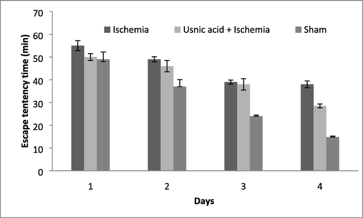
Escape latency of experimental animals during experimental period for four days.
3.6 Influence of usnic acid on MWM experiment
MWM analysis revealed that administered usnic acid effectively affect on the time spent in the target zone. MWM analysis revealed variations between experimental and treated Albino rats. Rats suffered with ischemia group spent little time (4 ± 1 s) than sham group (32 ± 2.2 s) and the difference was statistically significant (P < 0.05). Ischemic Albino rats administered with usnic acid showed an increase in the time spent during observation (27 ± 2.5 s) (Fig. 3).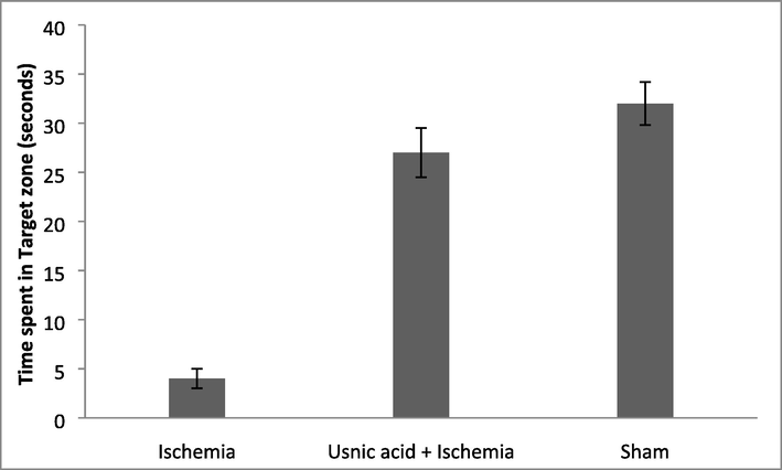
Albino rats time spent in target zone in the Morris water maze task analysis at various treatment times and different groups.
3.7 Usnic acid reduced MDA content in the experimental animal
Biochemical analysis revealed that the amount of MDA was found to be higher in the hippocampal region of ischemia group (59.4 ± 3.5 μM). Moreover, usnic acid treated ischemia group showed reduced MDA content and the sham group showed decreased MDA content (P < 0.05). In ischemia Albino rat treated with usnic acid showed 29.8 ± 3.5 μM MDA level and the result was described in Fig. 4. The analysis of brain tissue revealed effective inhibition of lipid peroxidation process.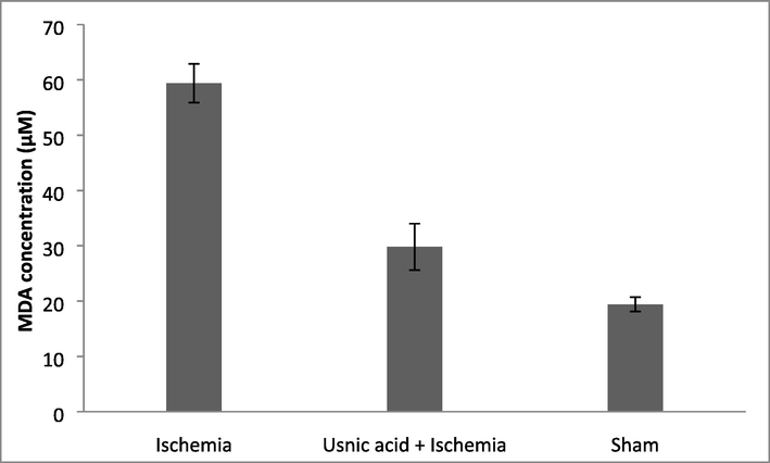
Influence of usnic acid on malondialdehyde concentration in the experimental albino rat after cerebral ischemia.
3.8 SOD and GSH activity of the Albino rats
SOD activity of brain hippocampus varied widely. In ischemic albino rat SOD activity reduced considerably. However, usnic acid treated ischemic albino rat showed improved SOD activity. The amount of SOD level was less in sham Albino rat and the result was described in Fig. 5. The amount of SOD activity increased significantly than control animals (P < 0.05). GSH level significantly decreased in the ischemia group Albino rat than control sham group. GSH content in the brain increased in the Sham and experimental animal treated with usnic acid.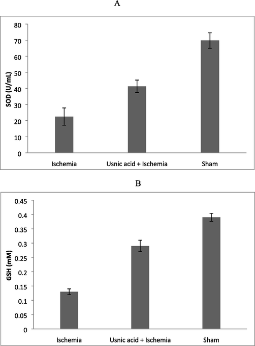
Influence of usnic acid on superoxide dismutase (SOD) (A) and glutathione (B) levels in experimental animals.
4 Discussion
Bioactive secondary metabolite producing endophytic fungi has been isolated and screened previously (Kaul et al., 2012; Lai et al., 2016). The endophytic fungi have been isolated previously from the medicinal plants (Baazeem et al., 2021a). Fungi isolated from the medicinal plants have potential biological properties; contribute to the pharmaceutical and therapeutic formulations. The endophyte, Taxus brevifolia has been produced an antitumour agent, taxol (Strobel et al., 2004). In recent years, immunosuppressive molecules have been identified from various endophytic fungi (Kumar et al., 2005; Song et al., 2013). Usnic acid has widely used in the preparation of deodorants, toothpaste, creams, sunscreen products, and mouthwash. In addition, its biological and ecological effects, anti-insect, anti-growth, and anti-herbivore properties had been previously described (Ingolfsdottir, 2002). The reducing property of the sample was analyzed using Fe3+ reducing system. The fungal extract acted as the reducing agent and it inactivated oxidant and reactive oxygen species. The steep increase in absorbance of the sample indicated increased reducing potential and increased of complex formation. The present finding revealed that usnic acid from the sample could donate electron and neutralize free radicals and generation of reactive oxygen species. The FRAP method of antioxidant assay has been performed in acidic medium to keep the iron more stable (Öztaskın et al., 2017). DPPH method has been widely used for the determination of radical scavenging property and antioxidant properties of the sample. ABTS scavenging activity has been widely used to test the antioxidant properties of plant samples and biological materials. This method facilitated to analyze the impact of pH on antioxidant properties for the biological samples (Oktay et al., 2003). The standard α-Tocopherol showed less scavenging activity in this antioxidant assay because of hydrophobic properties. Moreover, the used sample showed significant scavenging activity at the selected concentrations. The present finding revealed that usnic acid can effectively transfer hydrogen atom to DMPD.
Fungi produce various bioactive secondary metabolites (Baazeem et al., 2021b). With this available information, the present investigation was performed to study; cardiovascular-protective property of the fungi extract. Cardiovascular-protective property of usnic acid was observed in this study. Cardiovascular-protective activity was evaluated by determining ACE inhibition and HMGR inhibition, and fibrinolytic enzyme activity of the sample. At 0.1 mL concentration, HMGR inhibition activity was 18.4 ± 1.1% and it increased as 80.3 ± 2.4% at 0.5 mL concentration. ACE inhibitor activity was observed and was dose dependent. Usnic acid showed fibrinolytic property and was increased at higher concentrations. The bioactive potential of usnic and other related classes of compounds has been described previously. Fungi and lichens have usnic acid and depsides, and depsidone classes of chemical compounds have various physiological functions (Shukla et al., 2014). It has been previously reported that depsidones are considered as one of the important antioxidant molecules and are related to the incorporation into lipidic microdomains.
Cerebral ischemia causes highly complex pathological mechanisms that can lead to brain cell damage (Surapaneni et al., 2017). Inflammation, oxidative stress, and apoptosis are the important pathophysiological functions that are linked with cerebral ischemia (Khoshnam et al., 2017). The antioxidant properties of usnic acid have been reported previously. Usnic acid isolated from lichen species has been showed antidiabetic, anticholinergic and antioxidant activities. Antioxidant potential has been determined using various methods including, DPPH, ABTS, superoxide anion radical, scavenging activities using ferric ion, and cupric irons (Cakmak and Gülçin, 2019). Usnic acid has been showed antioxidant activities and showed cytotoxic potential (Araújo et al., 2015). In the present investigation, cardiovascular-protective properties of usnic acid were assayed in terms of ACE inhibition and fibrinolytic enzyme properties. Usnic acid showed ACE inhibition and fibrinolytic activity and was dose dependent. Behera et al. (2012) has been reported cardio-protective properties of usnic acid from the lichen, Usnea complanata. Usnic acid has been showed prooxidant and antioxidant properties (Kohlhardt-Floehr et al., 2010). Endoplasmic reticulum stress has been considered as one of the important cause for the pathogenesis of heart failure and ischemic heart diseases (Cornejo and Hetz, 2013). The cardio-protective properties of usnic acid have been reported previously. Usnic acid treated experimental animal has been showed reduced endoplasmic stress activation and significantly reduced the expression of pro-inflammatory cytokines through AMPK signalling pathway and this effect has been decreased in dose dependent manner (Li et al., 2014). Usnic acid has been showed ACE inhibition and HMGR inhibition activity (Behera et al., 2012).
5 Conclusions
The present findings showed improved antioxidant properties of usnic acid from the fungal strain. Antioxidant property of usnic was evaluated by various methods and usnic acid has potential Fe2+ chelating activity. The fungal extract showed HMGR inhibition and ACE inhibitor and fibrinolytic activity. Biochemical analysis revealed that the amount of MDA was found to be higher in the hippocampal region of ischemia group (59.4 ± 3.5 μM). Moreover, usnic acid treated ischemia group showed reduced MDA content (29.8 ± 3.5 μM). SOD activity was reduced in ischemic albino rat and improved SOD activity was observed in the treated animals. GSH content in the brain increased in the experimental animal treated with usnic acid. The experimental animal treated with usnic acid improved spatial memory and neural ischemia. Usnic acid is considered as a natural choice for the treatment of neurodegenerative disorders such as, cerebral ischemia and stroke.
Acknowledgement
The authors extend their appreciation to the deputyship for Research & Innovation, Ministry of education in Saudi Arabia for funding this research work through the project number (IFP-2020-29).
Declaration of Competing Interest
The authors declare that they have no known competing financial interests or personal relationships that could have appeared to influence the work reported in this paper.
References
- Blood-brain barrier dysfunction in ischemic stroke: targeting tight junctions and transporters for vascular protection. Am. J. Physiol. Cell Physiol.. 2018;315(3):C343-C356.
- [Google Scholar]
- Enhanced production, purification and biochemical characterization of therapeutic potential fibrinolytic enzyme from a new Bacillus flexus from marine environment. J. King Saud Univ. Sci.. 2020;32(7):3174-3180.
- [Google Scholar]
- Review of the biological properties and toxicity of usnic acid. Nat. Prod. Res.. 2015;29(23):2167-2180.
- [Google Scholar]
- Asteromine, a bioactive secondary metabolite from a strain of Mycosphaerella asteroma. Phytochemistry. 1995;38(3):595-597.
- [Google Scholar]
- The fibrin plate method for estimating fibrinolytic activity. Arch. Biochem. Biophys.. 1952;40(2):346-351.
- [Google Scholar]
- Essential oils of two medicinal plants and protective properties of jack fruits against the spoilage bacteria and fungi. Ind. Crop. Prod.. 2020;147
- [Google Scholar]
- In vitro antibacterial, antifungal, nematocidal and growth promoting activities of Trichoderma hamatum FB10 and its secondary metabolites. J. Fungi. 2021;7(5):331.
- [Google Scholar]
- Paecilomyces formosus MD12, a Biocontrol Agent to Treat Meloidogyne incognita on Brinjal in Green House. J. Fungi. 2021;7(8):632.
- [Google Scholar]
- Antioxidative and cardiovascular-protective activities of metabolite usnic acid and psoromic acid produced by lichen species Usnea complanata under submerged fermentation. Pharmaceut. Biol.. 2012;50(8):968-979.
- [Google Scholar]
- Anticholinergic and antioxidant activities of usnic acid-An activity-structure insight. Toxicol. Rep.. 2019;6:1273-1280.
- [Google Scholar]
- An update on inflammation in the acute phase of intracerebral hemorrhage. Transl. Stroke Res.. 2015;6(1):4-8.
- [Google Scholar]
- The unfolded protein response in Alzheimer’s disease. Semin. Immunopathol.. 2013;35(3):277-292.
- [Google Scholar]
- Paecilomyces sp. ZB is a cell factory for the production of gibberellic acid using a cheap substrate in solid state fermentation. Saudi J. Biol. Sci.. 2020;27(9):2431-2438.
- [Google Scholar]
- Future directions of acute ischaemic stroke therapy. Lancet Neurol.. 2015;14(7):758-767.
- [Google Scholar]
- Usnic acid and atranorin exert selective cytostatic and anti-invasive effects on human prostate and melanoma cancer cells. Toxicol. In Vitro. 2017;40:161-169.
- [Google Scholar]
- Endophytic fungi from medicinal plants: a treasure hunt for bioactive metabolites. Phytochem. Rev.. 2012;11:487-505.
- [Google Scholar]
- Emerging roles of microRNAs in ischemic stroke: as possible therapeutic agents. J. Stroke. 2017;19:166-187.
- [Google Scholar]
- Prooxidant and antioxidant behaviour of usnic acid from lichens under UVB-light irradiation–Studies on human cells. J. Photochem. Photobiol. B: Biol.. 2010;101:97-102.
- [Google Scholar]
- Immunomodulatory compounds from Pestalotiopsis leucothes, an endophytic fungus from Tripterygium wilfordii. Life Sci.. 2005;78:147-156.
- [Google Scholar]
- Bioactive dibenzo-α-pyrone derivatives from the endophytic fungus Rhizopycnis vagum Nitaf22. J. Nat. Prod.. 2016;79:2022-2031.
- [Google Scholar]
- Usnic acid inhibits ER stress activation through AMPK signaling pathway in rat cardiomyocytes. Eur. Rev. Med. Pharmacol. Sci. 2014;18:2538-2543.
- [Google Scholar]
- Five new Pseudophialophora species from grass roots in the oligotrophic pine barrens ecosystem. Fungal Biol.. 2015;119:1205-1215.
- [Google Scholar]
- Sugar for the brain: the role of glucose in physiological and pathological brain function. Trends Neurosci.. 2013;36:587-597.
- [Google Scholar]
- Behavior outcome after ischemic and hemorrhagic stroke, with similar brain damage, in rats. Behav. Brain Res.. 2013;244:82-89.
- [Google Scholar]
- Chemical constituents of the new endophytic fungus Mycosphaerella sp. nov. and their anti-parasitic activity. Nat. Prod. Commun.. 2011;6:835-840.
- [Google Scholar]
- Lichen-associated bacteria transform antibacterial usnic acid to products of lower antibiotic activity. Phytochemistry. 2021;181
- [Google Scholar]
- Determination of in vitro antioxidant activity of fennel (Foeniculum vulgare) seed extracts. LWT-Food Sci. Technol.. 2003;36(2):263-271.
- [Google Scholar]
- Novel antioxidant bromophenols with acetylcholinesterase, butyrylcholinesterase and carbonic anhydrase inhibitory actions. Bioorg. Chem.. 2017;74:104-114.
- [Google Scholar]
- Bioadhesive polymeric films based on usnic acid for burn wound treatment: Antibacterial and cytotoxicity studies. Colloids Surf.. 2019;B178:488-499.
- [Google Scholar]
- In vitro and in vivo properties of usnic acid encapsulated into PLGA-microspheres. J. Microencapsul.. 2004;21(4):371-384.
- [Google Scholar]
- Endophytic microbes: a novel source for biologically/pharmacologically active secondary metabolites. Asian J. Pharmacol. Toxicol.. 2014;2(3):1-6.
- [Google Scholar]
- Xanthone derivatives from Aspergillus sydowii, an endophytic fungus from the liverwort Scapania ciliata S. Lac and their immunosuppressive activities. Phytochem. Lett.. 2013;6(3):318-321.
- [Google Scholar]
- Physical exercise prevents motor disorders and striatal oxidative imbalance after cerebral ischemia-reperfusion. Braz. J. Med. Biol. Res.. 2015;48:798-804.
- [Google Scholar]
- European Stroke Organization guidelines for the management of intracranial aneurysms and subarachnoid haemorrhage. Cerebrovasc. Dis.. 2013;35(2):93-112.
- [Google Scholar]
- Natural products from endophytic microorganisms. J. Nat. Prod.. 2004;67(2):257-268.
- [Google Scholar]
- Neuroprotective effect of Clerodendron glandulosum against acute transient ischemia reperfusion cerebral injury in rats. J. Neurol. Neurorehabil. Res.. 2017;2:14-20.
- [Google Scholar]
- Tissue cultures of Usnea rubescens and Ramalina yasudae and production of usnic acid in their cultures. Agric. Biol. Chem.. 1985;49(11):3347-3348.
- [Google Scholar]
- Combination therapy with A1 receptor agonist and vitamin C improved working memory in a mouse model of global ischemia-reperfusion. Basic Clin. Neurosci.. 2013;4:111-116.
- [Google Scholar]
- Apigenin protects blood–brain barrier and ameliorates early brain injury by inhibiting TLR4-mediated inflammatory pathway in subarachnoid hemorrhage rats. Int. Immunopharmacol.. 2015;28(1):79-87.
- [Google Scholar]
- A simple method of genomic DNA extraction suitable for analysis of bulk fungal strains. Lett. Appl. Microbiol.. 2010;51:114-118.
- [Google Scholar]


