

Translate this page into:
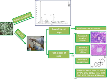
Consumption of sage (Salvia officinalis) promotes ovarian function by stimulating estradiol hormone release and controlling folliculogenesis, steroidogenesis, and autophagy
⁎Corresponding author at: Department of Zoology, College of Science, King Saud University, P.O. Box 2455, Riyadh 11451, Saudi Arabia. hharrath@ksu.edu.sa (Abdel Halim Harrath)
-
Received: ,
Accepted: ,
This article was originally published by Elsevier and was migrated to Scientific Scholar after the change of Publisher.
Peer review under responsibility of King Saud University.
Abstract
Abstract
Objectives
Salvia officinalis (sage) is widely sold in Saudi Arabia to make a popular herbal tea that women with reproductive disorders can drink daily. Despite the wide use of sage in traditional medicine, to our knowledge, little attention has been given to its effect on female reproduction. Our purpose was to understand the effects of sage extract on ovarian function to potentially shed light on its use in treating female infertility.
Methods
Forty female Wistar virgin female rats were randomly assigned to four groups, with 10 animals in each group. The control group received distilled water, and the three treatment groups received different concentrations of sage extract: 3.5, 15 or 60 mg/kg during 14 days. Ovarian tissues were cut into 5-μm sections and stained with hematoxylin and eosin. Plasma levels of reproductive steroids were measured using ELISA, and gene expression was examined via RT-PCR.
Results
Our results showed that low doses of sage (3.5 mg/kg and 15 mg/kg) have no effects on serum concentration levels of steroid hormone, the number of growing follicles, and on the transcripts of the steroidogenesis and folliculogenesis genes. In contrast, a high dose of the extract (60 mg/kg) significantly increased estradiol concentration levels. As a result, the number of growing follicles increased, while the number of abnormal follicles significantly decreased. Interestingly, sage extract also affected mRNA levels of genes involved in folliculogenesis and steroidogenesis processes. Two autophagy related genes, LC3 and ATG12, were not affected, but all of the investigated genes, INSL3, CYP17a, LHR, CCND2, IGF1, ESR1, ESR2, ACTB, GDF, and ATG5, significantly increased with the highest dose of sage.
Conclusion
As a remedy, sage extract is best used at a high dose when treating female infertility disorders, as sage extract may promote ovarian function by stimulating folliculogenesis and steroidogenesis.
Keywords
Ovary
Folliculogenesis
Hormone
Fertility
Ethnopharmacology

1 Introduction
Infertility is a major health and social problem that affects many people around the world (Lee et al., 2020b). There are differing opinions regarding the definition of infertility, but the World Health Organization (WHO) defines infertility as the inability to become or remain pregnant after one year of regular and unprotected sexual intercourse (WHO, 2017). Infertility affects approximately 48 million women globally and is caused by many factors. Twenty percent of infertility cases do not have an explanation. However, there are many known causes, including ovulation disorders, abnormal uterine function, obstruction of the fallopian tubes (Mascarenhas et al., 2012; Lee et al., 2020a), nutrition (Harrath et al., 2017), other diseases, and environmental effects (Lee et al., 2020b; Pivonello et al., 2020; Sirotkin et al., 2020). In Saudi Arabia and African cultures, infertility has a strong social meaning since marriage is considered to be successful only if a couple bears children. Thus, in most cases, women resort to complementary treatment with traditional therapies.
Salvia officinalis L. (Lamiaceae) has been used since ancient Egyptian civilization for increasing fertility in women, remedying sexual debility and treating menopausal and menstrual problems (Dweck, 2000). S. officinalis has been described in many ethnopharmacological reports (Martinez-Frances et al., 2017) and has been recommended for many gynecological diseases (Li et al., 2013). Therefore, this herb, known as “Maramia” in Saudi Arabia, is sold widely throughout the country and consumed as a tea known as Maramia tea. It is considered the “queen of herbs” since it is widely used in both medicinal and culinary preparations (Khan and Abourashed, 2010). The plant is well studied, and information on its biological activities has been widely reported (Martins et al., 2015; Ghorbani and Esmaeilizadeh, 2017; Ferreira Mendes et al., 2020). It has many antioxidant, anti-inflammatory, and antimicrobial activities against bacterial species, including Staphylococcus aureus and Candida albicans (Alizadeh, 2015). Sage essential oil has been traditionally used to treat many diseases in the nervous, circulatory, and respiratory systems (Radulescu et al., 2004; Loizzo et al., 2007), including bronchitis, persistent coughing, asthma, and angina (Khan et al., 2011; Walch et al., 2011).
Despite the widespread use of Salvia officinalis L. (Lamiaceae) in traditional medicine, to our knowledge, very little is known about the effect of this plant on female reproduction. Therefore, we studied the use of this plant as a traditional therapy for treating female reproductive disorders in Saudi Arabia and other countries to elucidate the effects of sage extracts on female ovarian functions. Specifically, we studied folliculogenesis and steroidogenesis as well as the potency of the plant extract in promoting female fertility. The findings of this study will be of significant interest as they will improve awareness regarding the effect of this plant on female fertility.
2 Materials and methods
2.1 Plant material
S. officinalis leaves were purchased in November 2018 from traditional medicine shops in Arar city, Saudi Arabia, and were then identified as being authentic by botanist colleagues at the Department of Faculty of Science in Arar, Saudi Arabia, under the voucher specimen code SO450420 and processed at the herbarium of the same department. At the laboratory, the leaves were weighed and then ground up.
2.2 Extraction
The S. officinalis leaves were ground, and the resulting powder (500 g) was successively extracted with hexane (to eliminate fixed oil) and then with dichloromethane (CH2Cl2) using a Soxhlet apparatus. The extracts were filtered with a Whatman filter. Then, the resulting solutions were evaporated using a rotary evaporator, which led to 12.36 g of n-hexane extract and 8.45 g of dichloromethane extract.
2.3 CH2Cl2 extract composition analysis
Gas chromatography (GC) and GC coupled with mass spectrometry (GC–MS/MS) (Thermo Trace GC Ultra/TSQ Quantum GC) were used to carry out CH2Cl2 analysis. The GC system was equipped with a split/splitless injector (220 °C), HP-5MS capillary column (TR5-MS, 60 m., 0.25 mm. id, and 0.25-µm film thickness), and flame-ionization detector (FID; 280 °C) and was coupled with an MS/MS detector, operating in the electron impact (EI) mode at 70 eV. We programmed the oven temperature to gradually increase from 50 °C to 280 °C in 10 °C/min increments and finally remain at 280 °C for 5 min (carrier gas = helium at 1.0 mL/min). The identification of the corresponding compounds was performed by using Wiley libraries and published literature (Adams, 2017) and by comparing their retention indices (RI) referenced from a series of n-alkanes and mass spectra from NIST/NBS.
2.4 Animal treatment
This study has been approved by the Scientific Research Ethics Committee at King Saud University (Reference No: KSU‐SE‐20‐76). We used forty female Wistar rats that weighed 200–250 g and were obtained from the Animal Care Centre. They were housed as one per cage (22 °C to 24 °C) with a 12-h light/dark cycle and were allowed food and water ad libitum. The females were randomly assigned to four groups with 10 rats in each group. Group 1 was the control group, and the rats in this group were fed 1 mL of distilled water. The rats in group 2 were orally fed 3.5 mg/kg b.w./day of sage extract. The rats in group 3 were orally fed 15 mg/kg b.w./day of sage extract. The rats in group 4 were orally fed 60 mg/kg b.w./day of sage extract.
2.5 Histology
After 14 days of treatment, the animals were weighed and sacrificed. Their ovaries were extracted, weighed, and fixed in neutral buffered formalin (NBF) for 24 h. Tissues were cut into 5-µm-thick sections and stained with hematoxylin and eosin. The total follicle number for each ovary was calculated using the method described in our previous study (Harrath et al., 2017).
2.6 Hormone concentration assay
Blood samples were collected into lithium heparin tubes that were centrifuged at 3000 rpm for 10 min to obtain plasma. Progesterone, estradiol, and testosterone levels were detected using competitive enzyme-linked immunosorbent assays (ELISAs) following the manufacturer's instructions (Vector Laboratories, NJ, USA).
2.7 Analysis of gene expression
RNA was extracted from ovarian tissues that were previously stored in RNAlater stabilization reagent (Qiagen, Westburg, The Netherlands). For the RNA extraction, we used an RNeasy Mini Kit (Qiagen, Westburg, The Netherlands) with on-column DNase treatment using an RNase-Free DNase Set (Qiagen). Real-time PCR (RT-PCR) was carried out with SYBR green and an Applied Biosynthesis 7500 Fast RT-PCR system (Carlsbad, CA) with the gene-specific primers shown in Table 1. cDNA from these samples was acquired by RT-PCR and multiple primer sets using an iScript™ cDNA synthesis kit (Applied Biosystem, Carlsbad, CA) (Table 1).
Gene Symbol
Sequences
INSL3
Forward:
CTTCCTCACCAGGCTTCTCA
Reverse:
CACCACCTGAGCCCTACAAT
CYP17a1
Forward:
ACTGAGGGTATCGTGGATGC
Reverse:
TCGAACTTCTCCCTGCACTT
LHR
Forward:
TTATTCCGCCATCTTTGAGG
Reverse:
ACAGGGGTTGAAAGCATCTG
CCND2
Forward:
CCTCACGACTTCATTGAGCA
Reverse:
GGTAGCACACAGAGCGATGA
IGF-I
Forward:
CCGCTGAAGCCTACAAAGTC
Reverse:
GGGAGGCTCCTCCTACATTC
ESR1
Forward:
CATCGATAAGAACCGGAGGA
Reverse:
AAGGTTGGCAGCTCTCATGT
ESR2
Forward:
GAAGCTGAACCACCCAATGT
Reverse:
CAGTCCCACCATTAGCACCT
ACTB
Forward:
AGCCATGTACGTAGCCATCC
Reverse:
ACCCTCATAGATGGGCACAG
GDF9
Forward:
GATGTGACCTCCCTCCTTCA
Reverse:
GCCTGGGTACTCGTGTCATT
ATG5
Forward:
CCTGAAGACGGAGAGAAGAAGAG
Reverse:
CGGGAAGCAAGGGTGTCAT
LC3
Forward:
TGTTAGGCTTGCTCTTTTGG
Reverse:
GCAGAGGAAATGACCACAGAT
ATG12
Forward:
CATTCTTACCTGGCGTTGAG
Reverse:
CACTTCAAACCCTGTAATCC
2.8 Statistical analysis
Data are expressed as the means ± standard error of the mean (SEM). We used GraphPad Prism version 5 to determine the statistical significance of the differences in the mean values between the treatment groups and control group. One-way analysis of variance (ANOVA) followed by Tukey’s multiple comparison was used for statistical comparisons. When the p-value was ≤ 0.05, the difference was considered statistically significant.
3 Results
3.1 Analysis of CH2Cl2 sage extract
In the chemical analysis of the CH2Cl2 extract from S. officinalis by GC–MS/MS, 43 different chemical constituents grouped into different classes of chemical groups were identified (Table 2). These groups included the following: oxygenated monoterpenes (58.1%), monoterpene hydrocarbons (10.36%), sesquiterpene hydrocarbons (8.2%), oxygenated sesquiterpenes (6.79%), oxygenated diterpenes (13-epimanool, 6.45%), and phenolic diterpenes (carnosol, 1.27%). This analysis revealed that the dominant constituents (>1%) were 1,8-cineole (47.40%), 13-epimanool, (6.45%), 4,4 dimethylandrost-5-ene (5.90%), caryophyllene (5.11%), camphene (4.88%), α-pinene (4.40%), camphor (4.32%), β-pinene (2.94%), and ledol (2.32%) (Table 2).
N°
Compound
MWa
RIb
Peak area (%)c
Identification
1
α-Pinene
136
938.2
4.40
RI, MS
2
Camphene
136
973.8
2.12
RI, MS
3
β-Pinene
136
965.6
2.94
RI, MS
4
E 2,3-Epoxycarane
152
1181
1.20
RI, MS
5
1,8-Cineole
154
1032
47.40
RI, MS
6
α-Thujone
152
1102
0.50
RI, MS
7
β-Thujone
152
1114
0.38
RI, MS
8
trans-Pinocarveol
152
1138
0.13
RI, MS
9
Camphor
152
1143
4.32
RI, MS
10
α-Terpineol
154
1192
0.82
RI, MS
11
Borneol
154
1188
0.35
RI, MS
12
Terpinen-4-ol
154
1187
0.32
RI, MS
13
β Fenchyl alcohol
154
1164
2.20
RI, MS
14
2-Hydroxycineole
170
1228
0.19
RI, MS
15
Linalyl acetate
196
1242
0.14
RI, MS
16
Thymol
150
1264
0.06
RI, MS
17
Bornyl acetate
196
1275
0.10
RI, MS
18
Carvacrol
150
1310
0.09
RI, MS
19
α-Terpinenyl acetate
196
1338
0.57
RI, MS
20
Caryophyllene
204
1436
5.11
RI, MS
21
Alloaromadendrene
204
1462
0.23
RI, MS
22
Calarene
204
1438
0.12
RI, MS
23
Aromadendrene
204
1435
1.53
RI, MS
24
α-Humulene
204
1442
0.78
RI, MS
25
Ledene
204
1484
0.43
RI, MS
26
Spathulenol
220
1557
0.36
RI, MS
27
Caryophyllene oxide
220
1575
0.55
RI, MS
28
Globulol
222
1582
0.08
RI, MS
29
Ledol
222
1586
2.32
RI, MS
30
Humulene-1,2-epoxide
220
1602
0.23
RI, MS
31
Tetracyclo[6.3.2.0(2,5)0.0(1,8)]tridecan-9-ol, 4,4-dimethyl-
220
2056
0.59
RI, MS
32
Alloaromadendrene oxide
220
1646
1.49
RI, MS
33
Arachidonic acid
304
2324
0.99
RI, MS
34
β-copaene
204
1430
0.12
RI, MS
35
Oleic Acid
282
2142
0.47
RI, MS
36
4,4,8Trimethyltricyclo[6.3.1.0(1,5)]dodecane-2,9-diol
238
2342
0.48
RI, MS
37
Biformene
272
1985
0.13
RI, MS
38
n-Hexadecanoic acid
256
1963
0.65
RI, MS
39
13-Epimanool
290
2036
6.45
RI, MS
40
5β,7βH,10α-Eudesm-11-en-1α-ol
222
1625
0.69
RI, MS
41
9,12-Octadecadienoic acid
280
2130
0.55
RI, MS
42
4,4 dimethylandrost-5-ene
286
–
5.90
MS
43
Carnosol
330
–
1.27
MS
3.2 Effect of sage extract on maternal body weight
We observed no differences in body weight between rats in the treatment groups and those in the control group (Fig. 1).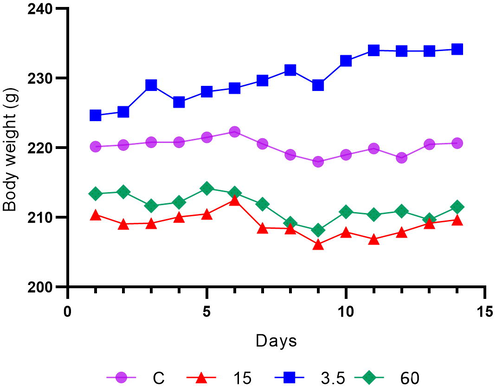
Body weight changes over 14 days in the different groups. The 15 and 60 mg/kg doses decreased body weight compared to the control, but the changes were not statistically significant. Values are given as mean ± SEM. *Significant (p < 0.05) when compared to the control.
3.3 Effect of sage extract on steroid hormone secretion
The serum progesterone, estradiol, and testosterone levels of the four groups are shown in Fig. 2. No significant differences in progesterone or testosterone levels were observed between the treatment groups. Estradiol levels in the control group were significantly different from those in the group receiving 60 mg/kg sage. However, estradiol levels in the control group were not significantly different from those in the groups low-doses (3.5 and 15 mg/kg) sage extract groups.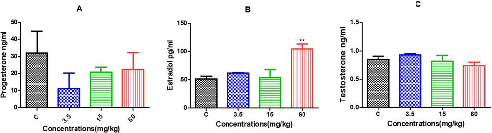
Serum hormone levels of female rats exposed to sage extracts compared to the control group. Compared to those in the control group, the levels of progesterone (pg/mL) (A), estradiol (pg/mL) (B), and testosterone (pg/mL) (C) in the 3.5 and 15 mg/kg/day groups did not change significantly. However, serum estradiol levels (ng/mL) significantly increased in females treated with high-dose sage extract (60 mg/kg/day) compared to those in the control group. Data are presented as mean ± SEM of triplicate measurements of plasma samples from at least 3 animals from each group.
3.4 Effects of sage extract on the number of follicles
The effects of aqueous sage extract on the number of ovarian follicles are presented in Fig. 3. While there were no significant differences in the number of primordial follicles in the treatment groups compared with that in the control group, there was a progressive increase in the number of growing follicles in the treatment groups compared to that in the control group. This increase was statistically significant only with the 60 mg/kg dose.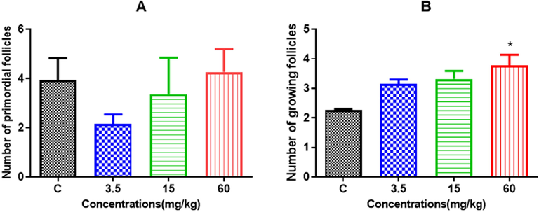
Effects of different doses of sage extract on the number of primordial (A) or growing follicles (B). All results show the mean ± S.E.M. of three sectioned ovaries for each group, each performed in triplicate. *p < 0.05 compared to control.
3.5 Effects of sage extract on the number of abnormal follicles
In this study, abnormal follicles in the ovary were defined as those containing multinucleate oocytes and/or multioocyte follicles (Fig. 4). We observed that sage extract significantly decreased the number of abnormal follicles in all treatment groups (3.5, 15 or 60 mg/kg) compared to the control group (Fig. 5).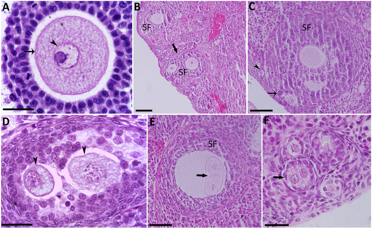
Ovarian tissue from the different groups showing abnormal follicles by H&E staining. (A) A typical normal secondary growing follicle. (B) Two secondary follicles in the process of merging (large arrow). (C) A secondary follicle merged with a primordial follicle (arrow) and a merging primordial follicle (arrowhead). (D) A secondary follicle containing two oocytes (arrowheads). (E) A secondary follicle with a large oocyte containing two merging oocytes. This is probably a result of two other primordial follicles after their fusion with the secondary follicle. (F) Primordial follicle with a binucleate oocyte (large arrow). SF: secondary follicle; Scale bar = 200 μm.
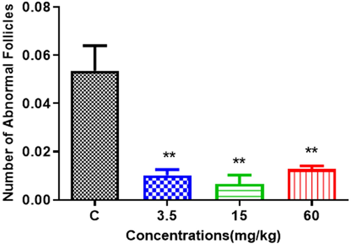
The number of abnormal follicles decreased significantly in the ovaries of females in the treatment groups (3.5, 15 or 60 mg/kg) compared to the control group. All results show the mean ± S.E.M. of three sectioned ovaries for each group, each performed in triplicate. *p < 0.005 compared to the control group.
3.6 Effect of sage extract on gene expression levels
We analyzed the effect of sage extract on ten genes involved in folliculogenesis, steroidogenesis, and autophagy (Fig. 6). Rats treated with a 60 mg/kg dose of sage extract had significantly increased mRNA levels of INSL3, CYP17a, LHR, CCND2, IGF1, ESR1, ESR2, ACTB, GDF, and ATG5 genes (Fig. 6 A, B, C, D, E, F, G, H, I and J). CYP17a mRNA levels were significantly increased in a dose-dependent manner (Fig. 6B), whereas mRNA levels of LHR, COND2 and ESR1 were significantly increased even at a low dose (3.5 mg/kg) (Fig. 6C, D and F).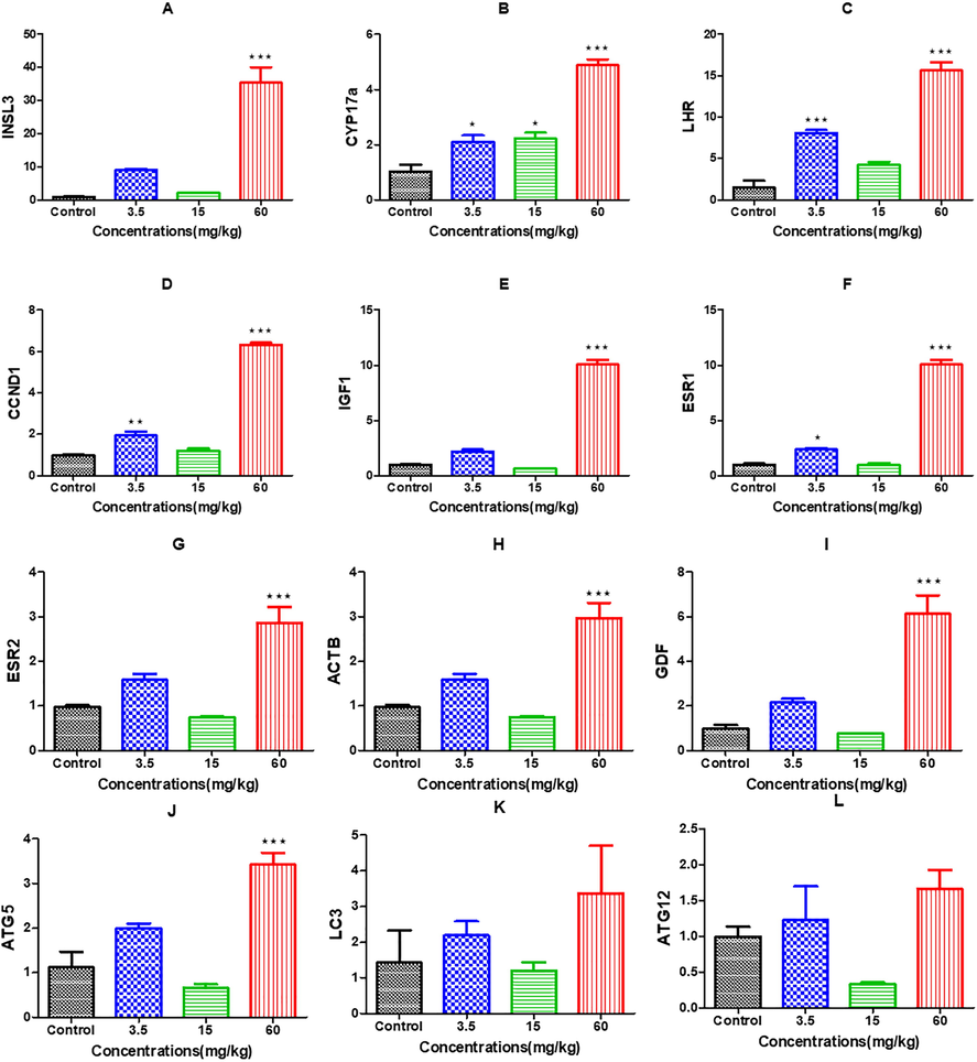
mRNA expression levels of different genes in the ovaries of rats in the treatment groups compared to the control group.
4 Discussion
Many studies have reported that consuming S. officinalis in traditional medicine has no side effects (Ghorbani and Esmaeilizadeh, 2017). In the present study, we evaluated the beneficial and/or potentially negative effects of different concentrations of sage extract on rat female reproduction. We found that sage extract had no effect on steroid hormone release when used at low doses (3.5 and 15 mg/kg), as evidenced by the fact that the levels of progesterone, estradiol, and testosterone did not change with treatment. However, at the higher dose (60 mg/kg), estradiol significantly increased. Similarly, the mRNA levels of different genes involved in folliculogenesis and steroidogenesis significantly increased with the high dose of S. officinalis. Interestingly, the number of growing follicles significantly increased, whereas the number of abnormal follicles decreased. Further research also revealed that S. officinalis promoted growth (Dadras et al., 2020). These effects may be related to the presence of several compounds in this plant. GC–MS analysis revealed that the dominant constituents (>1%) were 1,8-cineole (47.40%), 13-epimanool, (6.45%), 4,4 dimethylandrost-5-ene (5.90%), caryophyllene (5.11%), camphene (4.88%), α-pinene (4.40%), camphor (4.32), β-pinene (2.94%), and ledol (2.32%). No previous studies have reported the effects of these molecules on ovarian function. A previous study showed that the major component, 1,8-cineole, did not cause any toxicities or deaths among rats receiving repeated injections of doses up to 1000 mg/kg for 50 days (Caldas et al., 2016). Sage extract is widely used in the pharmaceutical and cosmetics industries since it has a variety of antitumor (Murata et al., 2013), hepatoprotective (Santos et al., 2001), and anti-inflammatory (Bastos et al., 2011) properties. Diterpenoids represented the second major component of the sage plant, and 13-epimanool and carnosol have various pharmacological properties, including antiproliferative activity due to their cytotoxic effects (Ruan et al., 2017). Generally, the effects of these compounds on the female reproductive system are not well known, but a few studies have indicated that some of these compounds have adverse effects on female reproduction by reducing the number of atretic follicles and increasing the number of developing follicles (Ruan et al., 2017). However, the effects of the two main components, 1,8-cineole and 13-epimanool, on ovarian function and the mechanisms involved need further exploration.
Since sage extract contains a rich collection of molecules, we hypothesized that high doses of the plant could improve ovarian function through estrogen signaling pathways, estrogen receptors, and ovarian steroidogenesis. Previous studies have reported that some molecules in plants, such as Senecio biafrae (Asteraceae), control female reproduction through the nervous system and are used widely in traditional medicine to cure female infertility (Lienou et al., 2020). Therefore, we hypothesize that sage extract may stimulate the release of gonadotropin-releasing hormone (GnRH) and follicle-stimulating hormone (FSH). FSH binding to granulosa cells leads to activation of FSH receptors that are frequently present on their surfaces. This upregulates the transcription of target genes involved in proliferation and differentiation (Escamilla-Hernandez et al., 2008; Hsueh and Rauch, 2012). Specifically, INSL3, CCND2, IGF-I, and GDF are essential for the proliferation and differentiation of granulosa cells during follicular growth and development (Yenuganti et al., 2016). Our data show that when we used the higher dose (60 mg/kg) of sage extract, the transcription of these genes increased, which promoted the proliferation of granulosa cells. Estradiol increased after treatment with 60 mg/kg sage extract and exerted its effects by binding to nuclear estrogen receptors (ESR1 and ESR2, also known as ERα and Erβ), consistent with an increase in mRNA transcripts of the corresponding genes. Interestingly, the number of growing follicles increased since estradiol is known to play a vital role in maintaining and developing follicles (Shi et al., 2019). Thus, our results are consistent with dozens of studies that have demonstrated that the estradiol/estrogen receptor pathway plays a key role during folliculogenesis (Liu et al., 2017). Moreover, estradiol activity is enhanced through autocrine signaling of the luteinizing hormone receptor (LHR) to maintain granulosa cell differentiation, leading to ovulation and corpus luteum formation (Kessel et al., 1985). This suggests that the LHR is upregulated in the ovaries of female rats treated with a high dose of 60 mg/kg sage extract. We also analyzed the expression of CYP17a, which encodes an enzyme involved in producing androgens (Miller, 2002). Our results showed that the mRNA levels of this gene significantly increased in a dose-dependent manner, suggesting its involvement in follicular development since decreased expression has been linked to reduced ovulation (Maganhin et al., 2014).
We also explored the expression of genes involved in autophagy since we know that many natural compounds are involved in multiple signaling pathways (Wang et al., 2017). Autophagy is an important process that supports cell survival and cell death pathways (Kobayashi et al., 2020). Our results showed that the high dose of sage (60 mg/kg) increased the transcription of all autophagy-related genes that we studied, including ATG5, LC3 and ATG12. However, the increase was only significant for ATG5, which is a key component in autophagy, and its expression is upregulated after DNA damage (Ye et al., 2018). We hypothesized that sage extract induced the expression of ATG5 to reduce ovarian pathogenesis by activating apoptotic signaling pathways. Recent evidence demonstrated that autophagy inhibition in bovine thecal cells induced the expression of cytochrome CYP17a, contributing to pathogenesis in polycystic ovarian syndrome (PCOS) (Kobayashi et al., 2020). Interestingly, abnormal follicles were sensitive to the effects of sage extract and were reduced in the ovary. Notably, the presence of more abnormal follicles is linked to reduced fertility (Perez-Sanz et al., 2013; Harrath et al., 2017). Additional studies are required to explore the precise molecular mechanisms that regulate the formation of abnormal follicles and to identify methods to reduce them.
One of the limitations of this study was that the estrogen activity could be attributed to the various flavonoid constituents present in sage plants (Michel et al., 2013; Sirotkin and Harrath, 2014; Zingue et al., 2016). These molecules are well-known phytoestrogens that are chemically analogous to mammalian 17β-estradiol and therefore match the binding domain of estrogen receptors. These factors could potentially affect all processes regulated by endogenous estrogens (Sirotkin and Harrath, 2014; Zingue et al., 2016). Due to technical problems, we could not proceed with high-performance liquid chromatography with ultraviolet detection (HPLC-UV) to determine the quantity of flavonoids present in our sage extracts. However, recent evidence has shown that S. officinalis extracts contain a total flavonoid content of 11.29 mg QE/mL (Gligor et al., 2020). This suggests that these phytoestrogens can bind to ER1 and/or ER2 in granulosa cells and promote cellular proliferation and cytodifferentiation (Rietjens et al., 2013) by activating proliferative and steroidogenic pathways.
Acknowledgements
The authors extend their appreciation to the Deputyship for Research & Innovation, “Ministry of Education” in Saudi Arabia for funding this research work through the project number IFKSURG-1442-164.
Declaration of Competing Interest
The authors declare that they have no known competing financial interests or personal relationships that could have appeared to influence the work reported in this paper.
References
- Essential oil composition, phenolic content, antioxidant, and antimicrobial activity of cultivated Satureja rechingeri Jamzad at different phenological stages. Zeitschrift Fur Naturforschung Section C-a. J. Biosci.. 2015;70(3–4):51-58.
- [Google Scholar]
- Inhaled 1,8-cineole reduces inflammatory parameters in airways of ovalbumin-challenged Guinea pigs. Basic Clin. Pharmacol. Toxicol.. 2011;108(1):34-39.
- [Google Scholar]
- Repeated-doses and reproductive toxicity studies of the monoterpene 1,8-cineole (eucalyptol) in Wistar rats. Food Chem. Toxicol.. 2016;97:297-306.
- [Google Scholar]
- Dietary administration of common sage (Salvia officinalis) and coneflower (Echinacea angustifolia) extracts affects growth, blood parameters and immune responses of Beluga, Huso huso. Turk. J. Fish. Aquat. Sc.. 2020;20(5):367-374.
- [Google Scholar]
- Dweck, A.C. 2000. Chapter The folklore and cosmetic use of various Salvia species, Sage-The Genus Salvia. In: Kintzios SE, editor. Amsterdam: Harwood Academic Publishers, pp. 1–25.
- Constitutively active protein kinase A qualitatively mimics the effects of follicle-stimulating hormone on granulosa cell differentiation. Molecular Endocrinology. 2008;22(8):1842-1852.
- [CrossRef] [Google Scholar]
- Antibacterial activity of salvia officinalis l. against periodontopathogens: an in vitro study. Anaerobe 2020:102194.
- [Google Scholar]
- Pharmacological properties of Salvia officinalis and its components. J. Tradit. Complement. Med.. 2017;7(4):433-440.
- [Google Scholar]
- Determination of a mixture of Plantago lanceolata L. and Salvia officinalis L. by high-performance liquid chromatography with ultraviolet detection (HPLC-UV) Anal. Lett.. 2020;53(9):1391-1406.
- [Google Scholar]
- Food restriction during pregnancy and female offspring fertility: adverse effects of reprogrammed reproductive lifespan. J. Ovar. Res.. 2017;10
- [Google Scholar]
- Ovarian Kaleidoscope database: ten years and beyond. Biol. Reprod.. 2012;86(8):192.
- [CrossRef] [Google Scholar]
- Autocrine role of estrogens in the augmentation of luteinizing hormone receptor formation in cultured rat granulosa cells. Biol. Reprod.. 1985;32(5):1038-1050.
- [Google Scholar]
- Pharmacological studies on Hypericum perforatum fractions and constituents. Pharm. Biol.. 2011;49(1):46-56.
- [Google Scholar]
- Leung’s Encyclopedia of Common Natural Ingredients Used in Food, Drugs, and Cosmetics (Third ed.). Hoboken, New Jersey: John Wiley & Sons Inc.; 2010.
- Inhibition of autophagy in theca cells induces CYP17A1 and PAI-1 expression via ROS/p38 and JNK signalling during the development of polycystic ovary syndrome. Mol. Cell. Endocrinol. 2020:110792.
- [Google Scholar]
- Complementary therapy for female infertility: protocol for an overview of systematic reviews. Integr. Med. Res.. 2020;9(1):62-64.
- [Google Scholar]
- Female infertility associated with blood lead and cadmium levels. Int. J. Environ. Res. Public Health. 2020;17(5)
- [Google Scholar]
- An ethnopharmacological investigation of medicinal Salvia plants (Lamiaceae) in China. Acta Pharm Sin B. 2013;3(4):273-280.
- [Google Scholar]
- Effect of different extracts and fractions of Senecio biafrae (Oliv. & Hiern) J. Moore on in vivo and in vitro parameters of folliculogenesis in experimental animals. J. Ethnopharmacol.. 2020;251:112571
- [Google Scholar]
- ZHANG, Z, Wang, C, Xu, Y, Duan, E, Xia, G: Estrogen receptors in granulosa cells govern meiotic resumption of pre-ovulatory oocytes in mammals. Cell Death Dis.. 2017;8(3)
- [Google Scholar]
- Cytotoxic activity of essential oils from Labiatae and Lauraceae families against in vitro human tumor models. Anticancer Res.. 2007;27(5a):3293-3299.
- [Google Scholar]
- Melatonin influences on steroidogenic gene expression in the ovary of pinealectomized rats. Fertil. Steril.. 2014;102(1):291-298.
- [Google Scholar]
- Ethnopharmacological and Chemical Characterization of Salvia Species Used in Valencian Traditional Herbal Preparations Front. Pharmacol.. 2017;8:467.
- [Google Scholar]
- Evaluation of bioactive properties and phenolic compounds in different extracts prepared from Salvia officinalis L. Food Chem.. 2015;170:378-385.
- [Google Scholar]
- National, regional, and global trends in infertility prevalence since 1990: a systematic analysis of 277 health surveys. PLoS Med.. 2012;9(12)
- [Google Scholar]
- New Concepts, Experimental Approaches, and Dereplication Strategies for the Discovery of Novel Phytoestrogens from Natural Sources. Planta Med.. 2013;79(7):514-532.
- [Google Scholar]
- Androgen biosynthesis from cholesterol to DHEA. Mol. Cell. Endocrinol.. 2002;198(1–2):7-14.
- [Google Scholar]
- Antitumor effect of 1, 8-cineole against colon cancer. Oncol. Rep.. 2013;30(6):2647-2652.
- [Google Scholar]
- Increased number of multi-oocyte follicles (MOFs) in juvenile p27Kip1 mutant mice: potential role of granulosa cells. Hum. Reprod.. 2013;28(4):1023-1030.
- [Google Scholar]
- Bisphenol A: an emerging threat to female fertility. Reprod. Biol. Endocrinol.. 2020;18(1):22.
- [Google Scholar]
- Capillary gas chromatography-mass spectrometry of volatile and semi-volatile compounds of Salvia officinalis. J. Chromatogr. A. 2004;1027(1–2):121-126.
- [Google Scholar]
- Mechanisms underlying the dualistic mode of action of major soy isoflavones in relation to cell proliferation and cancer risks. Mol. Nutr. Food Res.. 2013;57(1):100-113.
- [Google Scholar]
- The adverse effects of triptolide on the reproductive system of caenorhabditis elegans: Oogenesis impairment and decreased oocyte quality. Inter. J. Mol. Sci.. 2017;18(2)
- [Google Scholar]
- 1,8-cineole protects against liver failure in an in-vivo murine model of endotoxemic shock. J. Pharm. Pharmacol.. 2001;53(4):505-511.
- [Google Scholar]
- Efficacy of electroacupuncture in regulating the imbalance of AMH and FSH to improve follicle development and hyperandrogenism in PCOS rats. Biomed Pharmacother. 2019;113:108687
- [Google Scholar]
- Mechanisms of the direct effects of oil-related contaminants on ovarian cells. Environ. Sci. Pollut. Res. Int.. 2020;27(5):5314-5322.
- [Google Scholar]
- Antioxidant capacity and polyphenolic composition as quality indicators for aqueous infusions of Salvia officinalis L (sage tea) Front. Pharmacol.. 2011;2
- [Google Scholar]
- Natural products as modulator of autophagy with potential clinical prospects. Apoptosis. 2017;22(3):325-356.
- [Google Scholar]
- WHO. 2017. Sexual health and its linkages to reproductive health: an operational approach.
- Exploring the Role of Autophagy-Related Gene 5 (ATG5) Yields Important Insights Into Autophagy in Autoimmune/Autoinflammatory Diseases. Front. Immunol.. 2018;9:2334.
- [Google Scholar]
- Oleic acid induces specific alterations in the morphology, gene expression and steroid hormone production of cultured bovine granulosa cells. Gen. Comp. Endocrinol.. 2016;232:134-144.
- [Google Scholar]
- Estrogenic effects of Ficus umbellata Vahl. (Moraceae) extracts and their ability to alleviate some menopausal symptoms induced by ovariectomy in Wistar rats. J. Ethnopharmacol.. 2016;179:332-344.
- [Google Scholar]


