

Translate this page into:
Chrysanthemum morifolium extract mediated Ag NPs improved the cytotoxicity effect in A549 lung cancer cells
⁎Corresponding author at: Department of Marine Science, Bharathidasan University, Tiruchirappalli, Tamil Nadu, India. rajivgandhimicro@yahoo.com (Govindan Rajivgandhi)
-
Received: ,
Accepted: ,
This article was originally published by Elsevier and was migrated to Scientific Scholar after the change of Publisher.
Peer review under responsibility of King Saud University.
Abstract
In this study, the anti-cancer compound was extracted from Chrysanthemum morifolium (C. morifolium) by soxhlet apparatus method. The available anti-cancer compounds of Chrysanthemum morifolium extract were clearly detected by LC-MS analysis based on the retention time, percentages of height and occupied area. The anti-cancer compounds were separately screened from mixed compounds by NIST Wiley library interpretation. Further, the silver nanoparticle (Ag NPs) was synthesized from C. morifolium extract, and their surface plasmon resonance effect of the extract was scanned by UV–vis spectroscopy at 410 nm. Morphological identification of size and shape of the Ag NPs was confirmed by scanning electron microscope and transmission electron microscope images. Further, the anti-cancer effect of synthesized Ag NPs was exhibited more cytotoxicity effect at the concentration of 200 µg/mL. Furthermore, the morphological damages of A549 cells were clearly confirmed by fluorescence microscope using AO/EB fluorescence stain. Finally, the results were suggested that the biosynthesized Ag NPs has potential anti-cancer agent against A549 lung cancer cells.
Keywords
Plant material
LC-MS analysis
Spectroscopic analysis
Cytotoxic effect
Morphological damage

1 Introduction
Worldwide, the increasing mortality rate of lung cancer is increasing frequently due to the adaptation of cancer environment including continuous smoking, modernized culture, without exercise (Que et al., 2019). It is most dangerous disease for human for frequently increased death rate after blood cancer (He et al., 2016). It crossed at least 50,000 death per year compared with other cancer, and it is a leading cancer in developing and developed countries (Zhang et al., 2020). All the countries are struggling to prevent the lung cancer and their related infections. The current treatment methods are ineffective due to the routine use of unfavorable drug (Saravanakumar et al., 2019). The previous methods of surgical, chemotherapy and laparoscopy are not successful in control of lung cancer cells. These methods are also not effective for lifetime cancer patients due to the end stage. Due to these defects, all the countries are searching a new tool for improve the diagnosis of cancer in starting stage as well as new methods to improve the drugs against cancer cell cycles, cell signaling and target sites (Venugopal et al., 2017). Initially, if cancer cells enter into primitive stage, it will be difficult to eradicate due to the production of responsible gene expression. In medical field refereed that the initial stage of cancer cells inhibition is better choice to eradicate cancer cells completely. Recent years, all the developing and developed countries are motivated the research and development units for cancer cells to inhibit in initial stage (Tammina et al., 2017). More research activities, increased awareness program, decreased cancer environments, behavior changes are the best was to eradicate the cancer cells. Importantly, the alternative way is need to inhibit the cancer cells and it should be help to demolish the cancer cells in long term (Naveen kumar et al., 2018). Recent years, nanoparticles and their properties have enormous anti-cancer activities against cancer cell treatment long term with minimum toxicity. Also, this is one of the new routes to treat the cancer cells in initial stage and also prevent the cancer cells completely (Samuel et al., 2020).
Recent years, nanotechnology is entered into biomedical field and it given the solution to alternative drug choice in drug delivery process (Rajivgandhi et al., 2019aa, 2019bb, 2019bc). It is in anno size, so it is easy to success in biomedical field (Mortazavi-Derazkola et al., 2020). The nano sized particles are used in all the fields including soil, agriculture, food, pharmaceutical and biomedical with increased nature (Bello et al., 2017). Also, it has wide range of application in the entire field through the way of alternative agent (Sankar et al., 2013). In addition, the nano sized particles are used in environmental pollution control and material chemistry and astro physics (Jeyaraj et al., 2013). Usually, the nanoparticles have the sine of 1–1000 nm and recent years mostly used in biomedical field compared with other field (Esawy et al., 2019). It is an alternative agent for drug delivery, increased drug nature and inhibit the various infections effectively (Hashemi et al., 2020a, 2020b). Among the different nanoparticles, the most important and highly reported nanoparticle is silver nanoparticles (Ag NPs), and it is most exploited and effective biological characteristic agent compared with other nanoparticles (Rajasekharreddy and Usha Rani, 2014). Last ten years, the silver nanoparticles research is increased very high in the field of medical with enhanced bioactivities (Rao et al., 2020). It is synthesized form various routes such as chemical, physical and biological. Among the various route, the most reported biological routes are very efficient than other routes due to the unique properties with decreased toxicity (Korkmaz et al., 2020). Previously, more reports of biosynthesized Ag NPs has enormous biological activities like anti-microbial, anti-cancer, anti-viral, larvicidal and anti-biofilm activities (Manjunath Hulikere et al., 2019; Rajivgandhi et al., 2020a, 2020b; Bello et al., 2017; Sankar et al., 2013). At present, the biological mediated Ag NPs is very effective against various infections than other route of synthesis. In biological route, previously used plant, seaweed and microbes are best choice. Sometimes, the Ag NPs absorbed the source compositions, nutrients and other habitats and produced enhanced bioactivities compared with original one (Venugopal et al., 2017). Therefore, this study was concentrated on green synthesized nanoparticles using C. morifolium and their anti-cancer effect against A549 lung cancer cells which carried out by various invitro experiments.
2 Materials and methods
2.1 Needed chemicals
The Indian medicinal plant of C. morifolium was collected from Pottanam Village, Namakkal, Tamil nadu, India. Silver nitrate and nanoparticle synthesis materials were purchased from Suresh Scientific @ Co, Tiruchirappalli, Tamil Nadu, India. The cancer cells A549 were procured from King Institute, Guindy, Chennai, Tamil nadu, India. Other related chemicals and solutions were purchased from Ponmani @ Co, Tiruchirappalli, Tamil nadu, India.
2.2 Extraction of plant extract
The soxhlet apparatus was filled by 10 g of purely washed plant extract through powder nature and run the instrument continuously till solute fully. For the extraction, the methanol was acted as an effective solvent. After heating, the sample phases of the soxhlet were cooled 2 h and completely lyophilize after removal of the extract phases. The sample was lyophilized using lyophilizer for purify the active components of the extract (Rajivgandhi et al., 2020a, 2020b). Then, the available chemical compounds of the extract were scanned by LC-MS (Shimadzhu, Japan). Finally, the active biological components of the extract was purified separately and used to synthesis of Ag NPs for detection of anti-cancer activity.
2.3 Detection of anti-cancer compounds form LC-MS analysis
The presence of anti-cancer compounds in the C. morifolium was screened by using LC-MS instrument. This method was followed by previous report of Taskin et al. (2020). With some alteration. In LC-MS analysis, the instrument was attached with split ratio of 1:2 (Extract: dichloromethane) at 200 °C. The chemical components were purified by HP-4MS column of 20 m × 0.30 mm (dichloromethane + 5% phenyl). The 30S helium gas and injector temperature timing of start and end is 50–350 °C at 4 °C. It is oven temperature. The purified components materials were mixed with HCL and diluted with water with injected volume of 1:1 ratio.
2.4 Synthesis of Ag NPs
Sterile, clean100 mL test tube was taken and filled with 1:10 ratio of crude extract and silver nitrate. The test tube was maintained in the water bath, which previously set in 90 °C for proper heat. The test tube was allowed 1 h to change the color of the mixture samples. For adjustment of pH 1 N NaOH and 1 N H3PO4 was used After 1 h, the mixture of the solution color was gradually changed to yellow or orange or light brown color. The color changes were indicated that the Ag NPs was synthesized in the mixture solution. Consequently, the synthesized Ag NPs was accurate or not was proved by result of UV-spectrometer analysis at the wavelength of 200–800 nm. After initial confirmation, the morphology, size and shape of the green synthesized Ag NPs was confirmed by SEM at 0.1-30kv of accelerating voltage (Shimadzhu, Japan) and TEM instruments at 120–200 KV accelerated (Scavenging, Germany). The entire method was followed by Rajivgandhi et al. (2019a), Rajivgandhi et al. (2019b), Rajivgandhi et al. (2019c).
2.5 Anti-cancer activity
2.5.1 Cytotoxicity assay
Anti-cancer nature of green synthesized Ag NPs against A549 lung cancer cells were evaluated by 96-well micotitre plate assay for detection of cytotoxicity of the Ag NPs (Naveen kumar et al., 2018). Shortly, the fresh complete medium was filled by 24 h old ∼ 2 × 10⁴ cells of A549 through seeding method. The seeded plate was maintained at 37 °C for 24 h in CO2 incubator under reduced pressure with 5% related humidity. This stage was used to attach the cells into the plate. After attachment, the cells were treated with different diluted concentration (5–250 µg/mL) of Ag NPs and put into the CO2 incubator under reduced pressure with 5% related humidity at one day. Without addition of AgNPs containing A549 culture well was acted as a control. After, 100 μL MTT solutions were added into the wells and then incubate the plate at 37 °C for 4 h. Next, observe the formazan crytal formation in the treated wells and dissolved with 200 μL of fresh DMSO solution. Finally, the O.D of 590 nm was taken using microtitre reader plate (BioTek Instruments, Winooski VT) after the intracellular modified color changes wells. Finally, the color changes of the wells O.Ds were compared with control O.Ds and made to percentage of inhibition. Consequently, the IC50 concentration was noted by using following formula,
2.5.2 Detection of morphological variation (AO/EB) assay
The fluorescence absorption mediated morphological differentiation in the Ag NPs treated slide was proved by fluorescence microscope, with the following modification of Venugopal et al. (2017). The fresh 24 h old A549 culture was seeded into the 6-well adherent plate with inside the cover slide. Then, different concentration of Ag NPs was treated into the wells cultures and allowed to adherent or non-adherent the cells till 24 h. After incubation, the cells were trypsinized in all the wells for complete detachment of non-adherent cells. After, 10 μL of AO/EB was gradually added into all the wells including untreated control well. Then, mixed gently and excess stain was removed using Whatman No.1 filter paper. Finally, the morphological differentiation of treated or untreated cells were differentiated using fluorescence microscope analysis (Carl Zeiss, Jena, Germany) at magnification range of 40x.
3 Result
3.1 LC-MS report of Chrysanthemum morifolium extract
Totally, 55 peaks were exhibited in the extract of C. morifolium after take report of LC-MS (Fig. 1). All the peaks were carefully interpreted with Bharathidasan University using NIST Wiley Library for detection of available chemical derivatives in the extract. Also, the retention time, area of height, peak percentages were also discussed based on the NIST reports and identified 6 different anti-cancer compounds. Whether, the identified compounds were present in the plant materials or not were also checked in previous reported articles, and found that the articles have the identified compounds with anti-cancer compounds. The available peaks were effectively shown in Fig. 1. The available anti-cancer compounds of the extract and their retention time and occupied area, percentage of area were effectively screened such as Pyrrolo[1,2-a]pyrazine-1,4-dione, hexahydro-3, 7,9-Di-tert-butyl-1-oxaspiro(4,5)deca-6,9-diene-2,8-dione, trifluoroacetoxy hexadecane, heptadecyl trifluoroacetate, BIS(2-ethylhexyl) phthalate. The retention time of 11.30, 22.45, 30.16, 11.90 and 20.10, occupied area of 12, 7866, 19, 8453, 22, 8790, 20, 8976 and 19, 8079, area percentages of 3.6, 2.9. 3.0, 2.8 and 2. 6. The result was good agreement with previous report of Sixto et al. (2019), and anti-cancer compounds were present in plant extract. The similar study of anti-cancer activity was reported by Peng et al. (2020) and C. morifolium was excellent plant materials for inhibition of cancer cells. Recently, Rajasekharreddy and Usha Rani, 2014 reported that the plant C. morifolium increased anti-microbial and anti-cancer activities. Sometimes, the effect of compounds were identified based on the tropical and subtropical regions and it’s changed their potential activity also (Mighri et al., 2019). Some researchers are also reported that the plant C. morifolium was very effective anti-cancer compound producer and it alters the intracellular molecules in cancer cells (Ložnjak et al., 2020).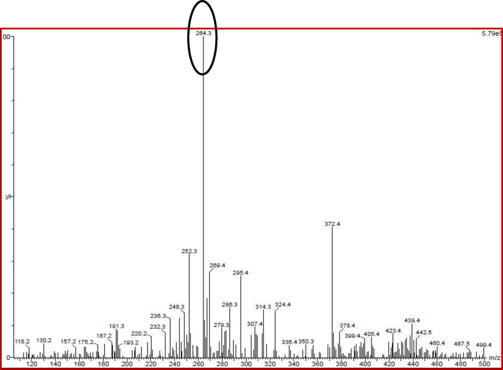
Available anti-cancer compound of Chrysanthemum morifolium extract by LC-MS analysis.
3.2 Characterization of Ag NPs
Based on the Mi theory, the exhibited Ag NPs was shown at the 410 nm, and the exhibited peak was confirmed that the Ag NPs was synthesized form C. morifolium extract (Fig. 2). The color changes were clearly observed in the C. morifolium iextract after 1 to 2 h time interval. Likewise, the Ag NPs formed solution was shown with greenish yellow color and it revealed the surface plasmon resonance effect of C. morifolium extract. After the O.D value of spectrophotometer result was also drawn between the 200–800 nm and it confirmed that the synthesized material was Ag NPs. The confirmed result was indicated that the extract has no any impurities and transferred the Ag NPs through the process of Ag + ion transferred into plant components containing extract for synthesis of Ag NPs. In addition, the spherical shaped morphology and separate colonies were also clearly observed in the SEM images. Exact Ag NPs was found with agglomerated spherical shape morphology by SEM morphology (Fig. 3a). Also, original Ag NPs size and shapes were clearly shown in the SEM and supported by TEM results. Further, the size and shape confirmations of TEM images were also effectively supports the result and it confirmed that the morphology was Ag NPs morphology. The SEM and TEM morphological results of Ag NPs size and shapes were shown in the Fig. 3b. The size and shape based SEM images were used to confirm the Ag NPs morphology. Previously, the spherical shaped morphology of Ag NPs was effectively indicated in reported evidence of Vimala et al. (2015). Recently, Rajivgandhi et al. (2020a), Rajivgandhi et al. (2020b) was also agreed the result, and green synthesized Ag NPs images were shown spherical morphology with agglomerated images. Finally, the characterizations of UV, SEM and TEM results were indicated that the plant of C. morifolium was important plant and used for synthesis of high volume ration and high surface area of Ag NPs.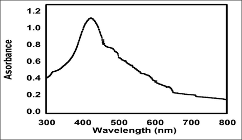
UV–vis spectroscopy analysis of Chrysanthemum morifolium extract mediated Ag NPs synthesis.
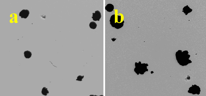
Morphological Differentiation of Ag NPs using SEM and TEM analysis of Chrysanthemum morifolium extract.
3.3 Anti-cancer studies
3.3.1 Cytotoxicity assay of cancer cells
After time interval of one day, the quantity of A549 cells were decreased and confirmed by O.D value calculation. The viability of the A549 cells was decreased at increasing concentration of Ag NPs. It shown more turbidity at the concentration of 300 µg/mL (Fig. 4). This result was suggested that the Ag NPs was transferred to surface of the cancer cells and extended the proliferation ability. Therefore, the cells were gone to minimum quantity and undergone to decline phase. In result, the 53% of cell viability was observed at the concentration of 200 µg/mL and it suggested the concentration dependent cell decrease. Initially, the decreased viability was started at the concentration of 25 µg/mL only. At this concentration was used for initial inhibition study and extended up to 300 µg/mL. All other invitro experiment, the IC50 concentration of 200 µg/mL was fixed. Based on the time and concentration, the inhibition concentration of 200 µg/mL was very effective compared to previous study of plant (Venugopal et al., 2017). This result was agreed by Hashemi et al. (2020), Hashemi et al. (2020b) and plant mediated Ag NPs is an excellent anti-cancer agent against A549 lung cancer cells. The inhibition was stimulated by morphology and mitochondrial region due to the effect of plant was reported by Khorrami et al. (2020). The more precipitation and increased concentration was observed against A549 cells after used plant and their color was changed due to the effect of formazan production. Our result was agreed by previous report of Naveen Kumar et al. (2018), the plant extract of C. morifolium is excellent plant for synthesis of Ag NPs with potential anti-cancer activity. Also, the phytochemical and bioactive derivatives were play a major role for improve the bioactivity of Ag NPs. Finally, our result was proved that the C. morifolium mediated Ag NPs was very effective anti-cancer agent for A549 lung cancer cells.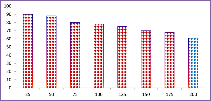
Cytotoxicity assay of Ag NPs treated A549 lung cancer cells by MTT microtitre model.
3.3.2 AO/EB stain usage of morphological differentiation
Consecutively, the confused morphological observation was observed in the IC50 dose of Ag NPs treated and untreated A549 cells by fluorescence microscope after using dual stains of AO/EB (Fig. 5). In this experiment, the emission of light intensity was shown in damaged cells compare with normal untreated cells. The condensation and necrotic cells were shown highly in the treated cells. In addition, the apoptosis was processed continuously and it leads to cell death. All the cells were undergone to decline phase and it confirmed more apoptosis was shown. The activator genes were processed successfully and induced more proliferation for damage. The intracellular membranes were shown more orange color compare with green color of control cells. Also, the necrotic cells were exhibited with orange color cells in the treated images and also shape and size of the cells were also changed. Instead, the smooth, normal and original morphology with A549 cells were viewed. AO is a dye which binds only in the normal cells, whereas, EB is a dye which has the ability to bind in damaged cells. In our result, the normal cells were observed the AO stain and it emitted green color intensity appearance. Whereas, the more amount of red color intensity with high proliferation ability of EB stain indicated that the cells were damaged due to the Ag NPs. Therefore, the AO/EB result was confirmed that the Ag NPs was very efficient against A549 lung cancer cells. In addition, the Ag NPs was acted as a building block against A549 cells in the cell cycle process, because, all the cells were undergone to decline phase. The more nutrient depletion and growth cell receptor damages were shown Saravanakumar et al. (2019). Recently, complete death cells due to the stimulation of responsible factors activation by biosynthesized Ag NPs reported by Venugopal et al. (2017). The nucleus leakages materials were observed the fluorescence dyes were clearly confirmed the Ag NPs ability against A549 lung cancer cells (Jeyaraj et al. (2013). This statement was agreed by Esawy et al. (2019), and biosynthesized Ag NPs has effective anti-cancer agents against various cancer cells and it proved against A549 cells previously. Previously, Lee et al. (2014) and Ukiya et al. (2002) were reported that the plant extract was shown anti-inflammatory, antiadipogenic and anti-cancer activity against various cancer cells. These are most supported to anti-cancer activity of C. morifolium extract. Therefore, the present result was suggested that the biosynthesized Ag NPs using marine algae C. morifolium as an important source for synthesis of potential anti-cancer agent.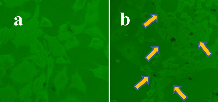
Detection of intracellular morphological damages of A549 lung cancer cells using AO/EB fluorescence dye by fluorescence microscope.
4 Conclusion
This study was concluded that the plant C. morifolium extract has excellent plant for synthesis of Ag NPs for inhibit the A549 lung cancer cells. Also, the phytochemical derivatives and their chemical components were influenced the Ag NPs activity against A549 cancer cells. Additionally, the IC50 concentration of the Ag NPs against A549 cells was shown at 250 µg/mL concentration. Further, the damaged morphology of A549 cells after influence of IC50 concentration was confirmed that the biosynthesized Ag NPs has excellent anti-cancer activity. Altogether, the biosynthesized Ag NPs is an excellent anti-cancer agent for A549 lung cancer cells.
Acknowledgement
The authors extend their appreciation to the Deputyship for Research & Innovation, ‘‘Ministry of Education” in Saudi Arabia for funding this research work through the project number IFKSURG-1438-091.
Declaration of Competing Interest
The authors declare that they have no known competing financial interests or personal relationships that could have appeared to influence the work reported in this paper.
References
- Antiproliferation and antibacterial effect of biosynthesized AgNps from leaves extract of Guiera senegalensis and its catalytic reduction on some persistent organic pollutants. J. Photochem. Photobiol. B: Biolog.. 2017;175:99-108.
- [Google Scholar]
- Evaluated bioactive component extracted from Punica granatum peel and its Ag NPs forms as mouthwash against dental plaque. Biocatal. Agricult. Biotechnol.. 2019;18:101073
- [Google Scholar]
- S.F. Hashemi, N. Tasharrofi, M.M. Saber, Green synthesis of silver nanoparticles using Teucrium polium leaf extract and assessment of their antitumor effects against MNK45 human gastric cancer cell line, J. Mol. Struct. 1208 (2020) 127889.
- S.F. Hashemi, N. Tasharrofi, M. Mahmoudi Saber, Green synthesis of silver nanoparticles using Teucrium polium leaf extract and assessment of their antitumor effects against MNK45 human gastric cancer cell line, J. Molecul. Struct. 1208, 2020, 127889.
- Effects of green-synthesized silver nanoparticles on lung cancer cells in vitro and grown as xenograft tumors in vivo. Int. J. Nanomed.. 2016;11:1879-1887.
- [Google Scholar]
- An investigation on the cytotoxicity and caspase-mediated apoptotic effect of biologically synthesized silver nanoparticles using Podophyllum hexandrum on human cervical carcinoma cells. Colloid. Surf. B: Biointerf.. 2013;102:708-717.
- [Google Scholar]
- Bacteriostatic activity of aquatic extract of black peel pomegranate and silver nanoparticles biosynthesized by using the extract. Biocat. Agricult. Biotech.. 2020;25:101620
- [Google Scholar]
- Biogenic silver nanoparticles synthesized from Rhododendron ponticum and their antibacterial, antibiofilm and cytotoxic activities. J. Pharmaceut. Biomed. Analy.. 2020;179:112993
- [Google Scholar]
- Evaluation of Compositae sp. plants for antioxidant activity, antiinflammatory, anticancer and antiadipogenic activity in vitro. Food Agricult. Immunol.. 2014;25:104-118.
- [Google Scholar]
- Quantification of folate in food using deconjugase of plant origin combined with LC-MS/MS: A method comparison of a large and diverse sample set. Food Chem.. 2020;305:125450
- [Google Scholar]
- Characterization, antioxidant and antimicrobial activity of silver nanoparticles synthesized using marine endophytic fungus- Cladosporium cladosporioides. Proce. Biochem.. 2019;82:199-204.
- [Google Scholar]
- LC/MS method development for the determination of the phenolic compounds of Tunisian Ephedra alata hydro-methanolic extract and its fractions and evaluation of their antioxidant activities. South Afr. J. Bot.. 2019;124:102-110.
- [Google Scholar]
- Facile green synthesis and characterization of Crataegus microphylla extract-capped silver nanoparticles (CME@Ag-NPs) and its potential antibacterial and anticancer activities against AGS and MCF-7 human cancer cells. J. Alloy. Compound.. 2020;820:153186
- [Google Scholar]
- Cytotoxicity effect of marine Sponge Alkaloid, Fascaplysin on HepG2 Hepatocellular carcinoma cell. Fron. Lab. Med.. 2018;2:41-48.
- [Google Scholar]
- A. Peng, L. Lin, M. Zhao, Screening of key flavonoids and monoterpenoids for xanthine oxidase inhibitory activity-oriented quality control of Chrysanthemum morifolium Ramat. ‘Boju’ based on spectrum-effect relationship coupled with UPLC-TOF-MS and HS-SPME-GC/MS, Food Res. Int. 137, 2020, 109448.
- Size dependent anti-invasiveness of silver nanoparticles in lung cancer cells. RSC Adv.. 2019;9:21134.
- [Google Scholar]
- Biofabrication of Ag nanoparticles using Sterculia foetida L. seed extract and their toxic potential against mosquito vectors and HeLa cancer cells. Mat. Sci. Engin. C. 2014;39:203-212.
- [Google Scholar]
- G.N. Rajivgandhi, G. Ramachandran, M. Maruthupandy, N. Manoharan, N.S. Alharbi, S. Kadaikunnan, J.M. Khaled, T.N. Almanaa, W.J. Li, Anti-oxidant, anti-bacterial and anti-biofilm activity of biosynthesized silver nanoparticles using Gracilaria corticata against biofilm producing K. pneumonia, Colloids and Surfaces A: Physicochem. Engin. Aspects, 600, 2020, 124830.
- Marine sponge alkaloid aaptamine enhances the anti-bacterial and anti-cancer activity against ESBL producing Gram negative bacteria and HepG 2 human liver carcinoma cells. Biocatal. Agric. Biotechnol.. 2019;17:628-637.
- [Google Scholar]
- Graphene/nickel oxide nanocomposites against isolated ESBL producing bacteria and A549 cancer cells. Mater. Sci. Eng.: C. 2019;102:829-843.
- [Google Scholar]
- Biosynthesized silver nanoparticles for inhibition of antibacterial resistance and biofilm formation of methicillin-resistant coagulase negative Staphylococci. Bioorg. Chemist.. 2019;89:103008
- [Google Scholar]
- Photocatalytic reduction and anti-bacterial activity of biosynthesized silver nanoparticles against multi drug resistant Staphylococcus saprophyticus BDUMS 5 (MN310601) Mat. Sci. Engin. C. 2020;114:111024
- [Google Scholar]
- Microwave-assisted rapid synthesis of silver nanoparticles using fucoidan: Characterization with assessment of biocompatibility and antimicrobial activity. Int. J. Biolog. Macromol.. 2020;163:745-755.
- [Google Scholar]
- Biosynthesized silver nanoparticles using Bacillus amyloliquefaciens; Application for cytotoxicity effect on A549 cell line and photocatalytic degradation of p-nitrophenol. J. Photochem. Photobiol. B: Biol.. 2020;202:111642
- [Google Scholar]
- Origanum vulgare mediated biosynthesis of silver nanoparticles for its antibacterial and anticancer activity. Colloid. Surf. B: Biointerf.. 2013;108:80-84.
- [Google Scholar]
- Unveiling the potentials of biocompatible silver nanoparticles on human lung carcinoma A549 cells and Helicobacter pylori. Sci. Rep.. 2019;9:5787.
- [Google Scholar]
- GC–MS and LC–MS/MS workflows for the identification and quantitation of pyrrolizidine alkaloids in plant extracts, a case study: Echium plantagineum. Revista Brasileira de Farmacognosia. 2019;29:500-503.
- [Google Scholar]
- Cytotoxicity study of Piper nigrum seed mediated synthesized SnO2 nanoparticles towards colorectal (HCT116) and lung cancer (A549) cell lines. J. Photochem. Photobiolog. B: Biol. 2017;166:158-168.
- [Google Scholar]
- Bioassay-guided isolation and antiproliferative efficacy of extract loaded in chitosan nanoparticles and LC-QTOF-MS/MS analysis of Achillea magnifica. South Afr. J. Botany. 2020;133:236-244.
- [Google Scholar]
- Constituents of Compositae plants: III. Anti-tumor promoting effects and cytotoxic activity against human cancer cell lines of triterpene diols and triols from edible chrysanthemum flowers. Cancer Let. 2002:7-12.
- [Google Scholar]
- The impact of anticancer activity upon Beta vulgaris extract mediated biosynthesized silver nanoparticles (ag-NPs) against human breast (MCF-7), lung (A549) and pharynx (Hep-2) cancer cell lines. J. Photochem. Photobiol. B: Biol.. 2017;173:99-107.
- [Google Scholar]
- Optimization of reaction conditions to fabricate nano-silver using Couroupita guianensis Aubl. (leaf & fruit) and its enhanced larvicidal effect. Spectrochimica Acta Part A: Mole. Biomolecular Spectros.. 2015;135:110-115.
- [Google Scholar]
- Biosynthesized silver nanoparticles using Caulerpa taxifolia against A549 lung cancer cell line through cytotoxicity effect/morphological damage. Saud. J. Biolog. Sci. 2020
- [CrossRef] [Google Scholar]


