

Translate this page into:
Biogenic synthesis of ZnO and NiO nanoparticles mediated by fermented Cocos nucifera. (L) deoiled cake extract for antimicrobial applications towards gram positive and gram negative pathogens
⁎Corresponding author at: Department of Physics, Sacred Heart College (Autonomous), Tirupattur 635601, Tamil Nadu, India. rramesh@shctpt.edu (R. Ramesh)
-
Received: ,
Accepted: ,
This article was originally published by Elsevier and was migrated to Scientific Scholar after the change of Publisher.
Peer review under responsibility of King Saud University.
Abstract
Background
Cocos nucifera (L) is a widespread tall plant and found to havelot of important pharmacological applications and it is very less toxic in nature. The water of the endocarp has high antioxidant activity, and its fiber was reported to have antibacterial, ant parasitic and anti-inflammatory properties. The oil derived from its edible parts have been used in cooking, in the preparation of the soaps and cosmetics etc. The water within can be processed to produce alcohol. Once the endocarp was crushed and the extract were removed it can used as a feed material and as a natural organic fertilizer. Herein, we report the novel method of utilizing the residue of the deoiled seed cake of Cocos nucifera (L) for preparing the metal oxides as a reducing agent and to test its antimicrobial level against human pathogens.
Methods
The method adopted in this investigation is the fermentation process of preparing the Cocos nucifera (L) extract by utilizing the water as the medium for the extraction process under room temperature.
Results
Structural nature and morphology of the synthesized samples were carried out using XRD and SEM analysis and the results revealed that ZnO belongs to hexagonal shape with primitive lattice cell arrangement and NiO had cubic structure with FCC lattice arrangements. The results of the average crystalline size of prepared nanoparticles using XRD and SEM were alike. Molecular bonding and stability were analyzed using FTIR instrumentation based on UV absorption and Tauc plot the bandgap of ZnO and NiO was calculated as 3.15 eV and 3.64 eV. Elemental composition was identified using EDAX analysis. Antibacterial assay was carried out against human pathogens including S.aureus, B.subtilis, E.coli and K.pneumoniae in comparison to the standard drug Gentamycin and the synthesized specimens ZnO and NiO showed the moderate activity with the zone of inhibition of about of 8 mm, 10 mm, 12 mm and 8 mm against the human pathogens.
Conclusion
Our results showed that the antimicrobial properties of the Cocus nucifera (L) mediated ZnO and NiO have moderate activity against the standard drug Gentamycin, three different doses were tested against the disease causing pathogens including S.aureus, B.subtilis, E.coli and K.pneumoniae. The zone of inhibition was found to be minimum. This may be attributed to the reason that even after the removal of the edible compounds from the seed of Cocos nucifera (L), the residue or the waste product found to have a moderate antimicrobial function against pathogens.
Keywords
Zinc Oxide
Nickel Oxide
Cocus nucifera (L)
Fermentation
Antimicrobial activity

1 Introduction
Synthesis of the nanoparticles by usingagrowastes, plant leaf extracts, fruit extracts becoming an interesting and fascinating in the field of nanotechnology. Chemical methods were harmful, and it may be cost effective (Barzinjy et al., 2020). Fermentation is one of the traditional methods that we use in our daily life for instance preparation of butter milk and curd from the raw milk is a perfect example for the fermentation process. Making beverages and fruit wines is also prepared by the fermentation process (Sharma et al., 2020). It may be aerobic or anaerobic method based on the need. During fermentation process active enzymes and bacteria were produced and these organisms have beneficial activity (Sunilkumar, 2021). For instance, supply of macronutrients can be enhanced by the fermented deoiled cakes, agro wastes in the field of agriculture (Guo et al., 2021). NPs were synthesized using sugarcane bagasse, straw, paddy husk etc., were reported by the researchers (Fernández-Cegrí et al., 2013). Deoiled cakes like coconut, peanut, neem cake is reported to be rich in nutrients, fibers, vitamins, and minerals in appreciable ratio andit contains a large group of phytochemicals including flavonoids, alkaloids, phenols, tannins, and other notable bioactive compounds (Amrutha, 2020). When the cake is fermented these bioactive compounds will be activated in the presence of beneficial microorganisms and become enzyme activated materials for metabolic activities (Anand et al., 2020). When metal oxide nanoparticles were prepared by using these fermented extracts, the bioactive compound will act as a good reducing or capping agents in the preparation of NPs (Angel Ezhilarasi et al., 2018; Bhuyan et al., 2015; Biswal and Misra, 2020). There are many more reports including plant, leaf, and waste extract published in this method of green synthesis. The green synthesis method is a cost effective and good yield of nanoparticles. Several researchers have demonstrated the production of metal oxides based on the leaf extracts, seed extracts, flower extracts, fruit peel extracts. These materials are eco-friendly and nontoxic in nature. For example, Rana et al., studied the nickel doped ZnO and they tested against disease causing pathogens and the results revealed that Ni doped Zn exposed to direct sunlight enhanced the antibacterial activity with lower concentration. Nanocomposites is another fascinating material in the field of nanotechnology, this may be prepared by any methods based on the precursor, doping material (Kaviyarasu et al., 2016; Kaviyarasu et al., 2020; Kaviyarasu et al., 2017; Magdalane et al., 2019; Maria Magdalane et al., 2018; Panimalar et al., 2020). Thambidurai et al., synthesized NiO-ZnO nanocomposites through altered molar ratios by co-precipitation method and showed a higher activity of the nanocomposites against pathogens (Thambidurai et al., 2020).
Recent interest on the synthesis of metal oxide nanoparticles was mainly focused on replacing the commercial drugs and curing the diseases by applying the NPs as in a specific way by means of targeted drug delivery becomes one of the emerging trends in the medical field. Even the short term and long-term diseases can be diagnosed and treated with the help of nanoparticles (Dayakar et al., 2017). Linguraju et al., Showed an effective activity of E.heterophylla (L.) leavesmediated NiO NPs with lung cancer lines and hepatocarcinoma cell lines and also they have tested the non-toxic ability of the sample on human erythrocytes and coagulation activity on PPP and PRP was tested (Lingaraju et al., 2020). Tyagi et al., showed the Ciprofloxacin conjugated ZnO NPs prepared by chemical method and they compared the activity of the pre proof oxides, nanoparticles and ciprofloxacin conjugated ZnO Nps against multiple bacterial pathogens and found that there was an 2.9 fold increase and 2.8 fold increase against E.coli and Streptococcus spp. when compared with the standard drug (Tyagi et al., 2020). Surwave et al., studied the modification in the outside sheath structure of the test bacteria haemophilus influenza by the application of ZnO NPs with two different sizes of morphology. Results indicated that the membrane was altered by the wild strain, and they arrived at a conclusion that the effect is concentration and size dependent and TEM results revealed that the antibacterial activity was decreased with increase of negative charge on the outer membrane is increased (Surwade et al., 2020). Sohail et al., demonstrated a multifaceted therapeutic agent zinc oxide using neemand the sample was tested against levofloxacin resistant strains and showed a better antioxidant and enzyme inhibition as compared with standard drugs (Sohail et al., 2020). Herein, we report the combined method ofgreen approach with fermentation process of preparing ZnO and NiO nanoparticles mediated by the aqueous extract of Cocos Nucifera (coconut de-oiled cake). The purpose of this study is to investigate theantimicrobial property of the metal oxides ZnO and NiO by the biogenic method of preparation.
2 Materials and method
For this experimental study nickel nitrate hexahydrate and zinc nitrate hexahydrate was used as the starting material and no precipitating chemicals were used in this study, deionized water was utilized for the preparation of precursor and other solutions which is needed (Elumalai and Velmurugan, 2015; Ezhilarasi et al., 2016; Haider et al., 2020). All the chemicals were used directly deprived of any purification. Method adopted here is the fermentation of the reducing agent Cocus nucifera (L). Coconut oil cake naturally contains very high fiber content and as well as nitrogen content (Helan et al., 2016). It was widely used as antibiotics and in the production of industrial enzymes (Isa khan et al., 2020; Pitchai and Rajeswari, 2017).
3 Experimental study
3.1 Preparation of Cocos nucifera (L) deoiled cake extract
Coconut deoiled cake was purchased from the local market and it was dried in the direct sunlight for 12 h to remove micro impurities present in the sample. Dried deoiled cake was crushed into a fine powder by mechanical grinding and the powder was sieved using an 80 μm wire mesh and the ungrained parts were removed (Zorkipli et al., 2016; Paulkumar et al., 2014; Suresh et al., 2015). About 15 g was weighed and mixed to 100 ml of deionized water and was continuously stirred well for about 6 h continuously at room temperature to get a blended mixture of the cake (Theophil Anand et al., 2019). The mixture was fermented for 48 h in an airtight container and kept undisturbed. Afterwards the fermented mixture was again agitated well and filtered with Whattmann filter paper. From this solution mixture about 50 ml was taken for the next step (Ramesh et al., 2021).
3.2 Preparation of ZnO and NiOnanoparticles
In this study about 50 ml of 1 mol zinc nitrate and nickel nitrate precursor solutions were prepared separately by using deionized water. For preparing ZnO, around 50 ml of the prepared Cocus nucifera (L) extract was mixed and continuously stirred for 4 h at ambient temperature, a pale white sample was obtained, and it was centrifuged at 5500 rpm and the sediment was separated and washed thrice with distilled water (Ramesh et al., 2021; Darrudi et al., 2013; Mishra and Sharma, 2015). Afterwards, the sample was heated in the microwave oven at 80 °C 4 h. Dried sample was calcinated at 400 °C in the muffle furnace for 3 h and ash colored the sample was obtained (Rajiv et al., 2013). A similar procedure was followed for the preparation of NiO, whereas the heating and calcination temperature was maintained as 110 °C and 500 °C as per the previous reports and the color of the sample was found to be greenish black which indicates the physical formation of nanoparticles (Bhuyan et al., 2015), experimental steps of preparation of NPs as given in the Fig. 1.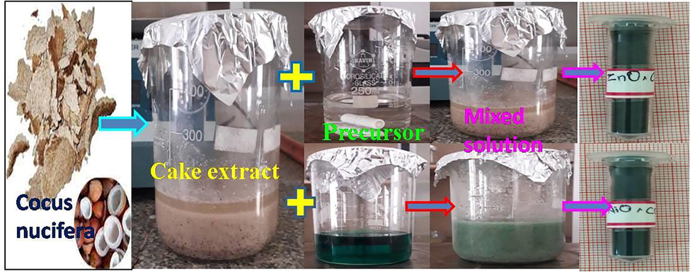
Experimental preparation of Cocus nucifera (L) mediated ZnO and NiO nanoparticles.
4 Results
4.1 Powder x-ray diffraction studies
The powder XRD study was utilized to recognize the shape, crystalline size, grid steady, and to discover the nature of the sample. In this investigation of the sample was recorded with the 2θ value range from 20° to 80° and is given in the Fig. 2(a-b). The (hkl) values were indexed and the prominent peaks were identified. The crystal plane (1 0 0), (0 0 2), (1 0 1), (1 0 3), and (1 1 2) was perfectly matched with the standard JCPDS card (89–1397). The structure was found as hexagonal with primitive lattice structure (Elamathi et al., 2020; Selvarajan and MohanaSrinivasan, 2013). From the calculation based on the lattice parameter the average crystalline size was founded 11.01 nm by using Debye-Scherer formula. Similarly, the (hkl) values of the NiO NPs were indexed and the prominent peaks were identified. The crystal plane (1 1 1), (2 0 0), (2 2 0), (3 1 1), and (2 2 2) was perfectly matched with the standard JCPDS card (78-0429) and the physical structure found to be cubic with FCC structure (DasS et al., 2018). From the calculation based on the lattice parameter the average size of the crystalline was calculated as 16.7036 by using Debye-Scherer formula.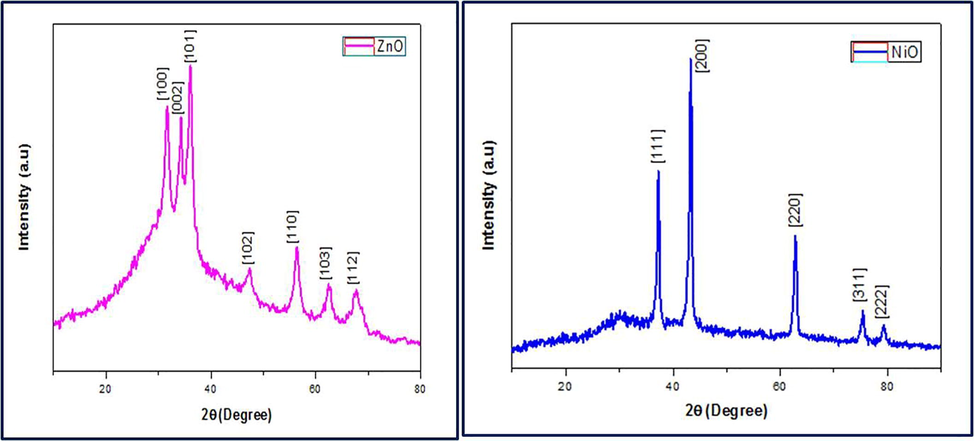
XRD pattern of Cocos nucifera (L) mediated ZnO and NiO nanoparticles.
4.2 FTIR spectroscopic analysis for molecular bonding
The FTIR investigation study was utilized to discover molecular bonding of the phytochemicals, bioactive compounds with the precursor molecule by the absorption of photon energy of the IR radiations (Sudhasree et al., 2014). The spectrum was investigated within the domain range 4500 cm−1 to 500 cm−1. FTIR image Cocos nucifera (L) extract mediated ZnO and NiO nanoparticles as shown in the Fig. 3(a-b). The extreme of the broadband uniting at the reach is 3680 cm−1 is because of the –OH vibration of phenols. Peaks at 2212 cm−1 and 1782 cm−1 was assigned the vibrational of C–H of the protein molecule (Bhuyan et al., 2015). The C-N bonding of aliphatic amines bunch is perceived at 1579 cm−1 and 1098 cm−1. The Zn-O molecular binding was witnessed at 478 cm−1 and 630 cm−1 (IndraPriyatharesini et al., 2020). In the spectrum of NiO, absorption peaks were detected at 3663 cm−1, 3385 cm−1, 1588 cm−1, 1740 cm−1 and these values are assigned to OH vibration of phenolic compounds, H–O–H stretching vibration of the molecules. C-N vibration was observed at 607 cm−1 and the molecular binding confirmation of Ni-O was obtained at 468 cm−1 and 580 cm−1 within the fingerprint region (Gordon et al., 2011).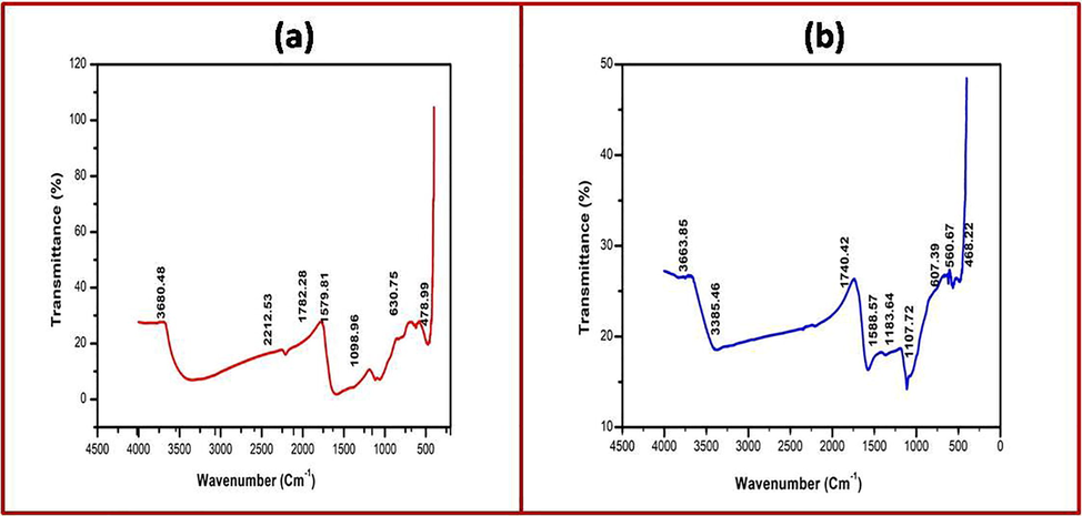
(a-b). FTIR spectrum of Cocus nucifera (L) mediated ZnO and NiO nanoparticles.
4.3 UV–visible spectroscopic studies
The development of zinc oxide and nickel oxide nanoparticles is confirmed by UV–visible spectroscopy investigation. Sample was irradiated with UV-light photons in the range 200 nm to 800 nm at room temperature (Rajith Kumar et al., 2020). Recored UV–visible spectrum and Tauc plot of ZnO and NiO is shown in the Fig. 4(a-b) & Fig. 5(a-b). Both the samples exhibited the absorption in the blue shift region and is mainly attributed to the reason that the decrease in the particles size due to quantum confinement of the photo generated electron and hole carriers caused the absorption of wavelength of radiation in the blue shift (Sudhasree et al., 2014) and the absorption peaks were identified at 395 nm and 308 nm confirmed the formation of metal oxides ZnO and NiO. Based on the UV absorption Tauc plot was drawn for both the samples and the calculated bandgap is 3.15 eV and 3.64 eV for the ZnO and NiO and it is corelated with the previous reports (Pillai et al., 2020).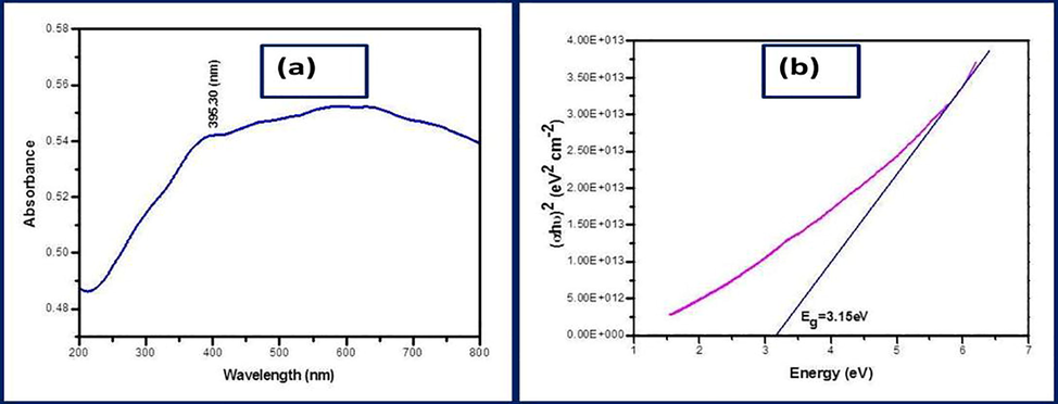
(a-b). UV–visible spectrum and Tauc plot of ZnO nanoparticles.
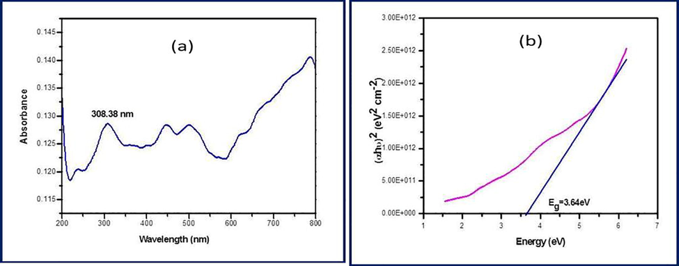
(a-b). UV–visible spectrum and Tauc plot of NiO nanoparticles.
4.4 Surface morphology and elemental composition
The morphological features of cocos nucifera (L) extract mediated zinc oxide and nickel oxide nanoparticles was examined by the scanning electron microscopy ambient. Range and size of the particle distribution was not uniform. The SEM image of both the samples exhibited inhomogeneous distribution of rock shaped and spheroid like particles with few agglomerates and this may be due to bioactive compounds of the cocos nucifera (L) cake interacted with the precursor during preparation effected in the modification of the morphology of the synthesized specimen (Ifeanyichukwu et al., 2020; Govarthanan et al., 2016). Avereage size of ZnO and NiO were calculated as 28 nm to 59 nm and 19 nm to 28 nm respectively. The prepared sample was examined by EDAX analysis and expected elements of Zn and O were present in the atomic ratio 77.67% and 2.33% and there is no impurities in the zinc oxide nanoparticles. Similarly in the case of NiO its about 70.38% of Ni and 29.62% of O were identified without any additional impurities. SEM image and EDAX spectrum of the synthesised samples were given in the Fig. 6(a-d).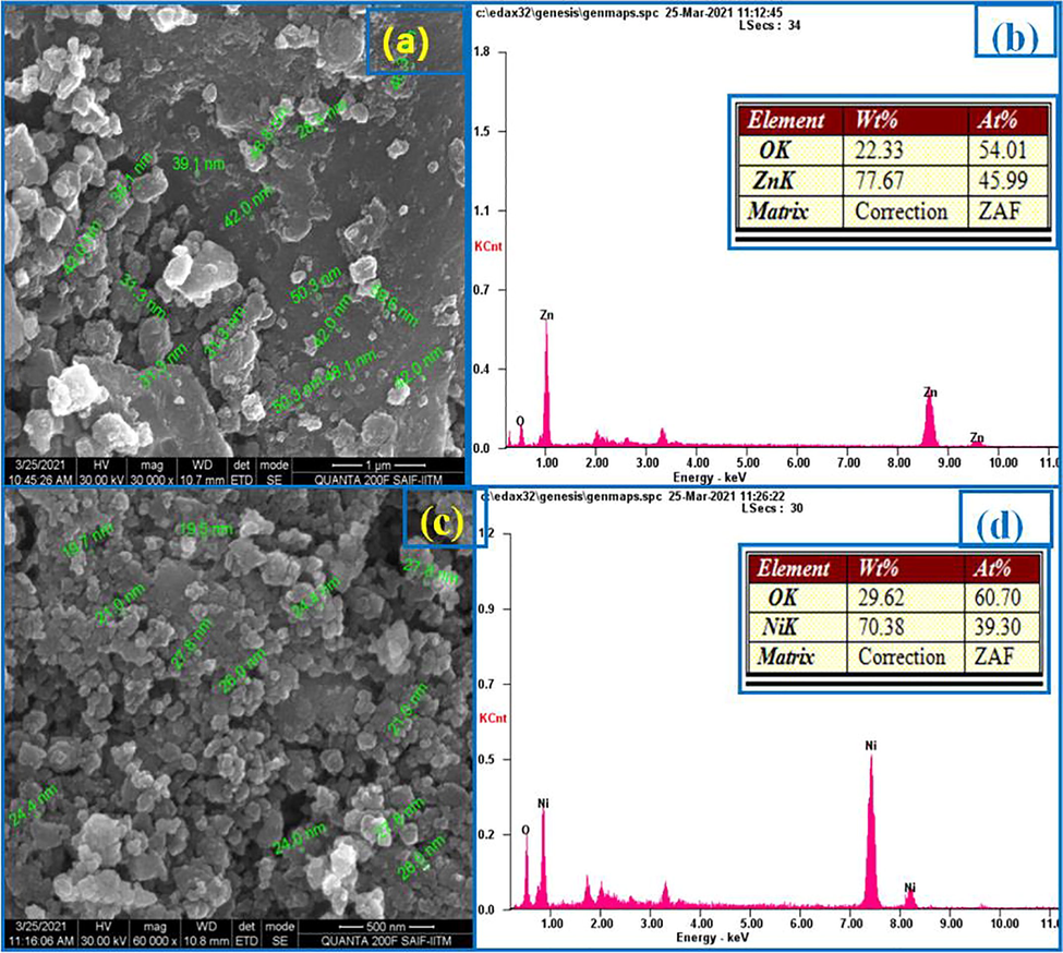
(a-b). SEM and EDAX of ZnO nanoparticles; and (c-d) are SEM and EDAX of NiO nanoparticles.
4.5 Antimicrobial assay
The main aim of the study is to examine the samples antimicrobial activity of the cocos nucifera (L) mediated ZnO and NiO nanoparticles. In general the derivatives which obtained from coconut is a good antibiotic and it has been used for curing many disorders (Lalithamba et al., 2018). Herein we tested the prepared metal oxides with disease causing bacterias by disc diffusion method (Roopan and Elango, 2015). The nanosized particle, easily penetrates the tough and rigid cell wall of bacteria to effects the vital molecular pathways (Elango et al., 2017). Here the sample was tested against human causing pathogens including S.aureus, B.subtilis, E.coli and K.pneumoniae with the reference drug Gentamycin (50 µg). The antibacterial activity of the test samples were carried out by disc diffusion method. The target microorganism were cultured in Nutrient broth and incubated for 24 hrs. The petri dishes containing Mueller Hinton agar (MHA) medium were cultured with diluted bacterial strain. The prepared discs were placed on the culture medium. The diameter of the clear zone around the disc was measured and expressed in millimeters as its antibacterial activity. The results revealed that average activity of the ZnO and NiO over the drug with the zone of inhibition of 8 mm,10 mm, 12 mm and 8 mm against the pathogens. Antimicrobial study of the sample is shown in Fig. 7(a-b) for the ZnO and NiO nanoparticles.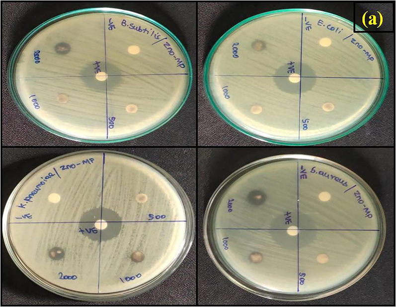
Antimicrobial analysis of ZnO against pathogens.
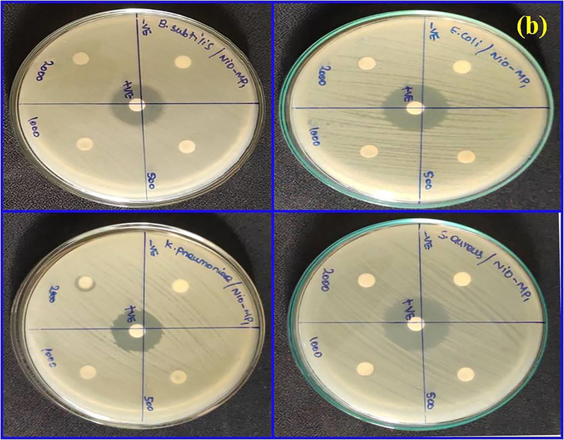
Antimicrobial analysis of NiO against pathogens.
5 Discussion
In this study, we tried to synthesis of metal oxides ZnO and NiO using the deoiled cake residue of the Cocos nucifera (L) by the process of fermentation with a greener approach. The unused deoiled cakes samples were collected, and it was fermented for 48 h for preparing the extracts. Cocos nucifera contains vitamins, minerals, bioactive compounds, antioxidant materials (Mariselvam et al., 2014). During the deoiling process the edible part of the seed is being crushed by mechanical means and the extracts was isolated as milk and oil according to our need. The milk and oil found to have medicinal properties and it can be mixed with other medicines to make embrocation’s. The composition of the oil contains the compounds like capric acid, caprylic acid, caproic acid, lauric acid, myristic acid, palmitic acid etc. Similarly, the edible part and the milk contains much more nutrients like proteins, carbohydrates etc., (Santos et al., 2013; Parasuraman et al., 2019). All these essential ingredients will be removed during the process of deoiling which involves boiling, heating, and pressurizing the edible parts and thus the cake residue is obtained as a byproduct and it have been used as a feed for the livestock in general. The high light of this experimental study is we synthesized the metal oxides by the process of fermentation of the cake residue. Once the oil was extracted from the edible part of any seeds like peanut, coconut, sesame etc., then it will be considered as a waste, and it contains only a minimum nutrient and may be used as feed for the cattles or as a fertilized in general. Here in this study the cake residue was fermented in a suitable condition in a dark and covered container was activated by the enzymes which modifies the nutrient level and the potential of the available nutrients and then the extract was filtered and used directly for the preparation of the metal oxides which acts as a bio reducing agent and hence the mechanism of the antibacterial activity was found to be present and moderate against the human pathogens even after the removal of the nutrients of the Cocus nucifera (L) (Naskar et al., 2013; Uddin et al., 2020; Renuka et al., 2020; Siddhardha et al., 2020). The graphical abstract of the mechanism is given in the below Fig. 8.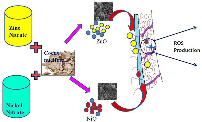
Mechanism of metal oxides ZnO and NiO using the deoiled cake residue of the Cocos nucifera (L) by the process of fermentation with a greener approach.
6 Conclusion
In this experimental work are successfully synthesized ZnO and NiO nanoparticles by using the green synthesis method of an eco-friendly approach by utilizing Cocus nucifera (L) seed cake extracts. The biomolecules present in the coconut deoiled cake extract act as the stabilizing agent formation of the ZnO and NiO nanoparticles. The physical structure of ZnO nanoparticle is a hexagonal system and primitive lattice structure and NiO nanoparticle is cubic system and face-centered lattice structure revealed by X-ray diffraction analysis. The FTIR investigation showed the functional groups, the optical bandgap energy was based on the UV studies. Morphological studies and elemental composition were carried out using SEM and EDAX instrumentation. Antimicrobial studies of the samples against bacteria revealeda moderate zone of inhibition with respect to standard drug gentamycin and this may be due to the reduced nutrients of the cake residue which modified the activity of the metal oxides.
Acknowledgments
The authors extend their appreciation to the Researchers supporting project number (RSP-2021/189), King Saud University, Riyadh, Saudi Arabia.
Declaration of Competing Interest
The authors declare that they have no known competing financial interests or personal relationships that could have appeared to influence the work reported in this paper.
References
- Ajayan, Hebsur NS: Green synthesis of Zinc oxide nanoparticle using Neem (Azadirachtaindica) and Tulsi (ocimumTenuiflorum) leaf extract and their characterization. Int. J. Curr. Microbiol. App. Sci.. 2020;9:277-285.
- [Google Scholar]
- Structural and optical properties of nickel oxide nanoparticles: Investigation of antimicrobial applications. Surface Interfaces. 2020;18:100460.
- [CrossRef] [Google Scholar]
- Green synthesis of NiO nanoparticles using Aeglemarmelos leaf extract for the evaluation of in-vitro cytotoxicity, antibacterial and photocatalytic properties. J. Photochemistry Photobiology, B: Biology. 2018;180:30-59.
- [Google Scholar]
- Green and eco-friendly synthesis of nickel oxide nanoparticles and its photocatalytic activity for methyl orange degradation. J. Mater. Sci.: Mater. Electron.. 2020;31(14):11303-11316.
- [Google Scholar]
- Bhuyan T, Mishra K, Khanuja M, Prasad R, Verma A: Biosynthesis of ZnONps from Azadirachtaindicafor antibacterial and photocatalytic applications. Mater Sci Semicond Process 2015,32:55–61.
- Biosynthesis of zinc oxide nanoparticles from Azadirachtaindica for antibacterial and photocatalytic application. Mater. Sci. Semiconductor Processing. 2015;32:55-61.
- [Google Scholar]
- Biosynthesis and characterization of silver nanoparticles for prospective application in food packaging and biomedical fields. Mater. Chem. Phys.. 2020;250
- [Google Scholar]
- Sol-gel synthesis, characterization and neurotoxicity effect of Zinc oxide nanoparticles using gum traganth. Ceram Int. 2013;40:4827-4831.
- [Google Scholar]
- Das S, Chatterjee S, Pramanik S, Devi PS, Kumar GS: A new insight into the interaction of ZnO with calf thymus DNA through surface defects. J Photochem Photobiol B 2018,178:339–47.
- Novel synthesis and structural analysis of zinc oxide nanoparticles for the non-enzymatic glucose biosensor. Mater. Sci. Eng. C. 2017;75:1472-1479.
- [Google Scholar]
- Investigation of structural and electrical properties of lithium cobalt oxide nanoparticles for optoelectronic applications. Surfaces Interfaces. 2020;20:100582.
- [CrossRef] [Google Scholar]
- Cocos nucifera coir-mediated green synthesis of Pd NPs and its investigation against larvae and agricultural pest. Artif. Cells Nanomed. Biotechnol.. 2017;45(8):1581-1587.
- [Google Scholar]
- Green synthesis, characterization and antimicrobial activities of zinc oxide nanoparticles from the leaf extract of Azadirachtaindica (L.) Appl. Surf. Sci.. 2015;345:329-336.
- [Google Scholar]
- Green synthesis of NiO nanoparticles using MoringaOleifera extract and their biomedical applications: cytotoxicity effect of nanoparticles against HT-29 cancer cells. J. Photochem. Photobiology B: Biology. 2016;164:352-360.
- [Google Scholar]
- V, Rubia F, Borja M: Effects of chemical and thermochemical pre-treatments on sunflower oil cake in biochemical methane potential assays. J. Chem. Technol. Biotechnol.. 2013;88(5):924-929.
- [Google Scholar]
- Synthesis and characterization of zinc/iron oxide composite nanoparticles and their antibacterial properties. Colloids Surf. A: Physicochemical and Engineering Aspects. 2011;374(1-3):1-8.
- [Google Scholar]
- Low-cost and eco-friendly synthesis of silver nanoparticles using coconut (Cocos nucifera)oil cake extract and its antibacterial activity. Artif. Cells Nanomed. Biotechnol.. 2016;44(8):1878-1882.
- [Google Scholar]
- Z, Luting Hu: Synthesis of well dispersed NiO ink for efficient perovskite solar cells. J. Alloy. Compd.. 2021;860:157889.
- [CrossRef] [Google Scholar]
- Green synthesized photochemically (Zingiberofficinale and Allium sativum) Reduced Nickel oxide nanoparticles confirmed Bactericidal and catalytic potential. Nanoscale Res. letters. 2020;15:50.
- [Google Scholar]
- Neem leaves mediated preparation of NiO nanoparticles and its magnetization, coercivity and antibacterial analysis. Results Phys.. 2016;6:712-718.
- [Google Scholar]
- Green Synthesis of Zinc Oxide Nanoparticles from Pomegranate (Punicagranatum) Extracts and Characterization of Their Antibacterial Activity. Molecules. 2020;25(19):4521.
- [Google Scholar]
- Synthesis of Zinc Oxide Nanoparticle using Cocos nucifera male flower extract and analysis their antimicrobial activity. Res. J. Pharm. Tech.. 2020;13:2151-2154.
- [Google Scholar]
- Isa khan M, Nawaz M, Bilal Tahir M, Iqbal T, Pervaiz M, Refique M,Aziz F, Yonas U, Alrobei H:Synthesis, characterization and antibacterial activity of NiO against pathogen. Inorganic chemistry communication 2020,122:108300.
- Photodegradation of organic pollutants rhb dye using UV simulated sunlight on ceria based TiO2 nanomaterials for antibacterial applications. Sci. Rep.. 2016;6(1):1-12.
- [Google Scholar]
- Elucidation of photocatalysis, photoluminescence and antibacterial studies of ZnO thin films by spin coating method. J. Photochem. Photobiol., B. 2017;173:466-475.
- [Google Scholar]
- High performance of pyrochlore like Sm2Ti2O7 heterojunction photocatalyst for efficient degradation of rhodamine-B dye with waste water under visible light irradiation. J. King Saud University-Sci.. 2020;32(2):1516-1522.
- [Google Scholar]
- Capsicum annuum Fruit Extract: A Novel Reducing Agent for the Green Synthesis of ZnO Nanoparticles and Their Multifunctional Applications. Acta ChimicaSlovenica 2018:354-364.
- [CrossRef] [Google Scholar]
- Biosynthesis of Nickel oxide Nanoparticles from Euphorbia heterophylla (L.) and their biological application. Arabian J. Chem.. 2020;13(3):4712-4719.
- [Google Scholar]
- Improved photocatalytic decomposition of aqueous Rhodamine-B by solar light illuminated hierarchical yttria nanosphere decorated ceria nanorods. J. Mater. Res. Technol.. 2019;8(3):2898-2909.
- [Google Scholar]
- Evaluation on La2O3 garlanded ceria heterostructured binary metal oxide nanoplates for UV/ Visible light induced removal of organic dye from urban wastewater. S. Afr. J. Chem. Eng.. 2018;26:49-60.
- [Google Scholar]
- RanjitsinghAJA, Usha Raja NanthiniA, KalirajanK, Padmalatha C, MosaeSelvakumar P: Green synthesis of silver nanoparticles from the extract of theinflorescence of Cocos nucifera (Family: Arecaceae) for enhancedantibacterial activity. SpectrochimicaActa Part A: Molecular and Biomolecular Spectroscopy. 2014;129:537-541.
- [Google Scholar]
- Green synthesis of zinc oxide nanoparticles using fresh peels extract of Punicagranatum and its antimicrobial activities. Int. J. Pharma Res. Health Sci.. 2015;3:694-699.
- [Google Scholar]
- Evaluation of antinociceptive and anti-inflammatory activity of hydro methanol extract of Cocos nucifera. J. Inflammopharm.. 2013;21:31-35.
- [Google Scholar]
- Studies of MnO2/g-C3N4 hetrostructure efficient of visible light photocatalyst for pollutants degradation by sol-gel technique. Surf. Interfaces. 2020;20:100512.
- [CrossRef] [Google Scholar]
- Synthesis and antimicrobial photodynamic effect of methylene blue conjugated carbon nanotubes on E. coli and S. aureus. Photochem. Photobiol. Sci.. 2019;18(2):563-576.
- [Google Scholar]
- Piper nigrumLeaf and Stem Assisted Green Synthesis of Silver Nanoparticles and Evaluation of Its Antibacterial Activity Against Agricultural Plant Pathogens. The Scientific World Journal. 2014;2014:1-9.
- [Google Scholar]
- VS, Rahdar A, Joseph J, Sadeghfar F, Anuf AR, Rajesh K, Kyzas, GZ: Green synthesis and characterization of zinc oxide nanoparticles with antibacterial and antifungal activity. J. Mol. Struct.. 2020;1211:128107.
- [CrossRef] [Google Scholar]
- Green synthesis of ZnO nanoparticles using Carica papaya leaf extracts for photocatalytic and photovoltaic application. J. Material Sci.: Mater. Electron.. 2017;28:10374-10381.
- [Google Scholar]
- Photocatalytic, nitrite sensing and antibacterial studies of facile bio-synthesized nickel oxide nanoparticles. J. Sci.: Adv. Mater. Devices. 2020;5(1):48-55.
- [Google Scholar]
- Rajiv P, Rajeshwari S, Venckatesh R: Bio-Fabrication of ZnONps using leaf extract of Partheniumhysterophorus L. and its size-dependent antifungal activity against plant fungal pathogens. Spectrochim Acta Part A Mol. Biomol. Spectrosc. 2013,112:384–387.
- R. Ramesh, V. Yamini, S.J. Sundaram, F.L.A. Khan, K. Kaviyarasu, Investigation of structural and optical properties of NiO nanoparticles mediated by Plectranthusamboinicus leaf extract, Materials Today: Proceedings 36, 268-272.
- R Ramesh, G Catherine, SJ Sundaram, FLA Khan, K Kaviyarasu, Synthesis of Mn3O4 nano complex using aqueous extract of Helianthus annuus seed cake and its effect on biological growth of Vigna radiata, Materials Today: Proceedings 36, 184-191.
- Biosynthesis of silver nanoparticles using Phyllanthus emblica fruit extract for antimicrobial application. Biocatalysis Agric. Biotechnol.. 2020;24:101567.
- [CrossRef] [Google Scholar]
- Exploitation of Cocos nucifera a non-food toward the biological andnanobiotechnology field. Ind. Crops Prod.. 2015;67:130-136.
- [Google Scholar]
- Evaluation of chemical constituents and antioxidant activityof coconut water (Cocusnucifera L.) and caffeic acid in cell culture. Anais daAcademia Brasileira de. Ciencias. 2013;85(4):1235-1247.
- [Google Scholar]
- Biosynthesis and characterization of ZnO nanoparticles using Lactobacillus plantarum. Mater. Lett.. 2013;112:180-182.
- [Google Scholar]
- Karanja (MilletiaPinnata (L.) panigrahi): a tropical tree with varied applications. Phytochem. Rev.. 2020;19(3):643-658.
- [Google Scholar]
- Chrysin-loaded chitosan nanoparticles potentiates antibiofilm activity against Staphylococcus aureus. Pathogens. 2020;9(2):115.
- [CrossRef] [Google Scholar]
- HumaZ, Shahnaz G, Qureshi O S, Khalid Q, Mirza S, Webster TJ, Hussain I: Green Synthesis of Zinc Oxide Nanoparticles by Neem Extract as Multi-facet Therapeutic Agents. J. Drug Delivery Sci. Technol.. 2020;59:101911.
- [CrossRef] [Google Scholar]
- Sudhasree S, ShakilaBanu A, Brindha P, Kurian GA:Synthesis of nickel nanoparticles by chemical and green routeand their comparison in respect to biological effect and toxicity, Toxicological and Environmental Chemistry 2014,96:743–754.
- SoundaryaTL, Udayabhanu B: Multifunctional applications of nickel oxide (NiO) nanoparticles synthesized by facile green combustion method by using, LimoniaAcidissima natural fruit juice. Inorganica Chem Acta. 2021;515:120059
- [Google Scholar]
- SharmaSC: Artocarpusgomezianus aided green synthesisofZnO nanoparticles: Luminescence, photocatalytic and antioxidant properties. SpectrchimiaActa Part A: Molecular and, Biomolecular Spectroscopy. 2015;141:128-134.
- [Google Scholar]
- Impact of the changes in bacterial outer membrane structure on the anti-bacterial activity of zinc oxide nanoparticles. J. Nanopart. Res.. 2020;22(2)
- [CrossRef] [Google Scholar]
- VenkatachalamM, SureshS: Enhanced bactericidal performance of nickel oxide-zinc oxide nanocomposites synthesized by facile chemical co-precipitation method. J. Alloy. Compd.. 2020;830:154642.
- [CrossRef] [Google Scholar]
- Green synthesis of ZnO nanoparticle using Prunus dulcis (Almond Gum) for antimicrobial and supercapacitor applications. Surf. Interfaces. 2019;17:100376.
- [CrossRef] [Google Scholar]
- Synthesis of Zinc oxide nanoparticles and its conjugation with antibiotic: antibacterial and morphological characterization.Environmental Nanotechnology. Monitoring Manage.. 2020;14:100391.
- [CrossRef] [Google Scholar]
- Rahat K:CocosnuciferaLeaf extract mediated green synthesis of silver nanoparticles for enhanced antibacterial activity. Journal of inorganic and organometalic polymers and materials. 2020;30(9):3305-3316.
- [Google Scholar]
- Nurul Nadia Mohd Zorkipli, Noor HaiderMohdKaur, Ahamed Azmin Mohamad: Synthesis of NiO nanoparticles through sol-gel method. Procedia chemistry 2016,19: 626-631.


