

Translate this page into:
Biogenic synthesis of silver nanoparticle using Cissus quadrangularis extract and its invitro study
⁎Corresponding authors at: School of Chemical and Energy Engineering, Faculty of Engineering, Universiti Teknologi Malaysia, Skudai 81310, Johor, Malaysia (Nur Izyan Wan Azelee). kanimozhi.vs@gmail.com (S. Kanimozhi), nur.izyan@utm.my (Nur Izyan Wan Azelee)
-
Received: ,
Accepted: ,
This article was originally published by Elsevier and was migrated to Scientific Scholar after the change of Publisher.
Peer review under responsibility of King Saud University.
Abstract
Objective
Green synthesis of metallic and metal oxide nanomaterial has received significant importance in various fields. Green synthesis of silver nanoparticles was performed using aqueous extracts of Cissus quadrangularis. Aqueous extracts of plants plays a major role to reduce the silver nitrate to silver nanoparticles and also aid as surface stabilizing material for silver nanoparticles production.
Methods
The synthesized nanoparticles was monitored by physicochemical techniques such as XRD, FT-IR, Uv–Vis, and SEM. Anti-arthritic activity of silver nanoparticles was examined by Egg albumin denaturation and albumin (BSA) denaturation method.
Results
XRD, FT-IR and Uv–Vis spectrum confirms crystalline nature of silver nanoparticles. SEM images show the spherical shape of the nanoparticles. Egg albumin denaturation method results revealed 81% inhibition and bovine serum albumin method shows 85% inhibition than standard drug.
Conclusion
It confirms that, the green synthesized silver nanoparticles would be useful in promoting research aiming at the development of new agent for arthritis applications. Hence, the utilization of natural harmless materials like plant extracts for nanoparticles synthesis offers various advantages like eco-friendliness and compatibility for drug and biomedical applications.
Keywords
Nanotechnology
Silver nanoparticles
Cissus quadrangularis
Anti-arthritic activity

1 Introduction
Nanotechnology has emerged worldwide of modern potentialities by fabricating nanomaterial with suitable size and shapes for various applications including medicine and agriculture field (Hasan, 2015; Khalil et al., 2013; Aref and Salem 2020; Badineni et al., 2021). Synthesis of nanoparticle has been extended to global attention due to their physicochemical properties. Various methods are used for nanoparticles synthesis. Among these physical and chemical mediated methods are toxic, expensive and dangerous to environment. This problem can be overcome by using nontoxic, economic and eco-friendly greener method (Dhuper et al., 2012; Anand et al., 2017). Therefore green synthesis method was adopted for synthesis of nanomaterial by plants and microorganism. Synthesis of nanoparticle from plants gives a special advantage compared to microorganism due to presence of various biomolecules in the plants which are responsible for stabilization and act as capping agents for nanoparticle synthesis (Iravani, 2011; Anand et al., 2019; Anand et al., 2020). Synthesis of silver nanoparticles using different plant extracts of Ficus carica, Cocous nucifera, Moringa oleifera, Carica papaya, Eucalyptus hybrid, Allium cepa, Aloe vera and Basella alba have been successful and also exhibited various significant biological activities (Geetha et al., 2014; Mariselvam et al., 2014; Prasad and Elumalai, 2011; Jain et al., 2009; Mani et al., 2021a; Dubey et al., 2009; Chandran et al., 2006; Ahmed et al., 2016; Mani et al., 2021b). Cissus quadrangularis is a vining plant which is found in India and also Africa. Cissus quadrangularis comes under the family of Vitaceae and also called as Vitis quadrangularis has been used medicinal purposes for centuries (Patil et al., 2017). It has been used for treating various diseases including helminthic, dyspeptic, ulcer, tonic seizure, irregular menses and respiratory diseases (Patil et al., 2017; Oben et al., 2007). Cissus quadrangularis was used as a reducing material for synthesizing zinc oxide nanoparticles that demonstrated potential inhibitory activity against various microbes like Pseudomonas aeruginosa, Eschericha coli, Acetobacter baumannii, Bacillus cereus, Mycobacterium smegmatis (Sathappan et al., 2021). Synthesis of silver nanoparticles from Cissus quadrangularis stem extract showed antibacterial activity (Kalpana et al., 2017a; 2017b). Valli and vaseeharan (2012) synthesized silver nanoparticles from Cissus quadrangularis that significantly inhibited the development gram positive and negative bacteria at low inhibitory concentrations of 10 μg/ml. Cissus quadrangularis mediated CaO nanoparticles has been examined for antibacterial activity. Highest zone of inhibition was obtained for Eschericha coli than other bacterial strains of Vibrio cholerae, Pseudomonas aeruginosa, Staphylococcus aureus, Klebsiella pneumoniae, Shigella dysenteriae and Salmonella sp. (Marquis et al., 2016). Among the other metal nanoparticles, silver nanoparticles plays a major role in various activities such as antibacterial, antioxidant, antifungal and cytotoxic activities. The silver nanoparticles were examined for different applications especially in medical fields such as photosensitizers, radio sensitizers, antiviral agents, and used as therapeutic agents to treat the skin, oral, breast, lung, and leukemia cancer due to their intrinsic therapeutic properties (Kumar et al., 2014; Wei et al., 2015; Nakkala et al., 2014). Rheumatoid Arthritis is an immune system illness characterized by chronic joint inflammation, infiltration of immune cells, destruction in bones and cartilage. There are several studies which proposed that, macrophages, B cells and T cells contribute to severe destruction in joints and damages occur in tissues by releasing various prostanoids and cytokines (Al-Zifzaf et al., 2015). A previous study reported that 82% of inhibition was obtained on BSA for anti-arthritic activity of zinc oxide nanoparticles prepared using Lagenaria siceraria extracts (Kalpana et al., 2017a). Luffa acutangula peel extract mediated ZnO nanoparticles were tested for their anti-arthritic property. The studies revealed that ZnO nanoparticles showed anti-arthritic activity by efficiently inhibiting the denaturation of both albumin and bovine serum albumin (Ananthalakshmim et al., 2021). Studies of Senthilkumar et al., 2017 suggested that, maximum inhibition of 90% was achieved for anti-arthritic activity of ZnO nanoparticles from leaf extracts of Tectona grandis. Hence, in the present study, aqueous extracts of Cissus quadrangularis was used as a reducing material for silver nanoparticles synthesis. The synthesized silver nanoparticles were monitored by XRD, FT-IR, Uv–Vis, and SEM techniques. The activity of silver nanoparticles was evaluated for anti-arthritic studies. This invitro anti-arthritic activity was determined by egg albumin and bovine serum protein (BSA) denaturation method.
2 Materials and methods
2.1 Synthesis of silver nanoparticles
Green synthesis of silver nanoparticles was performed using stem extracts of Cissus quadrangularis. Young stems of Cissus quadrangularis were washed and crushed with 100 ml of deionised water to prepare the extract. 5 mM silver nitrate solution was added to extract of Cissus quadrangularis. The solution was stirred in a magnetic stirrer for 10 h at room temperature. It was centrifuged to collect the silver nanoparticles.
2.2 Characterization
XRD analysis was performed using X-ray 7000, Shimadzu to confirm the amorphous/crystalline character of the materials. Diffraction patterns were measured using Cu, K-alpha radiation at 30 kV and 30 mA in, with 2θ range of 0° to 80°. The synthesized nanoparticle was characterized using Shimadzu FTIR spectrometer. Diffuse reflectance FTIR spectrum for silver nanoparticles was measured from 4000 to 400 cm−1. Surface morphology of nanoparticles was obtained from scanning electron microscopy (SEM). SEM analysis was performed using JEOL JM 5600 equipped with 20 kV voltage and wavelength range of 5–6 nm. UV–Vis spectrum was obtained from Jasco V650 Vis spectrophotometer with the wavelength ranging from 200 to 600 nm.
2.3 Anti-arthritic activity
The in-vitro study of the synthesized silver nanoparticles was tested by anti-arthritic activity. It was determined by two methods such as BSA denaturation and albumin denaturation method.
2.3.1 Albumin denaturation
Eggs albumin, phosphate buffered saline (PBS, pH6.4) were added to 1 ml of different concentrations of samples (100–500 µg) except blank. It was incubated and heated at 70 °C. Then the absorbance of the solution was measuresd at 660 nm. The protein denaturation (% inhibition) of the sample was calculated.
2.3.2 BSA denaturation
Different concentrations of samples (100–500 µg), phosphate buffer and 0.5% BSA were added in the test tubes and incubated in the incubator for 20 min at 37 °C. Absorbance was measured at 255 nm.
3 Results and discussion
3.1 XRd
Fig. 1 specifies the XRD patterns for silver nanoparticles from Cissus quadrangularis extract. XRD pattern revealed crystalline nature of silver nanoparticles with Bragg reflection 2θ values of 38.18°, 44.37°, 66.46° and 77.44° which could be indexed in (1 1 1), (2 0 0), (2 2 0), and (3 1 1) planes respectively. This confirmed the crystalline nature of silver nanoparticles. The same experimental results were reported by Nalvothu et al. (2014). The data was compared with JCPDS file no. 87-0720 which was good agreement with standard values. A high intensity diffraction peak at 38° which was indexed as (1 1 1) plane indicated the crystalline silver nanoparticles. Non indexed peak at 32° and 46° were related to bio organic molecules crystallization on the surface of nanoparticle (Gomathi et al., 2017; Mallikarjuna et al., 2014). The crystallite sizes of the nanoparticle are calculated by Debye- Scherrer formula.
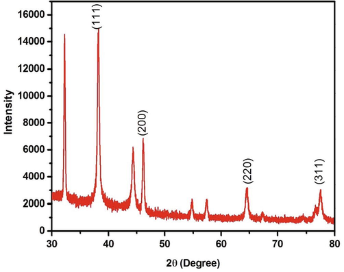
XRD pattern of Silver Nanoparticle from Cissus Quadrangularis Extract.
λ represents X-ray wavelength, β represents diffraction peak of full width at half maximum (FWHM) and θ represents the Braggs reflection peak. Particle size of the silver nanoparticles was calculated and tabulated (Table1). The average crystallite size of silver nanoparticles was 24 nm.
Miller indices (hkl) Plane
Peak position 2θ value
Height
width
Crystallite size
(1 1 1)
38.18
11564.25
0.37067
24.97
(2 0 0)
44.37
254.89
0.00339
28.68
(2 2 0)
66.46
1944.53
0.44756
22.23
(3 1 1)
77.48
1744.65
0.51389
20.15
3.2 FT-IR spectroscopy
FTIR spectrum of silver nanoparticles synthesized from Cissus quadrangularis is depicted in Fig. 2. FTIR spectrum was used to find the functional groups present in the nanoparticles. Broad band at 3378 cm−1 was related to water molecules of O–H stretching vibration which might be due to presence of phenols and alcohols; stretching vibration of C–H was found in the range of 2923 cm−1 arisen from aromatic compounds. The band at 1735 cm−1 attributed to C⚌O stretching vibration of carboxylic acid or ester, strong band at 1622 cm−1 was assigned to bending vibration of amide group in proteins. Lower bands occur at 1365 cm−1 and 1054 cm−1 denoted CH2 alkanes and stretching of C-O ester or ether in the silver nanoparticles (Raja et al., 2017).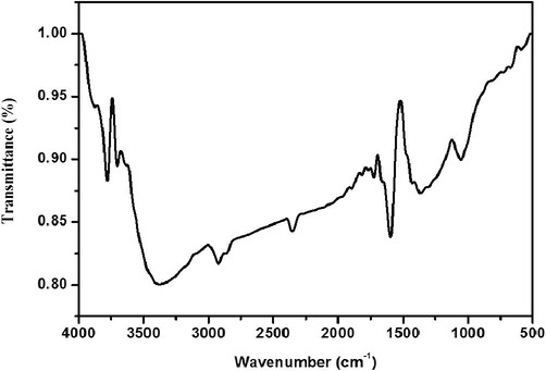
FTIR spectrum of Silver nanoparticle from Cissus Quadrangularis plant extract.
3.3 UV–Vis spectroscopy
Fig. 3 shows the UV–Vis spectrum of Cissus quadrangularis mediated silver nanoparticles. The metal nanoparticle contains free electron that provides surface plasmon resonance (SPR) absorption band which is due to the combination of electrons with light wave. A yellowish-brown colour can be evidenced for the reduced nature of silver ions from silver nitrate. The silver SPR band appeared in the range of 410 nm which confirmed the successful synthesis of silver nanoparticles.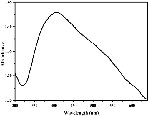
Uv–Vis spectrum of Silver nanoparticle from Cissus Quadrangularis aqueous extract.
3.4 Scanning electron microscopy
Morphological characteristics of synthesised silver nanoparticles were monitored using SEM (Fig. 4). SEM images of nanoparticles showed the spherical shape of poly dispersed nanoparticles with agglomeration. Agglomeration of nanoparticles might be due to presence of Cissus quadrangularis extracts.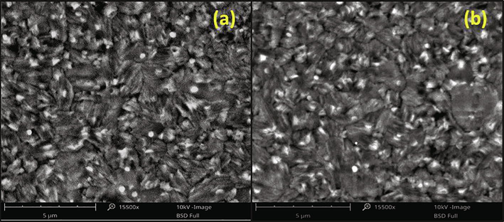
SEM images of Silver nanoparticle from Cissus Quadrangularis plant extract.
3.5 Evaluation of anti-arthritic activity
Synovial disease of arthritis is characterized by chronic inflammation in the joints and it could lead to destruction in the joints (Jayaprakasam and Ravi, 2012). Arthritic patients have the common problem of inflammation that occurs in the living tissues. Inflammation is caused by denaturation of protein. Steroids can decrease the inflammation in the tissues and also reduce immune activity which is responsible for inflammation. The anti-arthritic activities of silver nanoparticles of various concentrations 25, 50, 100, 150 and 200 μg/ml, were determined by albumin denaturation and BSA denaturation method. The anti-arthritic activity of synthesized samples of silver nanoparticles by albumin denaturation and BSA denaturation method are shown in Figs. 5 and 6 respectively.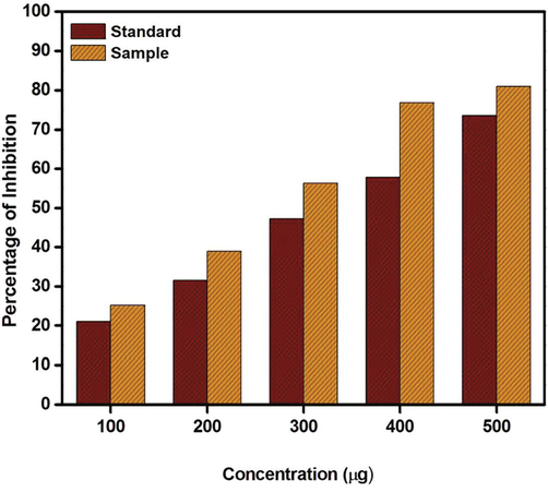
Anti-arthritic activity of Silver nanoparticle by albumin denaturation method (Standard- Diclofenac sodium drug and sample- Silver nanoparticle).
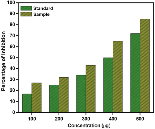
Anti-arthritic activity of Silver nanoparticle by BSA denaturation method (Standard- Diclofenac sodium drug and sample- Silver nanoparticle).
The silver nanoparticles displayed 25, 39, 56, 77, and 81% inhibition of denaturation of albumin and the standard powerful non-steroidal Diclofenac sodium drug (anti-inflammatory drug) exhibited 21, 31, 47, 57, and 73% inhibition of denaturation of albumin. Inhibition of protein denaturation by silver nanoparticles showed better result compared to reference standard drug. The present investigation demonstrated 81% inhibition of egg albumin denaturation and 85% inhibition of bovine serum albumin. Protein denaturation was decreased by increasing concentrations of silver nanoparticles. Therefore, the result showed that silver nanoparticles can inhibit the denaturation of BSA and albumin (Senthilkumar et al., 2017). Hence, the present study revealed that, silver nanoparticles are capable of inhibiting the denaturation of albumin and BSA in Rheumatic arthritic diseases.
4 Conclusion
A simple, eco-friendly, economic, non-toxic greener method was used for this study. Aqueous extracts of Cissus quadrangularis acted as stabilizing and reducing agents for synthesis of silver nanoparticles. The average size of the Silver nanoparticles was 24 nm and the assistance of plant extract in the reduction of silver ions to silver nanoparticles was evident from the functional groups detected in the FTIR spectrum. Uv–Vis spectrum displayed strong absorption band at 410 nm which confirmed the reduction of silver nanoparticles. SEM images displayed the spherical shape of the nanoparticles. The silver nanoparticles exhibited a remarkable efficacy for anti-arthritic activity. Hence, the result obtained from this study will be assisting the pathway to extend the performance of green synthesized silver nanoparticles from Cissus quadrangularis extracts in arthritis investigations and develop appropriate drugs.
Acknowledgements
The authors acknowledge the funding support from Researchers Supporting Project Number (RSP2022R465), King Saud University, Riyadh, Saudi Arabia for this research work.
Declaration of Competing Interest
The authors declare that they have no known competing financial interests or personal relationships that could have appeared to influence the work reported in this paper.
References
- A review on plants extract mediated synthesis of silver nanoparticles for antimicrobial applications: A green expertise. J. Adv. Res.. 2016;7(1):17-28.
- [Google Scholar]
- FoxP3+T regulatory cells in Rheumatoid arthritis and the imbalance of the Treg/TH17 cytokine axis. Egypt. Rheumatol.. 2015;37(1):7-15.
- [Google Scholar]
- Green synthesis of ZnO nanoparticle using Prunus dulcis (Almond Gum) for antimicrobial and supercapacitor applications. Surf. Interfaces. 2019;17:100376.
- [CrossRef] [Google Scholar]
- Structural and optical properties of nickel oxide nanoparticles: Investigation of antimicrobial applications. Surf. Interfaces. 2020;18:100460.
- [CrossRef] [Google Scholar]
- Biosynthesis of silver nanoparticles using agroforestry residue and their catalytic degradation for sustainable waste management. J. Cluster Sci.. 2017;28(4):2279-2291.
- [Google Scholar]
- Evaluation of Anti-inflammatory and Anti-arthritic activity of Luffa acutangula peel extract mediated ZnO nanoparticles. Res. J. Pharm. Tech.. 2021;14(4):2004-2008.
- [Google Scholar]
- Bio-callus synthesis of silver nanoparticles, characterization, and antibacterial activities via Cinnamomum camphora callus culture. Biocatal. Agricul. Biotechnol.. 2020;27:101689.
- [CrossRef] [Google Scholar]
- Effect of PVA/PVP protective agent on the formation of silver nanoparticles and its photo catalytic and antimicrobial activity. Mater. Today:. Proc.. 2021;36:121-125.
- [Google Scholar]
- Synthesis of gold nanotriangles and silver nanoparticles using Aloe vera plant extract. Biotechnol. Prog.. 2006;22(2):577-583.
- [Google Scholar]
- Green synthesis and characterization of zero valent iron nanoparticles from the leaf extract of Mangifera indica. Nano Trends: J Nanotech App.. 2012;13(2):16-22.
- [Google Scholar]
- Green synthesis of nanosilver particles from extract of Eucalyptus hybrida (safeda) leaf Dig J. Nanomater. Biostruct.. 2009;4:537-543.
- [Google Scholar]
- Green synthesis of silver nanoparticles using Cymbopogan Citratus(Dc) Stapf. Extract and its antibacterial activity. Aus. J. Basic Appl. Sci.. 2014;8(3):324-331.
- [Google Scholar]
- Green synthesis of silver nanoparticles using Datura stramonium leaf extract and assessment of their antibacterial activity. Resour.-Effic. Technol.. 2017;3(3):280-284.
- [Google Scholar]
- A Review on Nanoparticles: Their Synthesis and Types. Res. J. Recent. Sci.. 2015;4:1-3.
- [Google Scholar]
- Green synthesis of metal nanoparticles using plants. Green Chem.. 2011;13(10):2638.
- [CrossRef] [Google Scholar]
- Synthesis of plant-mediated silver nanoparticles using papaya fruit extract and evaluation of their antimicrobial activities. Dig. J. Nanomater. Biostruct.. 2009;4:557-563.
- [Google Scholar]
- Evaluation of anti-arthritic activity of the root extract of Acalypha indica linn. using In vitro techniques. Int. J. Phyto Pharm.. 2012;2:169-173.
- [Google Scholar]
- Lagenaria siceraria aided green synthesis of ZnO NPs: Anti-dandruff, Anti-microbial and Anti-arthritic activity. Res. J. Chem. Environ.. 2017;21(11):14-19.
- [Google Scholar]
- Green synthesis of zinc oxide nanoparticles using fresh stem of Cissus quadrangularis extract and its various invitro studies. Asian J. Chem.. 2017;29(6):1323-1327.
- [Google Scholar]
- Preparation and characterization of electrospun PLGA/silver composite nanofibers for biomedical applications. Int. J. Electrochem. Sci.. 2013;8:3483-4349.
- [Google Scholar]
- Green synthesis of silver nanoparticles using Alternanthera dentata leaf extract at room temperature and their antimicrobial activity. Spectrochim. Acta Part A Mol. Biomol. Spectrosc.. 2014;127:168-171.
- [Google Scholar]
- Phytochemical fabrication and characterization of silver nanoparticles by using Pepper leaf broth. Arab. J. Chem.. 2014;7(6):1099-1103.
- [Google Scholar]
- A novel biogenic Allium cepa leaf mediated silver nanoparticles for antimicrobial, antioxidant, and anticancer effects on MCF-7 cell line. Environ. Res.. 2021;198:1-9.
- [Google Scholar]
- Studies on the spectrometric analysis of metallic silver nanoparticles (Ag NPs) using Basella alba leaf for the antibacterial activities. Environ. Res.. 2021;199:111274.
- [CrossRef] [Google Scholar]
- Green synthesis of silver nanoparticles from the extract of the inflorescence of Cocos nucifera (Family: Arecaceae) for enhanced antibacterial activity. Spectrochim. Part A: Mol. Biomol. Spectrosc.. 2014;129:537-541.
- [Google Scholar]
- Evaluation of antiournal of Photochemistry and Photobiology B: Biology. 2016;155:28-33.
- Biological activities of green silver nanoparticles synthesized with Acorous calamus rhizome extract. Eur. J. Med. Chem.. 2014;85:784-794.
- [Google Scholar]
- Biogenic synthesis of silver nanoparticles using Tectona grandis leaf extract and evaluation of their antibacterial potential. Int. J. Chem. Tech. Res.. 2014;6(1):293-298.
- [Google Scholar]
- The effect of Cissus quadrangularis (CQR-300) and a Cissus formulation (CORE) on obesity and obesity-induced oxidative stress. Lipids Health Dis.. 2007;6:1-8.
- [Google Scholar]
- Sasa borealis leaf extract-mediated green synthesis of silver–silver chloride nanoparticles and their antibacterial and anticancer activities. New J. Chem.. 2017;41(3):1363-1371.
- [Google Scholar]
- Biofabrication of Ag nanoparticles using Moringa oleifera leaf extract and their antimicrobial activity. Asian Pac. J. Trop. Biomed.. 2011;1(6):439-442.
- [Google Scholar]
- Green biosynthesis of silver nanoparticles using Calliandra haematocephala leaf extract, their antibacterial activity and hydrogen peroxide sensing capability. Arabian J. Chem.. 2017;10(2):253-261.
- [Google Scholar]
- Green Synthesis of Zinc Oxide Nanoparticles (ZnO NPs) Using Cissus quadrangularis: Characterization, Antimicrobial and Anticancer Studies. Proc. Natl. Acad. Sci., India, Sect. B Biol. Sci.. 2021;91(2):289-296.
- [Google Scholar]
- Synthesis of ZnO nanoparticles using leaf extract of Tectona grandis (L.) and their anti-bacterial, anti-arthritic, anti-oxidant and in vitro cytotoxicity activities. New J. Chem.. 2017;41(18):10347-10356.
- [Google Scholar]
- Biosynthesis of silver nanoparticles by Cissus quadrangularis extracts. Mater. Lett.. 2012;82:171-173.
- [Google Scholar]
- Silver nanoparticles: synthesis, properties, and therapeutic applications. Drug Discov. Today. 2015;20(5):595-601.
- [Google Scholar]


