

Translate this page into:
Biogenesis of selenium nanoparticles and their anti-leukemia activity
⁎Corresponding authors. malsalhi@ksu.edu.sa (Mohamad S. AlSalhi), gsvelu@gmail.com (Ganesan Singaravelu)
-
Received: ,
Accepted: ,
This article was originally published by Elsevier and was migrated to Scientific Scholar after the change of Publisher.
Peer review under responsibility of King Saud University.
Abstract
Nanobiotechnology is an exuberant sphere of science that exhibits potential application in cancer therapy, targeted chemotherapy, molecular diagnosis and molecular imaging. Present investigation discloses green route synthesis of selenium nanoparticles leading to functionalization as anti leukemia nanomaterial. Emergence of resistance over cancer radiotherapy and chemotherapy is found to be the key factor behind the clinical treatment failure and recurrence. Selenium nanoparticles (SeNPs) with distinctive physicochemical features emerged as a novel nanocarrier and therapeutic agent with broad spectrum of medicinal properties. On the basis, biogenic technology has been proposed to synthesize selenium nanoparticles using Cassia auriculata and characterized using standard techniques. Newly synthesized selenium nanoparticles’ UV intense peak is achieved at 252 nm with crytalline surface morphology clear in SEM analysis. Cassia auriculata mediated selenium nanoparticles’ TEM analysis exhibits polydispersed with 10–20 nm. FTIR spectrum confirms the reduction of carboxyl group and capping of alkane group in Cassia auriculata synthesizes the selenium nanoparticles and its anti cancer potentiality has been explored by anti-leukemia activity using in vitro studies.
Keywords
Nanobiotechnology
Selenium nanoparticles
Biosynthesis
Cassia auriculata
In vitro anticancer activity

1 Introduction
Nanotechnology is the advancement of science deals with manipulation of molecules at the atomic level, or to generate materials and devices with unique properties (Daniel and Astruc, 2004; Saifi et al., 2019; Khan et al., 2019). The nanoparticles greatly influence its optical, catalytic, electronic properties regarding their size, shape, and crystalline structure (Fardsadegh and Jafarizadeh-Malmiri, 2019; Alam et al., 2019; Alfuraydi et al., 2019; AlSalhi et al., 2016; Devanesan et al., 2018). Nanomaterials fabrication can be achieved involving different physical and chemical techniques, apart from this biogenic synthesis was also employed (Devanesan et al., 2017).
Comparing the three fabrication, biological synthesis has upturned the active area of nano research to be more important than the physical and chemical synthesis of nanoparticle and paved way for new era i.e., Bio-nanotechnology (McKenzie and Hutchison, 2004; Devanesan et al., 2017). Noticeably, frequent physical and chemical synthesis methods may be produced high radiation, and toxic reductants are capable to affect both humans and other living organisms. In distinction, green synthesis route is a single step to synthesis the nanoparticles with low cost with eco-friendly (Elechiguerra et al., 2005; Blackman, 2009; Khan et al., 2015). In addition, biomolecule reductants are found to be more noteworthy than chemical reductants. Undeniably, bio-nanoscience concept was developed to design novel nanomaterials which are environmentally and biocompatible to human beings benign (Dahl et al., 2007; Devanesan et al., 2017; Elechiguerra et al., 2005). Such away formulated nanoparticals have anti fungal (Valsalam et al., 2019), anti bacterial (Valsalam et al., 2019; Junejo et al., 2019), anti insecticidal effect and anti microbial activity (Hariharan et al., 2012; Sowndarya et al., 2017).
The rationale behind the metallic origin nanoparticles is that nano materials have optical, magnetic, or structural features that are not available from bulk molecules (Elechiguerra et al., 2005; Devanesan et al., 2017). All metallic nanoparticles paved way for nano researcher to do advancement in medical investigation due to its individual specification and properties. The Present investigation discloses biogenic synthesis route for selenium nanoparticles and it is clear from the literature, only limited work has been conceded out on this aspect. It has been pointed out that, selenium is an important active center of large number of physiological enzymes in the human and it has been identified as an essential micro nutrient (Alagesan and Venugopal, 2019; Fardsadegh and Jafarizadeh-Malmiri, 2019) chiefly its role on defensive mechanism against diseases and immune modulatory role are worth mentioning and too helps in prevention of free radicals specifically reactive oxygen species (Loef et al., 2011; Santi et al., 2013; Guisbiers et al., 2016).
Plants extracts have been broadly used in the fabrication of the nanoparticles as compared to the microorganisms, which it can be correlated on the way to the exclusion of the tedious cell culture and isolation process (Fardsadegh and Jafarizadeh-Malmiri, 2019). In the present scenario, a simple green route synthesis of selenium nanoparticles using leaf extract of Cassia auriculata is addressed herein. The Cassia auriculata have worth medicinal properties, used for the treatments like antibacterial activity, antihelmintic potential, antipyretic Activity, free radical scavenging, anti diabetic Activity, anti-inflammatory, antiulcer activity, diuretic Activity and hepatoprotective actions (Perumal and Ignacimuthu, 2000; Joy et al., 2012).
Indeed, selenium nanoparticles synthesized using Cassia auriculata leaf might poses medicinal properties, for that reason, the newly genre selenium nanoparticle was subjected for anticancer studies. Cancer is a leading deadly disease worldwide next to cardiovascular disease in which leukemia cancer is a major form of cancer affecting a large size of the population. An estimated 60,140 new cases of leukemia are expected in 2016, estimated by global current Statistics adapted from the American Cancer Society's (ACS, 2016). To identify novel drugs with target delivery is the chore of the current research. Since, the current therapeutics has limitations associated with side effects, and drug resistance, and solubility nature of drugs (Huang et al., 2013; Chan et al., 2017; Xia et al., 2020). Consequently, findings of the present study very well reveal that, nanotechnology may facilitate to overcome the limitations.
2 Materials and methods
2.1 Collection and preparation of Cassia auriculata leaf extract
Fresh leaves of Cassia auriculata were collected from Thiruvalluvar University campus. Leaves were washed thoroughly with double distilled water, shade dried, and grind as a fine powder. Five gram of powdered leaf was soaked in 100 mL double distilled water and it was mixed well for 30 min without disturbing and then filtered.
2.2 Synthesis of selenium nanoparticles using Cassia auriculata
100 mL of 10 mM sodium selenite at concentration of 10 × 10−3 M was prepared by dissolving in double distilled water. Cassia auriculata leaves extract was added dropwise to sodium selenite solution under magnetic stirring condition and incubated in shaker for overnight at room temperature. The reduction of selenite ions into selenium nanoparticles was completed in 48 h.
2.3 Characterization of selenium nanoparticles
UV–visible spectrum was recorded by Techomp (UV 2300) spectrophotometry. FTIR measurements was analyzed by Thermo Nicolet Quator a resolution of 4 cm−1. XRD analysis was carried out using Siefert X-ray diffractometer with Cu-K∞ radiations. Scanning electron microscope analysis was undertaken to study the surface morphology of the newly formed selenium nanoparticles. The size and dispersion of the newly synthesized SNPS was determined using Transmission Electron Microscopy (JEOL JEM-1230 (JEOL, Tokyo, Japan).
2.4 Cell morphology assessment
HL60 cells were cultured in 24-well plates and incubated at 37 °C for 24 h before each well were individually treated with 0, 8, 16 μg/mL selenium nanoparticles for 48 h. Cell morphology was determined using a phase contrast microscope (Lu et al., 2010).
2.5 Apoptosis determination
Cells in 96-well plates were treated with or without 0, 8, 16 μg/mL selenium nanoparticles for 48 h. Cells were then stained by using Acridine orange/ethidium bromide (Ao/EtBr) dual staining (Alam et al., 2019; Alfuraydi et al., 2019; AlSalhi et al., 2016).
2.6 DNA fragmentation analysis
Cells in six-well plates were incubated with 0, 8, 16 μg/mL selenium nanoparticles 48 h. Cells were harvested by centrifugation and DNA was isolated before DNA fragmentation was determined by DNA gel electrophoresis (Ribble et al., 2005; Prasad and Selvaraj, 2014).
2.7 Effect of selenium nanoparticles on cytotoxicity
Human leukemia (HL-60) cell line was procured from the NCCS, Pune, India, and cultured in RPMI1640 medium and supplemented with a 2 mg mL−1 sodium bicarbonate, 4.5 mg mL−1 glucose, 100 μg mL−1 streptomycin sulfate, 40 μg mL−1 gentamycin, 100 U mL−1 penicillin as well as 10% heat-inactivated fetal calf serum. An environment of humidified air containing 5% CO2 was maintained at 37 °C. Vero cells also procured from NCCS, Pune. Influence of selenium nanoparticles on the proliferation of cells was analysed by MTT assay. Cells were suspended at 3 × 10⁵ Cell mL−1 and the cells were placed in 96-well microtiter plates (200 μl well−1) and incubated at 37 °C in a CO2 incubator in the presence of the selenium nanoparticles. After 5 days, cell viability was determined by MTT assay, from which cytotoxic dosage (CC50) was determined (Mosmann, 1983).
2.8 Statistical analysis
The experiments were carried out thrice and the data were expressed as mean ± standard deviation (SD). One way ANOVA, with Tukey HSD test were determined to find differences of significance level where P < 0.05 was considered.
3 Result and discussion
Generation of selenium nanomaterials with unique physic-chemical and opto electronic properties is a fascinating field in nanoscience. Current investigation opens up novel technology of selenium nanoparticles synthesis involving ethnopharmacological approach. Formation of selenium nanoparticles was accomplished with Cassia auriculata leaf extract. Pliot screening of large number medicinally important plant materials were subjected in this study and Cassia auriculata leaf extract, has found to have property of synthesing selenium nanoparticles. Cassia auriculata leaves extract’s bioreduction property of selenite ions into selenium nanoparticles was procured in 48 h reaction without maintaining any pH condition.
UV–vis spectra of the newly genere selenium nanoparticles was taken in different time intervals (i.e., 12 h, 24 h and 48 h), outcome discloses that the SPR band at 252 nm and at 48 h the maximum intensity was observed in Fig. 1. X-ray diffraction patterns of the biosynthesized selenium nanostructure produced by Cassia auriculata leaf extract was confirmed by the intense peaks observed in XRD image. Taking angular positions into account the Bragg peaks Fig. 2, a face centered cubic (FCC) structures was assigned to the selenium nanoparticles. The XRD pattern clearly shows that the selenium nanoparticles formed by Cassia auriculata are amorphous crystalline in nature. It is evident from the SEM analysis that the surface morphology of selenium nanoparticles synthesized utilizing Cassia auriculata leaf extract clearly shows the surface deposited selenium nanoparticles in Fig. 3. TEM analysis confirms the selenium nanoparticles formation noted that the newly synthesized nanoparticles are polydispersed in nature size are located between 10 and 20 nm shows in Fig. 4. TEM studies reveals that the newly formed of selenium nanoparticles.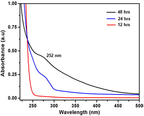
UV–Vis spectra of selenium nanoparticles synthesized using Cassia auriculata leaf extract. UV-analysis taken in the time interval of 12 h, 24 h, 48 h.
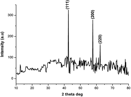
XRD spectrum of selenium nanoparticle synthesized using Cassia auriculata leaf extract.
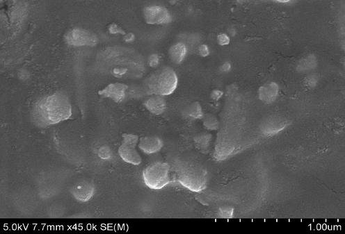
SEM micrographs of selenium nanoparticle solution formed using Cassia auriculata leaf extract.
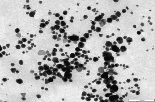
TEM micrographs of selenium nanoparticle solution formed using Cassia auriculata leaf extract.
In the FTIR spectrum of Cassia auriculata various peaks were observed between 4000 and 400 cm−1 corresponding to 1039 cm−1 – C–O stretch, alcohols, carboxylic acids, esters, ethers; 1128 cm−1 – C–N stretch, aliphatic amines; 1722 cm−1 – C = O stretch, aldehydes, saturated alkanes; 2860 cm−1 – C–H stretch and 3373 cm−1 – N–H stretch, 10, 20 amines in Fig. 5(a). The shift in peak from 1722 cm−1 to 1664 cm−1 indicates the formation of selenium nanoparticles by reduction of the carbonyl groups present in C. auriculata. Shift in peaks from 1039 cm−1 to 1106 cm−1 and disappearance of the peak at 2926 cm−1 indicates that alkanes, ether, esters and alcohols have involved in capping of selenium nanoparticles in Fig. 5(b).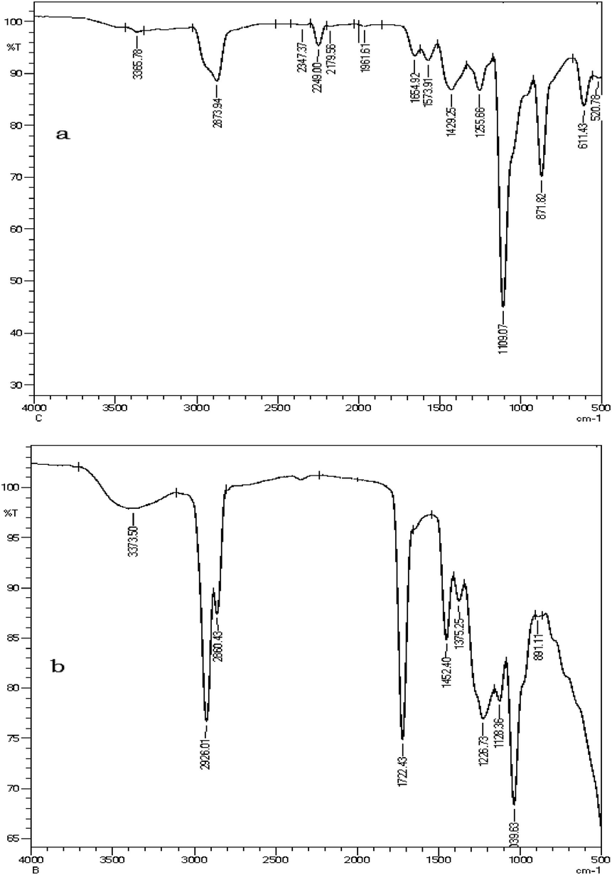
FTIR spectra of (a) nanoparticles synthesized from sodium selenite treated (b) Cassia auriculata leaf extract.
HL60 cells treated with various concentration of selenium in varying time intervals. The result shows in Fig. 6(a) and (b), nuclear morphology staining in leukemia cells due to selenium nanoparticle treatment and effects are mainly dependent on the dose exposed. Fig. 7(a) and (b), shows the selenium nanoparticles induced apoptosis in leukemia cell and results shows bright green for early apoptotic cells and red color with condensed and fragmented nuclei. The increased exposure of selenium nanoparticles enhances the DNA gel electrophoresis of DNA fragmentation is shown in Fig. 8. Furthermore, results clearly proves the newly genera selenium nanoparticles bring about anti-cancer activity, which is been well explored. The effect of newly formed selenium nanoparticles using Cassia auriculata leaf extract on HL60 leukemic cancer cells and vero cells were analyzed using MTT assay. Cell viability was determined (Fig. 9); process was conducted for 120 h. Cassia auriculata leaf extract generated selenium nanoparticles caused a significant cytotoxicity in leukemia cells in a concentration dependent manner with CC50 value of 7.01 µg/ml where as for vero cells it is 109.13 µg/ml in Fig. 9.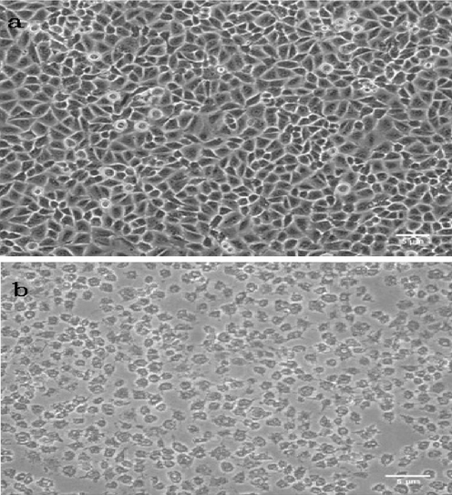
Morphological analysis of HL60 cells (a) control and (b) Cassia auriculata synthesized selenium nanoparticles treated with HL60 cells.
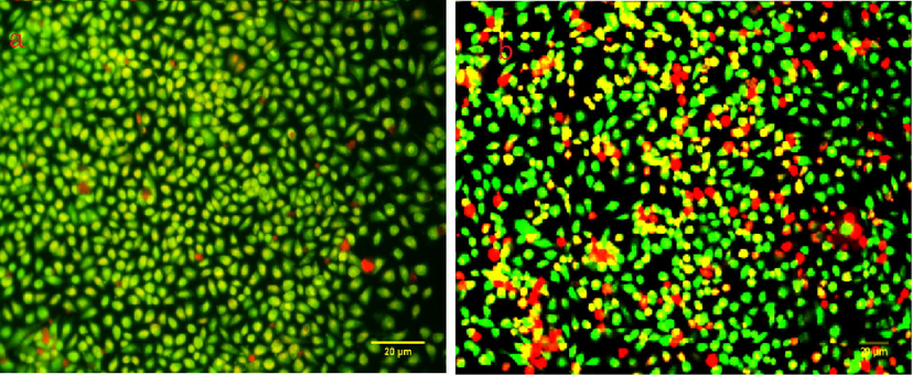
(a) Control and (b) selenium nanoparticles induced apoptosis (Dual staining).
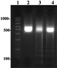
DNA fragmentation analysis using agarose gel electrophoresis. Lane 1: marker; Lane 2: control (HL60 cells); Lane 3: Cassia auriculata synthesized Selenium nanoparticles treated with HL60 cells (8 µM); Lane 4: Cassia auriculata synthesized Selenium nanoparticles treated with HL60 cells (16 µM).
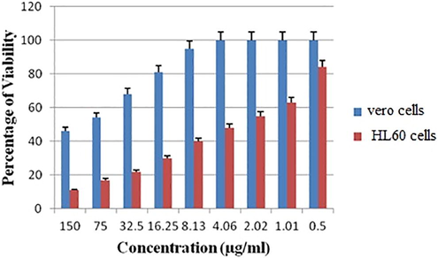
Differential cytotoxic effects of Cassia auriculata synthesized selenium nanoparticles on cancerous HL60 leukemic cells and Vero cells from a representative experiment are presented, n = 3.
Generation of metal nanoparticles mainly used up at the present is to reduce the lethal effects of injurious chemical reducing agent. The various biomimetic approaches are being explored for the production of biocompatible nanoparticles. Significantly the discovery of new efficient nanoparticle with low toxicity is in emergency need in medicinal side, this focus of attention, enhanced the green route synthesis to develop benign nanomaterials, the other conventional methods are not advantageous over biological methods due to drawbacks like time consuming, high cost higher toxic grade and solubility, etc., Green synthesis of nanomaterial deserve merit to overcome over the limitations of microbial synthesis of nanomaterial. The result addressed herein is on the greener synthesis of selenium nanoparticles using medicinally valuable plant Cassia auriculata leaf extract. Other scientists reported on the synthesis of colloidal selenium nanoparticles using Terminalia arjuna leaf extract (Prasad and Selvaraj, 2014). Another selenium nanoparticles synthesized using lemon leaf was reported by the same authors Prasad and Selvaraj, it prevents UV exposed cells from DNA damage (Prasad et al., 2013). Using gum-arabic, Kong and coworker synthesis the selenium nanoparticles sized 35 nm (Kong et al., 2014). Sharma and coworkers reported on Vitis vinifera synthesized selenium nanoballs with size ranging from 3 to 18 nm (Sharma et al., 2014). Li and his colleagues, using Capsicum annum synthesized selenium nanorods and nanoballs (Li et al., 2007). Ramamurthy and coworkers have synthesized new selenium nanoparticles using fenugreek seed extract, achieved oval shaped nanoparticles and it works better against cancerous cell (Ramamurthy et al., 2013). All these above synthesized nanoparticles are biogenic that can full fill medical need.
Additionally, in vitro anticancer results clearly reveal that the newly synthesized selenium nanoparticles are having promising bustle in medical field. Previous studies have attributed that, Selenium nanoparticles synthesized with Spirulina polysaccharides delay the growth of cancer cells by inducing apoptosis confirmed by raise in sub G1 cell cycle arrest, DNA fragmentation, and chromatin condensation. Equally, the Selenium nanoparticles-doxorubicin nanoconjugates ease the absorption of the antibiotic by cellular uptake and thereby influencing its cytotoxic effects against tumour cell (Yang et al., 2012). It has been proved that biogenic synthesized spherical sized 54 nm selenium nanoparticle, exhibits cytotoxicity at 40 µg/ml, inhibits cancer cell growth (HeLa cells) to about 72.5% (Li et al., 2019) where as 40.5% viability was observed in about 2 µg/ml in HT-20 cells (Ranjitha and Ravishankar, 2018).
Recently, at the concentration of 4 µM Se@Ga nanoparticles was treated in HepG2, the cell numbers reduced with cytoplasm shrinkage efficiently inhibited the proliferation (Li et al., 2019). Additionally selenium nanocomposite prepared by means of selenious and ascorbic acid obtained HL60 cell death at the concentration of 200 µmol/1 (Valueva et al., 2007) while in the current research at 7 µg/ml cell shrinkage and induction of apoptosis happen. Apoptosis is one of the most important mechanism of the anticancer agent, Newly genre selenium nanoparticles, while subjected for dual staining reveals apoptotic early stages by yellowish green staining and orange color with late apoptotic stages, apoptotic rate were higher in 7 µg/ml and dye does not enter the intact cell membrane structure, appropriately HE-SeNPs synthesized using hawthorn fruit induced HepG2 cells apoptosis, Annexin V and PI was applied in combination together found proportions of higher rate of apoptosis in 20 µg/ml (Cui et al., 2018).
Selenium nanoparticles, in breast cancer mice model shows hypersensitivity delayed-type response and also stimulate the defensive mechanism via enhancing the interferon production (Yazdi et al., 2012). It has been reported that aromatase inhibitor anastrozole found to be a drug of choice for the breast cancer however it found to have serious side effects, like osteoporosis and bone fracture (Goss et al., 2003; Thürlimann et al., 2005; Vekariya et al., 2013), but conjugation of anastrozole with selenium nanoparticles exhibit minimal side effects. It is worth mentioning that the newly synthesized selenium nanoparticles with organic molecules could induce cytotoxic effect and damage the cancer cell with minimal toxicity (Vahidi et al., 2020; Anand et al., 2020).
4 Conclusion
The present study highlights the importance of selenium nanoparticles, results addressed herein very well reveal that functionalization of selenium nanoparticles as anti-cancerous nanomaterial has been achieved with biomolecule reductant without any capping molecules. Consequently, newly genre selenium nanoparticle’s is stable due to the capping of phytoconstituent as the evident from FTIR. Moreover, serves as a potential anti-proliferative agent signifying the immense growth control against cancer cell. The combination of phytocompounds and nanoparticle resolve the unique platform for the drug and gene delivery in cancer therapy. Biogenic nano-formulation may have increased solubility, less toxicity and enhanced clinical efficiency. So, it is reasonable to infer that green chemistry approach on the fabrication of selenium nanoparticles is promising on the interface of nanoscience and medicinal science in developing nanotherapeutics against cancer disease. At a halt, extensive clinical translational studies need to be exploited for safe full pledge nanotherapeutics practices.
Acknowledgement
The authors extend their appreciation to the Deanship of Scientific Research at King Saud University for funding the work through the research group project number RGP-023.
Declaration of Competing Interest
The authors declare that they have no known competing financial interests or personal relationships that could have appeared to influence the work reported in this paper.
References
- Synthesis of selenium nanoparticles using probiotic bacteria lactobacillus acidophilus and their enhanced antimicrobial activity against resistant bacteria. J. Clust. Sci. 2019
- [CrossRef] [Google Scholar]
- Green synthesis of selenium nanoparticle using leaves extract of Withania somnifera and its biological applications and photocatalytic activities. BioNanoSci.. 2019;9:105-116.
- [CrossRef] [Google Scholar]
- Eco-friendly green synthesis of silver nanoparticles from the sesame oil cake and its potential anticancer and antimicrobial activities. J. Photochem. Photobiol. B. 2019;192:83-89.
- [CrossRef] [Google Scholar]
- Green synthesis of silver nanoparticles using Pimpinella anisum seeds: antimicrobial activity and cytotoxicity on human neonatal skin stromal cells and colon cancer cells. Int. J. Nanomed.. 2016;11:4439-4449.
- [CrossRef] [Google Scholar]
- Human serum albumin interaction, in silico and anticancer evaluation of Pine-Gold nanoparticles. Process Biochem.. 2020;89:98-109.
- [CrossRef] [Google Scholar]
- Metallic nanoparticles. In: Misra P., Black J.A., eds. Handbook of Metal Physics. Amsterdam: Elsevier BV; 2009. p. :4-6.
- [Google Scholar]
- Cancer-targeted selenium nanoparticles sensitize cancer cells to continuous γ radiation to achieve synergetic chemo-radiotherapy. Chem. Asian J.. 2017;12(23):3053-3060.
- [CrossRef] [Google Scholar]
- Green synthesis of selenium nanoparticles with extract of hawthorn fruit induced HepG2 cells apoptosis. Pharm. Biol.. 2018;56(1):528-534.
- [CrossRef] [Google Scholar]
- Gold nanoparticles: assembly, supramolecular chemistry, quantum size-related properties, and applications towards biology, catalysis, and nanotechnology. Chem. Rev.. 2004;104:293-346.
- [CrossRef] [Google Scholar]
- Antimicrobial and cytotoxicity effects of synthesized silver nanoparticles from Punica granatum peel extract. Nanoscale Res. Lett.. 2018;13:315.
- [CrossRef] [Google Scholar]
- Rapid biological synthesis of silver nanoparticles using plant seed extracts and their cytotoxicity on colorectal cancer cell lines. J. Clust. Sci.. 2017;28:595-605.
- [CrossRef] [Google Scholar]
- Interaction of silver nanoparticles with HIV-1. J. Nanobiotechnol.. 2005;3:6.
- [CrossRef] [Google Scholar]
- Aloevera leaf extract mediated green synthesis of selenium nanoparticles and assessment of their In vitro antimicrobial activity against spoilage fungi and pathogenic bacteria strains. Green Proc. Syn.. 2019;8s(1):399-407.
- [CrossRef] [Google Scholar]
- A randomized trial of letrozole in postmenopausal women after five years of tamoxifen therapy for early-stage breast cancer. N. Engl. J. Med.. 2003;349:1793-1802.
- [CrossRef] [Google Scholar]
- Inhibition of E. coli and S. aureus with selenium nanoparticles synthesized by pulsed laser ablation in deionized water. Int. J nanomed.. 2016;11:3731-3736.
- [CrossRef] [Google Scholar]
- Microbial synthesis of selenium nanocomposite using Saccharomyces cerevisiae and its antimicrobial activity against pathogens causing nosocomial infection. Chalcog. Let.. 2012;9:509-515.
- [Google Scholar]
- Selective cellular uptake and induction of apoptosis of cancer-targeted selenium nanoparticles. Biomaterials. 2013;34(29):7106-7116.
- [Google Scholar]
- Medicinal values of avaram (Cassia auriculata linn.): a review. Int. J. Curr. Pharm. Res.. 2012;4:1-3.
- [Google Scholar]
- Synthesis of tobramycin stabilized silver nanoparticles and its catalytic and antibacterial activity against pathogenic bacteria. J. Inorg. Organomet. Polym.. 2019;29:111-120.
- [CrossRef] [Google Scholar]
- Green approach for the effective reduction of graphene oxide using Salvadora persica L. root (Miswak) extract. Nanoscale Res. Lett.. 2015;10:1-9.
- [CrossRef] [Google Scholar]
- Nanoparticles: properties, applications and toxicities Arabian. J. Chem.. 2019;12:908-931.
- [CrossRef] [Google Scholar]
- Synthesis and anti-oxidant properties of gum Arabic-stabilized selenium nanoparticles. Int. J. Biol. Macromol.. 2014;65:155-162.
- [CrossRef] [Google Scholar]
- Rapid, room-temperature synthesis of amorphous selenium/protein composites using Capsicum annuum L. extract. Nanotechnology. 2007;18:405101-405109.
- [CrossRef] [Google Scholar]
- Synthesis and cytotoxicity of selenium nanoparticles stabilized by α-D-glucan from Castanea mollissima Blume. Int. J. Biol. Macromol.. 2019;129:818-826.
- [CrossRef] [Google Scholar]
- Selenium and Alzheimer's disease: a systematic review. J. Alzheimers Dis.. 2011;26:81-104.
- [CrossRef] [Google Scholar]
- Danthron induces DNA damage and inhibits DNA repair gene expressions in GBM 8401 human brain glioblastoma multiforms cells. Neurochem. Res.. 2010;35:1105-1110.
- [CrossRef] [Google Scholar]
- Rapid colorimeteric assay for cellular growth and survival: application to proliferation and cytotoxicity assays. J. Immunol. Methods. 1983;65:55-63.
- [CrossRef] [Google Scholar]
- Biosynthesis of Se nanoparticles and its effect on UV-induced DNA damage. Colloids Surf. B. 2013;103:261-266.
- [CrossRef] [Google Scholar]
- Biogenic synthesis of selenium nanoparticles and their effect on As (III)-induced toxicity on human lymphocytes. Biol. Trace Elem. Res.. 2014;157:275-283.
- [CrossRef] [Google Scholar]
- Antibacterial activity of some folklore medicinal plants used by tribes in Western chats of India. J. Ethanopharmacol.. 2000;69:63-71.
- [CrossRef] [Google Scholar]
- Green synthesis and characterization of selenium nanoparticles and its augmented cytotoxicity with doxorubicin on cancer cells. Bioprocess Biosyst. Eng.. 2013;36:1131-1139.
- [CrossRef] [Google Scholar]
- Extracellular synthesis of selenium nanoparticles from an Actinomycetes Streptomyces griseoruber and evaluation of its cytotoxicity on HT-29 cell line. Pharm. Nanotechnol.. 2018;6(1):61-68.
- [CrossRef] [Google Scholar]
- A simple technique for quantifying apoptosis in 96-well plates. BMC Biotechnol.. 2005;10:5-12.
- [CrossRef] [Google Scholar]
- Protective effect of nanoceria on cisplatin-induced nephrotoxicity by amelioration of oxidative stress and pro-inflammatory mechanisms. Biol. Trace Elem. Res.. 2019;189:145-156.
- [CrossRef] [Google Scholar]
- Selenium containing compounds from poison to drug candidates: a review on the GPx-like activity. Curr. Chem. Biol.. 2013;7(1):25-36.
- [CrossRef] [Google Scholar]
- Biomolecule-mediated synthesis of selenium nanoparticles using dried Vitis vinifera (raisin) extract. J. Mol.. 2014;19:2761-2770.
- [CrossRef] [Google Scholar]
- Green synthesis of selenium nanoparticles conjugated Clausena dentata plant leaf extract and their insecticidal potential against mosquito vectors. Artif. Cells Nanomed. Biotechnol.. 2017;45(8):1490-1495.
- [CrossRef] [Google Scholar]
- A comparison of letrozole and tamoxifen in postmenopausal women with early breast cancer, Breast International Group (BIG) 1-98 Collaborative Group. N. Engl. J. Med.. 2005;353:2747-2757.
- [CrossRef] [Google Scholar]
- Emerging selenium nanoparticles to combat cancer: a systematic review. J. Clust. Sci. 2020;31:301-309.
- [CrossRef] [Google Scholar]
- Biosynthesis of silver and gold nanoparticles using Musa acuminata colla flower and it pharmaceutical activity against bacteria and anticancer efficacy. J. Photochem. Photobiol. B Biol.. 2019;201:111670.
- [CrossRef] [Google Scholar]
- Structural-morphological and biological properties of selenium nanoparticles stabilized by bovine serum albumin. Russ. J. Phys. Chem.. 2007;81:1170-1173.
- [CrossRef] [Google Scholar]
- Alleviating anastrozole induced bone toxicity by selenium nanoparticles in SD rats. Toxicol. Appl. Pharm.. 2013;268:212-220.
- [CrossRef] [Google Scholar]
- Functionalized selenium nanoparticles for targeted siRNA delivery silence Derlin1 and promote antitumor efficacy against cervical cancer. Drug Deliv.. 2020;27(1):15-25.
- [CrossRef] [Google Scholar]
- The immunostimulatory effect of biogenic selenium nanoparticles on the 4T1 breast cancer model: an in vivo study. Biol. Trace Elem. Res.. 2012;149:22-28.
- [CrossRef] [Google Scholar]
- Surface decoration by Spirulina polysaccharide enhances the cellular uptake and anticancer efficacy of selenium nanoparticles. Int. J. Nanomed.. 2012;7:835-844.
- [CrossRef] [Google Scholar]


