

Translate this page into:
Assessment of cyto-nephrotoxicity and growth performance in Labeo rohita induced by fluorescein dye Y and B
⁎Corresponding authors. profbilal@yahoo.com (Bilal Hussain), mushahid@ksu.edu.sa (Shahid Mahboob)
-
Received: ,
Accepted: ,
This article was originally published by Elsevier and was migrated to Scientific Scholar after the change of Publisher.
Peer review under responsibility of King Saud University.
Abstract
The cyto-nephrotoxicity in fishes induced by dyes is a global issue. Fluorescein dye has a wide range of applications in industry and biological laboratories. In vivo experiment was planned to assess the sublethal effects of Fluorescein dye, Eosin Y, and Eosin B in Labeo rohita. This freshwater fish was exposed to increasing concentrations (0.0011, 0.0023, and 0.0047 mg/L) of both dyes in the aquarium for definite time intervals. A dye-free trial was set as a control. Fish was anesthetized and dissected by cutting on the ventral side to remove a kidney. Cyto-nephrotoxicity was detected through histopathology. The fish's significant mortality was observed when exposed to 0.0023 mg/L (LC50) and 0.0047 mg/L concentration of Eosin Y dye. These trial groups showed many kidney tissues alterations such as glomerular, tubular epithelial cell's degeneration, a reduced lumen of tubules, vacuolation, absence of bowmen's space, necrosis, glomerulonephritis with increased per tubular space, shrinking of the glomerulus, congestion, clogging, and degeneration of tubules. Eosin B showed less mortality and tissue damage, including glomerular contraction and increased space between glomerulus and capsule. ANOVA showed a highly significant reduction in weight gain of fish at different treatments levels of Eosin Y, but less or none in the case of Eosin B. Increase in fish weight was found to be 66.3 ± 0.38 g when exposed to Eosin Y at 0.0047 mg/L, but it was 69.05 ± 0.63 g when exposed to Eosin B while it was almost identical 69.2 ± 0.23 g for both dye-free trial groups. The pair-wise comparison showed that exposure of the Eosin Y at treatment level 0.0047 mg/L was found to be highly toxic for the kidneys of Labeo rohita. Although both classes of dye are cytotoxic at small concentrations, Eosin Y is acute cyto-nephrotoxic and was found to be involved in growth retardation. To prevent society from fatal consequences, usage of dyes having acute toxicity must be banned or switched to alternates having no effects.
Keywords
Cytotoxicity
Growth
Labeo rohita
Lethal
Fluorescein

1 Introduction
Exposure to biological and industrial dyes causes serious health issues in organisms. Significant sources of dyes are textile industries, pharmaceutical industries, tannery, etc. They are disposed of untreated into natural water bodies causing severe aquatic pollution (Sinha and Hanamghar, 2019; Robinson et al., 2001). Dyes and their derivatives like benzidine, naphthalene, and other aromatic compounds are highly mutagenic and carcinogenic (Forgacs et al., 2004). Fish are exceptional model organisms to absorb such xenobiotics. Fish is the best model to study these contaminants' carcinogenic or mutagenic potential and cytological alterations (Strzyzewska et al., 2016). Fish organs like kidneys and liver having a role in the filtration of body fluids and metabolism are essential in determining the health of fish in the freshwater ecosystem. The biomarkers study of such vital organs has led to marvelous environment risk assessments (Dai et al., 2020). Histopathological studies are considered helpful to identify sub-lethal and chronic effects of freshwater pollutants (Hussain et al., 2019).
Pollutants enter into fish organs (Kondera et al., 2014) and damage metabolic pathways. The kidney and liver are specific organs to perform essential functions like excretion and have a role in maintaining the volume and pH of body fluids and erythropoiesis. They are also involved in osmoregulation to provide fish a stable internal environment. Kidneys produce hormones, excrete toxins, and ensure adequate plasma for its proper flow to vital organs. Toxicants like dyes can damage nephrons, resulting in kidney dysfunction (Nordberg et al., 2012). Thus kidneys are excellent organs to reflect environmental stress to fishes, and tissue damage acts as a tremendous biomarker to estimate a load of pollutants (Perera and Pathiratne, 2010; Nordberg et al., 2012). This project was designed to detect in-vivo cyto-nephrotoxicity induced in Labeo rohita due to fluorescein dye Y and B.
2 Material and methods
2.1 Fish procurement and water sampling
Labeo rohita in the weight range of 60–85 g was procured from fish seed hatchery, Satiana Road, Faisalabad. Fish were acclimatized in glass tanks for eight days in fisheries laboratory with continuous aeration before exposure to the fluorescein dye Y and B. After four days, water in an aquarium was replaced with fresh water, and fish were fed with commercial feed 5% of body weight. The water was replaced daily with fresh water to remove fish excreta. All necessary fish morphometric measurements were performed before and after the application of both classes of dye. Fish was exposed to both categories of dye for 15 days.
2.2 Experimental plan
Healthy fish specimens were grouped into four major groups having twenty specimens. Both dyes were dissolved in distilled water, and three dilutions viz., 0.0011 mg/L, 0.0023 mg/L, and 0.0047 mg/L of Eosin Y and Eosin B dye were prepared. Standard water quality parameters such as temperature, pH, and dissolved oxygen were kept normal during the experiment (Ben Ameur et al., 2012). Each experimental trial group was exposed to these increasing concentrations in triplicate. LC50 value was determined by exposing fish for both classes of dyes for 24, 48, 72, and 96 h. A dye-free fish group on similar conditions was set as a control. Morphometric measurements of fish specimens were performed weekly to note growth performances.
2.3 Histopathology
Fish specimens from each trial group were anesthetized by chloroform, dissected to remove kidneys, and immediately preserved in formalin (10% solution). These tissues (2 mm) were subjected to histopathological assessment by the paraffin wax method. Dehydration of kidney tissues was conducted by isopropyl alcohol grading, with 50%, 70%, 90%, and absolute isopropyl alcohol for 1 h and then cleaned in xylene. These tissues were solidified and inserted in a paraffin cube. Tissue specimens were mounted on microtome for cutting it into sections. The optimal thickness of the sections was 5 μm. Paraffin was cleared by xylene (1–2 min) before section staining. Dehydrated slides were placed in water. These rehydrated sections were placed in Hematoxylin stain solution for 30 min. These slides having sections were washed in tap water for 3 min. The sections on the slides were turned bluing. Then sections were treated with 70% ethanol + 1% HCl for few seconds. This step removed the excess stain and exposed nuclear contents. Slides were washed in tap water for 3–5 min until they attained clear blue color. The slides were placed in eosin stain solution for 10–12 min and washed in tap water for 1–5 min. Slides were dehydrated, cleared, and mounted (Ben Ameur et al., 2012; Chavan and Muley, 2014). Photomicrographs of these stained sections were taken through a microscope at 400× and 600×.
2.4 Statistical analysis
Fish mortality data were used to calculate LC50 values by Probit analysis. ANOVA was used to compare the LC50 values for both classes of dyes exposed to fish. All experiments were repeated three times, and data were presented average means of these replicates. Mean values were compared and evaluated with the DMR test (p < 0.05). Growth performance data were analyzed through one-way analysis of variance through SPSS 9 software. Data was plotted to prepare charts through MS Excel (2007).
3 Results
The fish Labeo rohita exposed to increasing concentrations (0.0011, 0.0023, 0.0047 mg/L) of Eosin dye Y and B. LC50 was detected as 0.0023 mg/L, which showed almost 50% instant mortality of fish after exposure to Eosin Y dye while it was far greater, i.e., 0.039 mg/L for the Eosin B dye. All necessary water quality parameters such as pH, DO, alkalinity, BOD, COD, conductivity, salinity, etc. were normal in all trial groups. Although fish specimens in each trial group were fed with the same commercial feed according to 5% of their body weight but significant differences were found among the weight of fish at different levels of treatments indicating the more toxic effect of the Eosin dye Y. The highest weight reductions were noticed for trial group 0.0023 and 0.0047 mg/L even at these lower concentrations of Eosin dye Y. Increase in fish weight was found to be 66.3 ± 0.38 g when exposed to Eosin Y at 0.0047 mg/L but it was 69.05 ± 0.63 g when exposed to Eosin B while it was almost same 69.2 ± 0.23 g for both dye free trial groups. Significant weight reduction was observed with increase in the concentrations of Eosin dye Y but not in the case for Eosin dye B (Fig. 1).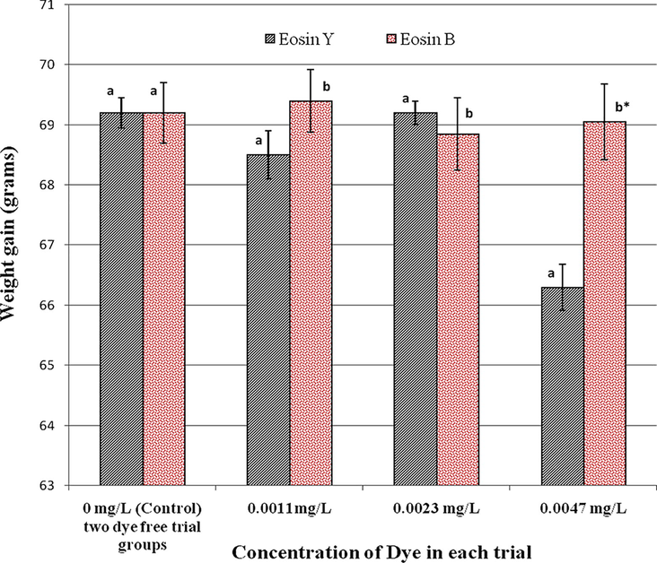
Comparative analysis of the effect of exposure of both types of fluorescein dye on the weight gain of fish during experimental trials. Different alphabets in each trial group indicate significant (p < 0.05) differences. *Highly significant differences.
Histopathology of kidney tissues indicated normal structural arrangements of cells in control groups. Average normal arrangements of chromaffin cells, epithelial cells, normal structures of glomeruli, tubules, bowmen’s capsule and thin inter capsular space were observed (Fig. 2). Fish treated with different grading concentrations of dye Y showed various tissue alterations in kidney tissues. The fish treated with Eosin dye B treated with 0.0011 mg/L and 0.0023 mg/L concentration showed mild necrosis in the tubules and slight degeneration of glomerulus (Fig. 3). Kidney tissues of fish treated with 0.0047 mg/L concentration of Eosin dye B showed shrinkage of the glomerulus, mild tissue degeneration and increase in space between glomerulus and capsule (Fig. 4). Photomicrograph for the kidney of Labeo rohita exposed to lower concentration (0.0011 and 0.0023 mg/L) of Eosin Y dye showed the tubular, epithelial cell’s and glomerular degeneration, necrosis in the kidney, and absence of bowmen's space (Fig. 5). Photomicrograph for the histopathology of kidney exposed to higher concentration (0.0047 mg/L) showed the vacuolation, necrosis, reduced lumen of tubules (Fig. 6), absence of bowmen’s space, shrinking of a glomerulus, congestion, glomerulonephritis with increased per tubular space, clogging and degeneration of tubules (Fig. 7). From histopathological examination of kidney tissues of fish exposed to both dyes, Eosin dye Y was found to be a highly toxic dye responsible for the induction of many alterations in tissues of the kidney, an organ of prime importance in the body. Eosin dye B did not significantly influence the fish's weight gain or loss and was less toxic than Eosin dye Y.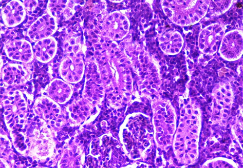
Histopathology of fish Labeo rohita shows the normal kidney structure in the control group (400×).
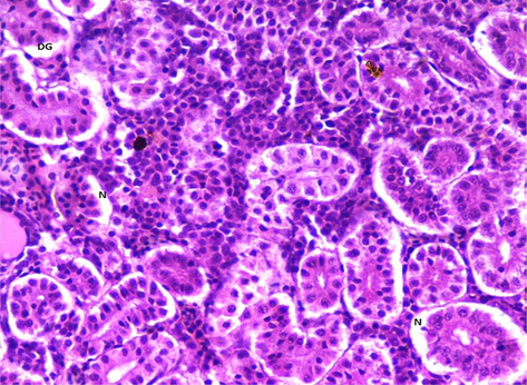
Histopathological examination showed the structure of the kidney of Labeo rohita treated with 0.0011 mg/L and 0.0023 mg/L concentration of dye showed mild necrosis (N) in the tubules and slight degeneration of glomerulus (DG) (400×).
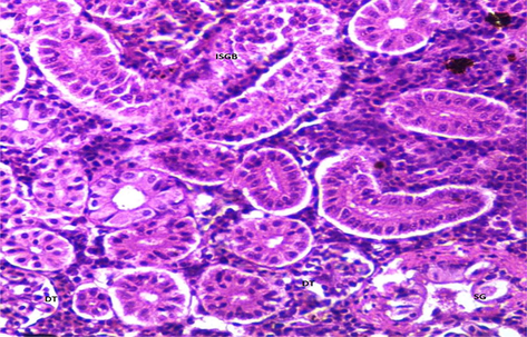
Histopathological examination showed the structure of the kidney of Labeo rohita treated with 0.0047 mg/L concentration of Eosin dye B showed mild degeneration of tissue (DT), shrinkage of the glomerulus (SG), and increase in space between glomerulus and bowmen's capsule (ISGB) (600×).
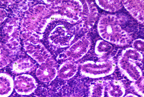
Histopathological examination showed the structure of the kidney of Labeo rohita treated with 0.0011 mg/L and 0.0023 mg/L concentration of Eosin Y dye showed the tubular (TD), epithelial cell’s (ED) and glomerular degeneration (GD) and necrosis (N) in the kidney (600×).
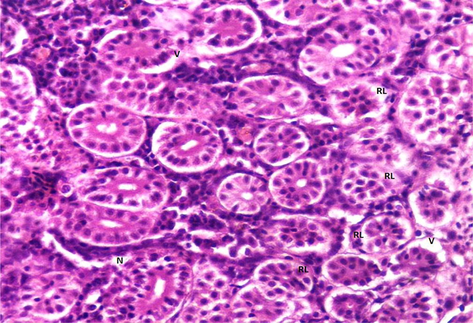
Histopathological examination showed the structure of the kidney of Labeo rohita treated with higher concentration (0.0047 mg/L) showed the vacuolation, necrosis, reduced lumen of tubules (600×).
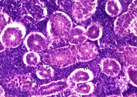
Histopathological examination showed the structure of the kidney of Labeo rohita treated with higher concentration (0.0047 mg/L) showed the shrinking of the glomerulus (SG), absence of bowmen's space, glomerulonephritis with increased per tubular space (GITP), clogging of tubules (CT), and degeneration of tubules (DT) (600×).
4 Discussion
Industry discharges dyes directly into the freshwater ecosystems. These dyes directly induce harmful or toxic effects on aquatic life and indirectly on human beings. Such detrimental effects are genotoxic, nephrotoxic, and cytotoxic (Villela et al., 2006). Such textile dyes diminish water quality and make it unsuitable for aquatic fauna (Verma, 2011) by damaging vital organs such as the kidney, liver, brain, and intestine. Histopathological evaluation provides a complete estimation of the health of aquatic organisms and acts as a valuable tool to monitor the effects of environmental toxicants (Drishya et al., 2016). Such biomarkers were found to be very useful in toxicant exposure studies. The kidney of fish is a primary organ to be affected by freshwater pollution (Paulo et al., 2012; Oliveira et al., 2013) due to its filtration of body fluids. Paulo et al. (2012) and Vijay et al. (2015) used histopathology as a tool to determine the effects of textile dyes and environmental contaminants on kidney tissues of fish Cirrhinus mrigala. Histopathological modifications in fish organs are mainly due to toxicants and stressors and in the vicinity of the fish (Abdel-Moneim et al., 2012; Paulo et al., 2012; Neelima et al., 2015; Drishya et al., 2016; Hussain et al., 2020).
The present study's findings of the induction of tissue alterations in fishes corroborate the conclusions of Sreedevi and Chitra (2014) and Drishya et al. (2016). Goel and Garg (1980) also observed similar findings in the kidney of C. punctatus when exposed to triaminoazobenzene dye. Barot and Bahadur (2011) also found tubular degeneration and glomerular contraction when they exposed the Labeo rohita to the low concentration of acid orange dye. Vacuolation has been found in the kidney of Labeo rohita after exposure to Eosin Y, corroborating the findings of Javed et al. (2017) who also found vacuolation in fish kidney tissues. There was degeneration, congestion, and necrosis in the kidney of Labeo rohita when exposed to the eosin Y dye. Hadi and Alwan (2012) and Bijoy Nandan and Nimila (2012) also noticed the many degenerative changes, congestion, and necrosis in the kidney of fish during a histopathological examination. Barot (2015), de Oliveira et al. (2016), Rocha et al. (2017), and Aksu et al. (2017) also confirmed that prolonged exposure to industrial dyes stimulates toxicity and induces alterations in fish tissues. Only glomerular contraction and increase in space between glomerulus and bowmen’s capsule was observed in the fish kidney after exposure to lower to a higher concentration of Eosin B dye. Saenphet et al. (2009) also observed similar alterations in the kidney tissues. Karthikeyan et al. (2006) researched the impacts of dyes in textile effluents on Mastacembelus armatus fish. This fish species is widely consumed due to its protein profile and was found with tissue degradation, corroborating the current study's findings.
Water quality parameters such as pH, DO, alkalinity, conductivity, and salinity, etc. were kept normal, and feeding was the same in all trial groups because changes in the quality of the water cause behavioral abnormalities (Jacquin et al., 2020) may affect feeding. Sharma et al. (2008) performed research to determine the harmful or toxic effects of tomato red dye on fish and reported similar findings. Birhanlı and Ozmen (2005) researched the hazardous or poisonous effects of six different textile dyes on frog embryos and reported similar results. Tissue alterations increase with rising concentrations of fluorescein dye. Kaur and Kaur, (2015) said identical findings. Decreased lumen, degeneration of nephrons, and glomerulus contraction were due to the intervention of dye in the kidney filtration process (Barot and Bahadur, 2013; Oliveira et al., 2013; Dai et al., 2020). Barot (2015), in his study, described similar tissue alterations in Indian major carp Catla catla confirming the findings of this research work that fluorescein dye induces nephrotoxicity in Labeo rohita like shrinkage glomerulus and degeneration of renal tubules etc. Results of this study, such as glomerular degeneration, necrosis, and shrinkage of the glomerulus were due to environmental pollution (Abdel-Moneim et al., 2012; Paulo et al., 2012; Massar et al., 2014; Drishya et al., 2016; Brraich and Kaur, 2017; Hussain et al., 2019). The findings of the current study will be helpful in environmental monitoring strategies and minimize the eco-toxicological impacts of freshwater pollution.
5 Conclusion
Fluorescein dye has two classes, Eosin Y and Eosin B. While Eosin Y showed acute vacuolation, necrosis, reduced lumen of tubules, absence of bowmen's space, shrinking of the glomerulus, clogging and degeneration of tubules, congestion, and glomerulonephritis. It is concluded that the application of eosin dye B showed mild tissue degeneration, glomerular shrinkage, and increased space between glomerulus and capsule. The present study recommends the use of less toxic Eosin dye B. The study also suggests and insists on its removal before releasing in the effluent as a small amount of fluorescein dye released untreated is quite lethal to aquatic life. The current study will help in environmental screening strategies to minimize the toxicological impacts of freshwater contamination.
Acknowledgements
“The authors (SM and KAG) express their sincere appreciation to the Researchers Supporting Project number (2021/93), King Saud University, Riyadh, Saudi Arabia”.
Declaration of Competing Interest
The authors declare that they have no known competing financial interests or personal relationships that could have appeared to influence the work reported in this paper.
References
- Histopathological biomarkers in gills and liver of Oreochromis niloticus from polluted wetland environments, Saudi Arabia. Chemosphere. 2012;88(8):1028-1035.
- [Google Scholar]
- Biochemical impacts of the textile dyes Remazol Brillant Blue R and Congo Red on the crayfish Astacus leptodactylus (Decapoda, Astacidae) Crustaceana. 2017;90(13):1563-1574.
- [Google Scholar]
- Oxidative stress, genotoxicity and histopathology biomarker responses in mullet (Mugil cephalus) and sea bass (Dicentrarchus labrax) liver from Bizerte Lagoon (Tunisia) Mar. Pollut. Bull.. 2012;64(2):241-251.
- [Google Scholar]
- Histotoxicity of Acid Orange 7 on organs of freshwater fish Labeo rohita. Res. Revi. A. J. Toxicol.. 2011;1:1-9.
- [Google Scholar]
- Evaluation of azo dye toxicity using some haematological and histopathological alterations in fish Catla catla. IJFSB. 2015;9(5):415-418.
- [Google Scholar]
- Behavioural and histopathological effects of azo dye on kidney and gills of Labeo rohita fingerlings. J. Environ. Biol.. 2013;34(1):147-152.
- [Google Scholar]
- Evaluation of the toxicity and teratogenity of six commercial textile dyes using the frog embryo teratogenesis assay–Xenopus. Drug Chem. Toxicol.. 2005;28(1):51-65.
- [Google Scholar]
- Histopathological alterations in the kidneys of Labeo rohita due to lead toxicity. J. Environ. Biol.. 2017;38(2):257-262.
- [Google Scholar]
- Effect of heavy metals on liver and gill of fish Cirrhinus mrigala. Int. J. Curr. Microbiol. Appl. Sci.. 2014;3(5):277-288.
- [Google Scholar]
- Dai, C., Liu, Q., Li, D., Sharma, G., Xiong, J., Xiao, X., 2020. Molecular Insights of Copper Sulfate Exposure-Induced Nephrotoxicity: Involvement of Oxidative and Endoplasmic Reticulum Stress Pathways. Biomolecules, 10 (2020) 1010; doi:10.3390/biom10071010
- Textile dyes induce toxicity on zebra fish early life stages. Environ. Toxicol. Chem.. 2016;35(2):429-434.
- [Google Scholar]
- Histopathological changes in the gills of fresh water fish, Catla catla exposed to electroplating effluent. Int. J. Fish. Aquat.. 2016;4:13-16.
- [Google Scholar]
- Removal of synthetic dyes from wastewaters: a review. Environ. Int.. 2004;30(7):953-971.
- [Google Scholar]
- Histopathological changes produced in the liver and kidney of Channa punctatus after chronic exposure to 2, 3′, 4-triaminoazobenzene. Bull. Environ. Contam. Toxicol.. 1980;25(1):330-334.
- [Google Scholar]
- Histopathological changes in gills, liver and kidney of fresh water fish, Tilapia zillii, exposed to aluminum. Int. J. Pharm. Life Sci.. 2012;3(11):2071-2081.
- [Google Scholar]
- Environmentally induced nephrotoxicity and histopathological alternations in Wallago attu and Cirrhinus mrigla. Saudi J. Biol. Sci.. 2019;26(4):752-757.
- [Google Scholar]
- Assessment of DNA integrity through MN bioassay of erythrocytes and histopathological changes in Wallago attu and Cirrhinus mrigala in response to freshwater pollution. Saudi J. Biol. Sci.. 2020;27(1):251-260.
- [Google Scholar]
- Effects of pollution on fish behavior, personality, and cognition: Some research perspectives. Front. Ecol. Evol.. 2020;8:86.
- [CrossRef] [Google Scholar]
- Multiple biomarker responses (serum biochemistry, oxidative stress, genotoxicity and histopathology) in Channa punctatus exposed to heavy metal loaded waste water. Sci. Rep.. 2017;7(1):1-11.
- [Google Scholar]
- Impact of textile effluents on fresh water fish Mastacembelus armatus (Cuv & Val) E-J. Chem.. 2006;3(4):303-306.
- [Google Scholar]
- Variability in antioxidant/detoxification enzymes of Labeo rohita exposed to an azo dye, acid black (AB) Comp. Biochem. Physiol. C. Toxicol. Pharmacol.. 2015;167(1):108-116.
- [Google Scholar]
- High affinity of cadmium and copper to head kidney of common carp (Cyprinus carpio L.) Fish Physiol. Biochem.. 2014;40(1):9-22.
- [Google Scholar]
- Structural Changes in Kidneys of Common Carp (Cyprinus carpio L.) Inhabiting a Polluted Reservoir, Umiam in Meghalaya, India. J. Adv. Microsc. Res.. 2014;9(2):105-109.
- [Google Scholar]
- Lindane toxicity: Histopathological, behavioural and biochemical changes in Etroplus maculatus (Bloch, 1795) Mar. Environ. Res.. 2012;76:63-70.
- [Google Scholar]
- Histopathological alterations in Gill, Liver and Kidney of Cyprinus carpio (Linn.) exposed to Cypermethrin (25% EC) Int. J. Adv. Res. Biol. Sci. 2015;2(2):34-40.
- [Google Scholar]
- Kidney dysfunction and cadmium exposure factors influencing dose-response relationships. J. Trace Elem. Med. Biol.. 2012;26(2-3):197-200.
- [Google Scholar]
- Single and combined effects of microplastics and pyrene on juveniles (0+ group) of the common goby Pomatoschistus microps (Teleostei, Gobiidae) Ecol. Indic.. 2013;34:641-647.
- [Google Scholar]
- Histopathological alterations observed in the liver of Poecilia vivipara (Cyprinodontiformes: Poeciliidae) as a tool for the environmental quality assessment of the Cachoeira River, BA. Braz. J. Biol. Sci.. 2012;72(1):131-140.
- [Google Scholar]
- Multiple biomarker responses of Nile tilapia (Oreochromis niloticus) exposed to textile industry effluents reaching Bolgoda North Lake, Sri Lanka. SLJAS. 2010;15(1):1-11.
- [Google Scholar]
- Remediation of dyes in textile effluent: a critical review on current treatment technologies with a proposed alternative. Bioresour. Technol.. 2001;77(3):247-255.
- [Google Scholar]
- Ecotoxicological risk assessment of the “Acid Black 210” dye. Toxicology. 2017;376(1):113-119.
- [Google Scholar]
- Histopathological alterations of the gills, liver and kidneys in Anabas testudineus (Bloch) fish living in an unused lignite mine, li district, Lamphun province, Thailand. Southeast Asian J. Trop. Med. Public Health. 2009;40(5):1121-1126.
- [Google Scholar]
- Toxicity of tomato red, a popular food dye blend on male albino mice. Exp. Toxicol. Pathol.. 2008;60(1):51-57.
- [Google Scholar]
- Acute toxicity of triarylmethane dye, crystal violet to Indian major carp fish, Labeo rohita. IJRAR. 2019;6(2):288-300.
- [Google Scholar]
- Biochemical and genotoxic effects of octylphenol in hepato-mitochondrial fractions of freshwater fish, Oreochromis mossambicus. J. Cell Tiss. Res.. 2014;14(2):4211-4217.
- [Google Scholar]
- Morphologic evaluation of the gills as a tool in the diagnostics of pathological conditions in fish and pollution in the aquatic environment: a review. Vet. Med.. 2016;61(No. 3):123-132.
- [Google Scholar]
- Toxicity assessment of dye containing industrial effluents by acute toxicity test using Daphnia magna. Toxicol. Ind. Health. 2011;27(1):41-49.
- [Google Scholar]
- Elecrophoretic pattern and histopathological study of muscle tissue tilapia, Oreochromis mossambicus exposed textile dyes. Int. J. Sci. Res.. 2015;5(10):2319-7064.
- [Google Scholar]
- DNA damage and repair in haemolymph cells of golden mussel (Limnoperna fortunei) exposed to environmental contaminants. Mutat. Res. Genet. Toxicol. Environ. Mutagen.. 2006;605(1-2):78-86.
- [Google Scholar]


