

Translate this page into:
An open-source software for calculating 1D gamma index in radiation therapy
⁎Corresponding authors. dttai@ntt.edu.vn (Duong Thanh Tai), hbomer@iau.edu.sa (Hiba Omer), abdelmoneim_a@yahoo.com (Abdelmoneim Sulieman),
-
Received: ,
Accepted: ,
This article was originally published by Elsevier and was migrated to Scientific Scholar after the change of Publisher.
Peer review under responsibility of King Saud University.
Abstract
Purpose
This study was developed to create computer software for performing the gamma index comparison between measurement and Monte Carlo (MC) simulation for the percentage depth dose (PDD) and beam off-center ratio profile (OCR).
Materials and methods
The gamma software was built in the matrix laboratory (MATLAB) software environment. The developed software was compared with ScanDosematch and Bistromath software’s gamma evaluation to assess its accuracy. A set of reference and evaluated dose distribution, which were obtained from measurement and MC simulation, was input to the software to calculate the 1D gamma index using different criteria (i.e. 3%/3 mm, 2%/3 mm, and 2%/2 mm).
Results
We compared the two results of gamma index at 3%/3 mm, 2%/3 mm, and 2%/2 mm criteria, one calculated by the proposed software and one manually. The comparison showed high agreement between the proposed software and theoretical calculation.
Conclusions
Based on the results, we concluded that our developed software has high accuracy, compared to theoretical calculation. This software could serve as a non-commercial and open-source tool for researchers and students.
Keywords
Gamma index
Matlab
Open-source software
Monte Carlo simulation
Radiation therapy
Treatment planning system
ScanDosematch
Bistromath

1 Introduction
The clinical use of Monte Carlo (MC) simulation techniques is developing rapidly due to its ability in providing a great accuracy in the radiation dose calculations, beam modelling, and patient geometry. Those are critical components for an efficacious patient treatment planning verification. MC simulation of a clinical linear accelerator are difficult and time-consuming task and has numerous challenges to be encountered to improve the accuracy of MC calculations for patient treatments (Seco and Verhaegen 2013). The current challenges include precise development of interactions, radiobiological modelling, improvements of treatment planning (TP) tools based on the MC simulation, and developments of fast MC codes (Seco and Verhaegen 2013, Mauraro et al., 2020). Once the simulation is complete, the results are compared together to measured data. There are many ways to evaluate the result of simulation like as point to point difference, 1D, 2D, and 3D gamma index (Tai et al., 2017).
The gamma index is among the most popular methods used in evaluating MC simulation (Low 2010). It has traditionally been used to compare a 2D measured plane against a 2D or 3D dose distribution. It combines dose difference and distance difference to calculate a dimensionless metric for each point in the evaluated distribution. (Hussein et al., 2017) It is being used increasingly at oncological facilities as well as educational institutions for radiation therapy verification (Al Sa'd et. al 2013, Low and Dempsey 2003). However, because of a lack of non-commercial and open-source software, the application of gamma index evaluation for educational purposes is still limited. Although there is some free software for gamma testing method (Tabrizi et al., 2020, Scherman 2009) they may not be fully capable of calculating one-dimensional (1D) gamma index. BistroMath software (Bistromath.nl) can compare the measurement and simulation data by using gamma index. However, this software is not easy for beginners and only read *.txt input files. ScanDosematch software (QXRay Consulting) can also be used for gamma evaluation purpose but the result of comparison has not showed the percentage of gamma passing rate.
To solve the issue, we developed a free-of-charge and open-source software which enables calculating 1D gamma index from measurement and simulating data. The proposed software is implemented in MATLAB (The MathWorks Inc.). Our software provides an analysis interface accepting Digital Imaging and Communications in Medicine (DICOM) images or simulated three-dimensional depth dose (3ddose files) as well as *.txt input files. This software is supposed to serve students and researchers as a non– commercial and open-source tool for calculating of 1D gamma index.
2 Materials and methods
2.1 The gamma index
The gamma index (Γ) is defined as the minimum Euclidean distance between reference dose (Dr) and evaluated (or simulated) dose (De) in the reference and evaluated dose distributions, respectively. A particular De is compared to all Dr and Γ is calculated as follows:
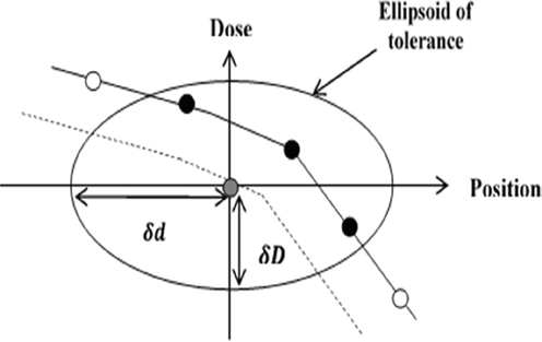
Schematic representation of the gamma index method in 1D (Low and Dempsey, 2003).
2.2 Workflow of the proposed software
The proposed software in this study can accept inputs (i.e. reference and evaluated dose distributions) of three types (*.txt, *.dcm, or *.3ddose). The gamma index is calculated using two main steps: interpolation and calculation. Fig. 2 illustrate of the flowchart of calculating gamma index in this software. The software is available at: https://github.com/thanhtaiphys/An-open-Source-Software-for-Calculating-1D-Gamma-Index-in-Radiation-Therapy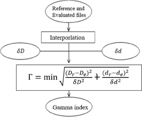
Flowchart of calculating gamma index in the proposed software.
2.2.1 Interpolation
In the case of dose comparison between simulation and measurement, it is possible that the number of points of the two distributions is not the same. To solve that problem, this software applies a linear interpolation for the two datasets before further calculations.
2.2.2 Calculation of gamma index
When conducting a gamma index calculation, the value of δd and δD can be chosen flexibly (e.g. δD/δd = 3%/3 mm). Low et al (Low et. al 1998) recommend that the pixel spacing should be 1/3δd, and Wendling et al (Wendling et al. 2007) concluded that the spacing should be of the order of 1/10δd. Hence, in this software, we designed so that δD and δd can be input arbitrarily by users, instead of some fixed values.
3 Results
3.1 Validation of the proposed software
The accuracy of this software was investigated by using two mathematical tests (namely two horizontal parallel lines with known distance and two inclined parallel lines with known distance tests) and one self-validated test.
3.2 Two horizontal parallel lines with known distance test
In this test, one reference dose distribution was chosen arbitrarily as (Dr = 2). Another evaluated dose distribution De was created by the formula Dr + 0.01Dr (De = 2.02). The two datasets were generated in such a way that for each point in Dr there exists a corresponding point in De. These two dose lines were used to simulate a uniform profile region.
Gamma index can be calculated using Eq. (1). Moreover, because the difference between Dr and De is 1%, Eq. (1) can be rewritten as follow.
From Eq. (2), it can be seen that the gamma index only depends on δD.
3.3 Two inclined parallel lines with known distance test
This test was used to simulate the PDD or OCR. In this test, reference and evaluated dose distributions are described Dr = (100 – d) and De = (100 – d) + s, respectively, where s is the percentage separation of the two distributions.
At every point
, let us define
as a real shift around d but small enough compared to
. As a result,
can be expressed as follow:
The above assumption is valid for points with
lower than
and
in the order of one or two DTAs which is a practical condition. In this test, Eq. (1) can be rewritten as follow.
For
and
,
has a first derivative and it is zero for the following
3.4 Validation using a self-validated test
In this test, the experiment and simulated profiles were chosen to be the same. Because the same file was input, there are no dissimilarity, and the results of gamma test should be zero. The gamma result of our software in this test is exactly zero (Fig. 3). The agreement between mathematical calculation and the result of this software can be observed.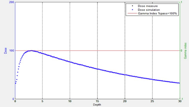
Gamma results.
Fig. 4 exhibits the interface of the proposed software in this study. By observing Fig. 4, it can be seen that dose difference (δD) and DTA (δd) could be freely adjusted. A graph showing reference dose, simulated (evaluated) dose, and gamma index is available.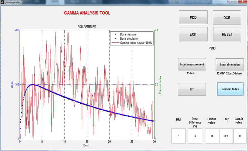
Interface of the proposed software in this study.
In the two horizontal parallel lines with known distance test, the calculated gamma index is only depending on dose difference as in Eq. (2). The results of this test with the simulated dose distributions at three value of δD/δd (3%/3 mm, 2%/3 mm, and 2%/2 mm) are shown in Fig. 5.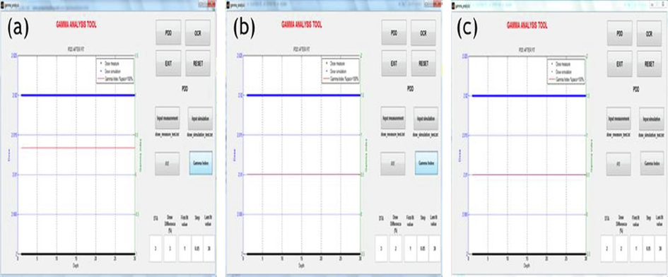
Analysis results of the two horizontal parallel dose lines at (δD/δd) of a) 3%/3 mm, b) 2%/3 mm, c) 2%/2 mm.
Fig. 6 summarizes the results. According to Fig. 6, it can be seen that the gamma index calculated by the proposed software only correlated with δD, the same the prediction of Eq. (2).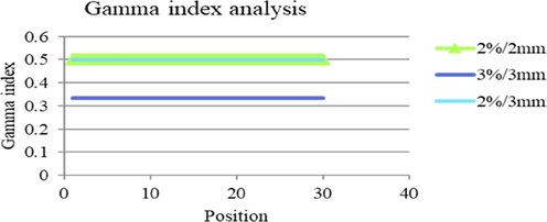
Summary of gamma index analysis at (δD/δd) of (3%/3 mm, 2%/3 mm, and 2%/2 mm).
In the two inclined parallel lines with known distance test, a reference point at position di together with an evaluated point at position di + lmin will give the minimum gamma index. The result of this test with the simulated dose distributions at 3 criteria of δD/δd (3%/mm, 2%/3 mm, and 2%/2 mm) is illustrated in Fig. 7.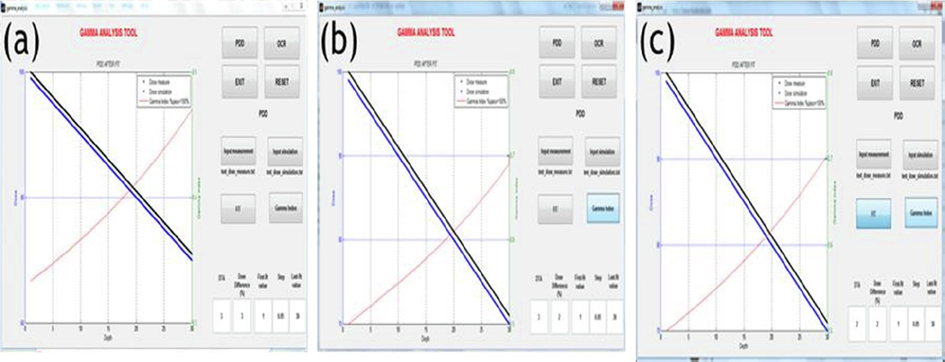
Gamma analysis results of the two inclined parallel dose lines at (δD/δd) of a) 3%/3 mm, b) 2%/3 mm, and c) 2%/2 mm.
Fig. 8 summarizes the results. According to Fig. 8, the agreement between the mathematical gamma index calculation and the results of this software can be observed. To test the accuracy of our proposed sofware, it was compared to the gamma evaluations of ScanDosematch and Bistromath software. Figs. 9 and 10 show gamma index comparison of our Software with other Software for PDD and OCR respectively. It can be seen that our proposed software not only show the gamma histogram but also the average gamma passing rate.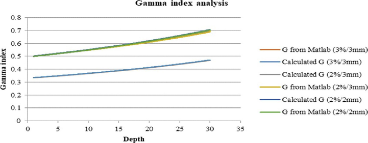
Summary of gamma analysis at (δD/δd) of 3%/3 mm, 2%/3 mm, and 2%/2 mm.
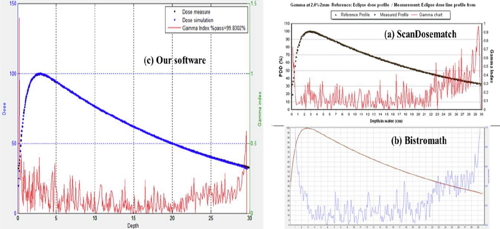
Gamma index comparison of the PDD from Software with other Software for PDD.
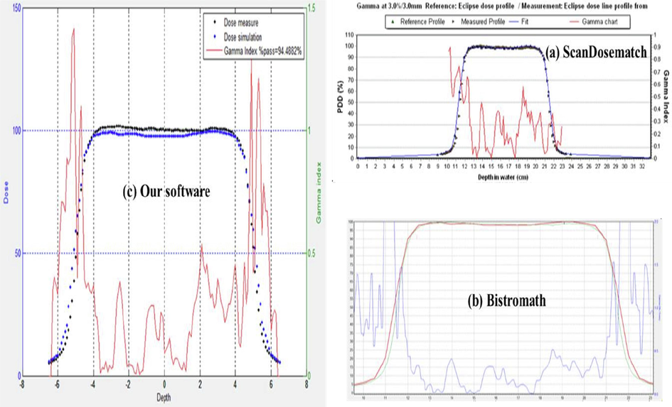
Gamma index comparison of our Software with other Software for OCR.
4 Discussion
Gamma index method is one of the standard validations for quality assurance in radiation therapy, and its usage is increasing at oncological facilities as well as institutions. At schools and universities, the lack of non-commercial and open-sourced software may limit the application of gamma-evaluation test for educational purposes. In this work, a free and modifiable software for conducting gamma test is proposed.
This software is expected to be a reliable toolkit which can facilitate educational as well as research purposes. There are commercial codes for the calculation of gamma evaluation for instance myQA software (IBA Dosimetry, Schwarzenbruck, Germany). Verisoft software (PTW, Frieburg, Germany) is commonly used for patient-specific quality assurance, not use for the analysis of profiles and depth dose curves. Therefore, the free program was introduced. (Tabrizi et al. 2020) introduced three software for the calculation of the gamma index. They used the measured and calculated dose profiles provided by (Low, 2010) to calculate the gamma index. The input type of their programs is still not easy to use. The new thing in our program is three types (*.txt, *.dcm, or *.3ddose) are supported. Snyder et al. (Snyder et al., 2019) used the Scandosematch software to perform gamma analyses between modeled and measured data. Like as Bistromatch software, the limitation of Scandosematch has not shown the percentage of gamma passing rate in interface of software.
As we can see in Fig. 9 and Fig. 10, we solved this issue. Our software is easy to use, support three types of input file, and efficient. However, there are some limitations to the present study. First, although this proposed software was carefully tested, the validation tests are purely theoretical calculation, and no other commercial software were used. Second, the structure of this software is very simple and only 1D gamma calculation is applicable. In the future, more development (2D and 3D gamma) and validated tests with other commercial software would further improve the reliability of this software. Jayamani et al. (Jayamani et al., 2022) used 2D gamma index to determine and quantify the dose delivery accuracy between dose measurements and calculation for the whole brain radiotherapy using pencil beam PB and Collapsed Cone CC algorithms.
Dose delivery accuracy in brain treatment is very crucial due to the presence of critical organs in the proximity of a treatment region that has irregular features with sharp density gradients.
5 Conclusions
The current study presented a development and validation of a non-commercial and modifiable software for conducting 1-D Gamma test. The developed software can be used to perform gamma analyses between modeled and measured data. The current software is a reliable, easy to use, has ability to support three types of input file, and efficient tool to be used at educational facilities and institutions for research purposes.
Acknowledgement
The authors express their gratitude to Princess Nourah bint Abdulrahman University Researchers Supporting Project (Grant No. pnursp2023R12), Princess Nourah bint Abdulrahman University, Riyadh, Saudi Arabia
Declaration of competing interest
The authors declare that they have no known competing financial interests or personal relationships that could have appeared to influence the work reported in this paper.
References
- Al Sa'd, M., Graham, J., Liney, G., 2013. A software tool for 3D dose verification and analysis. In: Journal of Physics: Conference Series. IOP Publishing.
- Assessment of using a gamma index analysis for patient-specific quality assurance in Japan. J. Appl. Clin. Med. Phys.. 2022;23:e13745.
- [Google Scholar]
- Bistromath.nl. Pretreatment QA for IMRT available at https://bistromath.kegge13.nl/index.php?i=intro.htm retrieved 12 August, 2021.
- Gamma index analysis as a patient-specific quality assurance tool for high-precision radiotherapy: a clinical perspective of single institute experience. Cureus. 2022;14(10)
- [Google Scholar]
- Challenges in calculation of the gamma index in radiotherapy – Towards good practice. Phys. Med. (36-11):1-11.
- [Google Scholar]
- Whole brain 3D conformal radiotherapy plan verification using gafchromic film in 2D gamma analysis. Phys. Technol. Med.. 2022;3(1):29-40.
- [Google Scholar]
- Evaluation of the gamma dose distribution comparison method. Medical Phys.. 2003;30(9):2455-2464.
- [Google Scholar]
- A technique for the quantitative evaluation of dose distributions. Med. Phys.. 1998;25(5):656-661.
- [Google Scholar]
- Challenges in Monte Carlo simulations as clinical and research tool in particle therapy: a review. Front. Phys.. 2020;8:567800
- [Google Scholar]
- QXRay Consulting Introducing ScanDoseMatch software Radiation Beam Data validation done faster. Available from: https://www.qxrayconsulting.com/sdm/.
- Development and evaluation of methods for comparison of dose distributions in radiotherapy using calculated, synthetic and simulated measured dose distributions. Medicine 2009
- [Google Scholar]
- Monte Carlo techniques in radiation therapy. Boca Raton, FL: CRC Press; 2013. p. :334.
- The commissioning and validation of monaco treatment planning system on an Elekta VersaHD linear accelerator. J. Appl. Clin. Med. Phys.. 2019;20(1):184-193.
- [Google Scholar]
- Introduction of a reliable software for the calculation of the gamma index. Iranian J. Med. Phys./Majallah-I Fīzīk-I Pizishkī-i Īrān. 2020;17(3):133-136.
- [Google Scholar]
- Tai D., Son N., Loan T., Tuan H., 2017. A method for determination of parameters of the initial electron beam hitting the target in Linac. In: Journal of Physics: Conference Series. IOP Publishing.


