

Translate this page into:
Synthesis and characterization of silver nanomaterial from aqueous extract of Commelina forskaolii and its potential antimicrobial activity against Gram negative pathogens
⁎Corresponding authors. rathi.muthaiyan@kahedu.edu.in (M.A. Rathi), vkgopalakrishnan@gmail.com (VK. Gopalakrishnan)
-
Received: ,
Accepted: ,
This article was originally published by Elsevier and was migrated to Scientific Scholar after the change of Publisher.
Peer review under responsibility of King Saud University.
Abstract
Abstract
Aim
Green synthesis of silver nanoparticles from medicinal plants have been progressively acquiring attractiveness to the researchers due to its sustainable nature, nontoxic and economically beneficial. The present study was to synthesize silver nanoparticles (AgNPs) from aqueous extract of Commelina forskaolii Vahl and exhibit its potential antimicrobial and cytotoxic activity.
Material and Methods
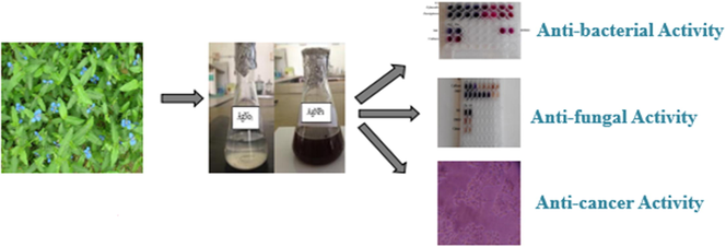
The whole plant of Commelina forskaolii was used to synthesize AgNPs. The synthesized AgNPs was then characterized by UV – visible spectroscopy, Fourier Transform Infrared Spectroscopy (FTIR), Scanning Electron Microscopy (SEM) and Transmission Electron Microscopy (TEM). The AgNps are widely tested for antibacterial, antifungal and cytotoxic property.
Results
The phytochemical screening of the aqueous extract showed the presence of secondary metabolites such as alkaloids, flavonoids, tannins, phenols, saponins, steroids, glycosides and proteins. The UV – vis absorption spectrum exhibited key peaks at 425 nm. FTIR spectrum revealed that the biochemical compounds are responsible for the reduction and capping material of AgNPs. SEM analysis showed, the average size of synthesized AgNPs ranged from 18 to 27 nm. TEM micrographs revealed that the particle size was to be 30–40 nm. The AgNPs exhibited potential antimicrobial activity against bacterial species (Enterococcus fecalis, Pseudomonas aeruginosa) showed MIC at about 62.5 µg/ml and 125 µg/ml respectively and fungal species (Candida albicans and Aspergilus niger) 250 µg/ml and 31.2 µg/ml respectively. The synthesized AgNPs showed potential cytotoxic activity against human breast cancer cell line (MCF-7) with the IC50 value of 50.2 µg/ml. The present investigation concludes the effectiveness of confirmed AgNPs might be used in pharmacological field for the treatment of bacterial, fungal and breast cancer.
Keywords
Commelina forskaolii
MTT
TEM
Silver nanoparticles
Antimicrobial
Cytotoxic activity

1 Introduction
Nanotechnology is one of the fast growing field and it has begun as an outstanding field with surplus of applications in plenteous areas including medicine, pharmacology, sensing devices, microelectronics and drug delivery etc. (Saini et al., 2010). The physiochemical properties of nanomaterials such as its nano size, high thermal conductivity they are widely used in the medical field for diagnosis and drug delivery approaches (Zhang et al., 2008). Physical and chemical approaches of nanoparticles are quite cost expensive and harmful to the environment because of the use of hazardous chemicals (Gurunathan et al., 2015). Green synthesis of silver nanoparticle production holds less cost effectiveness, ecofriendly and bulk scale synthesis without the utilization of high pressure, energy, temperature and hazardous chemicals (Singh et al., 2010). The synthesis of nanoparticles from plant based materials, bacteria, fungi and enzymes possesses numerous benefits in pharmaceutical and biomedical applications whereas toxic chemical are not involved for the synthesis procedure. Due to the unique properties of nanoparticles synthesized from biological origin the young researchers showed their interest to do research on nanoparticle worldwide (Verma et al., 2010). Numerous metals were used to synthesize nanoparticle such as silver, copper, gold, iron, platinum, silica and nickel (Ittiyavirah and Paul, 2016). Among these metals silver has the capability to cleave bacterial cell walls and inhibit the bacterial cell growth and interrupts the cell metabolism and cause cell death. It is a naturally occurring metal which is identified to be one of the antimicrobial constituents (Mohanta et al., 2017a,b; Logeswari et al., 2015). Nanoparticles can be synthesized from all the above mentioned metals. Among all these, silver nanoparticles possess distinctive properties such as chemical stability, good conductivity and showed potent antibacterial, antiviral, antifungal activity (Ahmad et al., 2003; Klaus-Joerger et al., 2001). In ancient days onwards medicinal plants were serving as a companion to humankind to treat against various disorders. Most commonly medicinal plants and microorganisms are used to synthesis nanoparticles (Mulfinger et al., 2007). The eco synthesis of nanoparticles from medicinal plants is due to the presence of secondary metabolites as reducing agents (Palaniselvam et al., 2016). The medicinal plant selected for the present investigation was Commelina forskaolii, belong to the family Commelina. It is an herbaceous plant commonly known as rat’s ear also known as rat's ear, native to Africa, Arabia, and India. Since this plant is perennial, fast growing, and small herbs, we utilized the whole plant for synthesis of nanoparticles. Commelina diffusa, belongs to this family as used by the traditional people for wounds, joint pains and burns. In Africa and Southern part of Asia it has been used to treat urinary tract infection, respiratory tract infections, diarrhea, and hemorrhoids (David, 1998). Sultana et al., (2018) reported that this plant also also used to treat fever, influenza, rheumatoid arthritis. The other species of Commelina such as Commelina benghalensis, used for skin softening, firing inflammation, smoothing emptying of bowel and also in treatment of leprosy. Since no any scientific investigation has been reported in Commelina forskaolii, and the above mentioned medicinal plants belong to this family exhibited potential activity in treating various illness prompt us to perform the current investigation to synthesis AgNPs from Commelina forskaolii, and carry out its potent antimicrobial and cytotoxic activity.
2 Materials and methods
2.1 Chemicals and reagents
Silver nitrate (AgNo3), MTT, Fetal Bovine Serum (FBS) were purchased from Sigma – Aldrich. Ferric chloride, hydrochloric acid, DMSO, Nutrient Agar, Nutrient broth were purchased from Himedia, Pvt.ltd. All other chemicals used for the study were purchased of analytical grade.
2.2 Preparation of extract
The whole plant of Commelina forskaolii was collected in the month of June 2018 from Western Ghates, Coimbatore, Tamil Nadu and authenticated by Botanical Survey of India, Coimbatore. The plant materials were washed in sterile water to remove any dirt or other unwanted objects, shade dried at room temperature and powdered using mixer. 300 g of plant powder were weighed and mixed with 1.5 L of water and boiled at 70 °C for 20 min. After cooling the extracts were filtered with Whatman filterpaper No 1 and dried by using rotatory evaporator and deposited at 20 °C for further use.
2.3 Phytocemical screening
The dried aqueous extract was diluted with water and screened qualitatively for the analysis of phytochemicals such as alkaloids, flavonoids, tannins, phenols, saponins, terpenoids, steroids and glycosides by the method of (Evans, 2002).
2.4 Biosynthesis of silver nanoparticles (AgNPs)
Silver nanoparticles (AgNPs) from aqueous plant extract were executed according to Geethalakshmi and Sarada (2012) with slight modifications. Aqueous plant extract was mixed with freshly prepared AgNO3 solution in the ratio 1:1 and incubated the mixture at room temperature. The appearance of brownish color was noticed after 24 hrs of incubation at room temperature. After that the mixture were centrifuged at 18,000 rpm for 20 min. The deposited pellet was washed with double distilled water and dried at room temperature.
2.5 Characterization
The synthesized AgNPs were characterized using various spectroscopic and microscopic techniques. The UV – visible spectrum of AgNPs were characterized by UV – visible spectrophotometer (Shimadzu 1700 Pharm spec) at the wavelengths ranges from 200 nm to 600 nm. Fourier Transform Infrared (FTIR) analysis of AgNPs was performed using Fourier Transform Infrared Spectroscopy (Thermo Fisher Scientific). SEM determine the surface morphology of nanoparticles Size, shape. TEM analysis was used to determine the qualitative measurement of synthesized nanoparticles such as, particle size, distribution and morphology (Pyrz and Buttrey,2008). Energy Dispersive Microanalysis techniques (EDX) was carried to confirm the presence of silver and other metal elements in the synthesized nanoparticles.
2.6 Antimicrobial activity
The pure culture of bacteria such as Enterococcus faecalis, Pseudomonas aeruginosa and fungi such as Candida albicans and Aspergillus niger were used to assess the antimicrobial activity of the AgNPs. The Minimum Inhibitory Concentration (MIC) of AgNPs was evaluated by broth dilution (resazurin) method Balachandran et al., (2015). Test was carried out in a 96 well plates under aseptic conditions separately for both bacterial and fungal species. A volume of 100 μl of sample was pipetted into the first well of the plate. To all other wells 50 μl of nutrient broth was added and serially diluted it and added 10 μl of resazurin indicator solution. The experiment was performed in different concentrations (7.8, 15.6, 25, 31.2, 50, 62.5, 100, 125 µg/ml) of AgNPs.
2.7 Cytotoxic activity
The human breast cancer cell lines MCF − 7, were procured from National Center for Cell Science, Pune and cultured in MEM (Minimal Essential Media) supplemented with 10 % Fetal Bovine Serum, 1 % penicillin - streptomycin at 37 °C atmosphere in 5 % CO2. MTT (3-(4, 5 dimethylthiazol-2-yl)-2,5-diphenyltetrazolium bromide) assay was performed to evaluate the in vitro cytotoxic activity of AgNPs on MCF-7 by the method of Mosmann (1983). Cells were seeded on 96 well plates (5000 cells/well) cultured for a day and then treated with different concentration of extract for 48 h at 37 °C in 5 % CO2. At the end of the incubation, medium was removed and MTT (5 mg/ml) was added and the cells were further incubated for 4 h after which the media was removed. DMSO was added in each well to solubilize the formazan crystals. The absorbance was read at a wave length 595 nm using a microtitre ELISA plate reader. Experiments for extract were carried out in triplicate including untreated cell control and blank cell – free control. Cell viability was expressed as percentage over the control.
2.8 Statistical analysis
All the assays were carried out in triplicates (n = 3) and the results represented as Mean ± S.D.
3 Results and discussion
3.1 Phytochemical screening
Results of qualitative phytochemical screening showed the presence of alkaloids, flavonoids, phenols, tannins, saponins, steroids, glycosides and proteins in aqueous extract of Commelina forskaolii (Table. 1). The presence of secondary metabolites such as alkaloids and flavonoids of plant origin are known for their numerous pharmacological significances. So far phytochemical screening was not assessed from this plant species. Similarly, Zhang and Dai, 2009 reported that one of the species of Commelina ie. Commelina communis showed the presence of phytochemicals such as flavonoids, glycosides, phenolic acid, alkaloids, pyrimidine, sterols and polysaccharides. Bioactive compounds such as orientin, vitexin, rutin, apigenin, vanillic acid, caffeic acid, quercetin and isorhamnetin were present in Commelina communis (Xia Zhang et al., 2018). Ezeabara et al. (2019) reported that Commelina diffusa and Commelina benghalensis possessed alkaloids, ash, fat, flavonoids and carbohydrates. +: the components are present.
S. No
Contents
Aqueous extract
1
Alkaloids
+
2
Flavonoids
+
3
Tannins
+
4
Phenols
+
5
Saponins
+
6
Steroids
+
7
Glycosides
+
8
Proteins
+
3.2 UV- analysis
The maximum absorption was found with a broad peak at 425 nm which indicates the presence of AgNPs in the sample solution owing to the surface plasmon resonance effects (Fig. 1). According to the literature survey it was mentioned that UV – vis spectroscopy reveals the maximum UV–visible absorption in the range of (400 nm–500 nm) for the initial characterization of synthesized nano particles and exhibit a surface plasmogen resonance effects (Amendola et al., 2010). The AgNPs strongly interact with specific wave lengths of light is one of its unique optical property. Depending upon the size and morphology of synthesized nanoparticles varies from yellow – green to brown (Raza et al., 2016). This recommends that the phytochemicals present in the Commelina forskaolii acts as a reducing agent.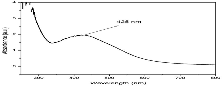
UV– vis spectrum of AgNPs.
3.3 FTIR analysis
The FTIR spectra of AgNPs is shown in Fig. 2. The band values obtained at 3289.50, 1617.18, 1377.66, 1030.27, and 464.86 cm−1, which revealed the presence of hydroxyl, amino and disulfide groups. The functional groups are responsible for the surface coating and effective stabilization of synthesized nanoparticles (Akintelu et al., 2020). The phytochemicals present in the plant extract is responsible for the the reduction and capping process during the synthesis of AgNPs.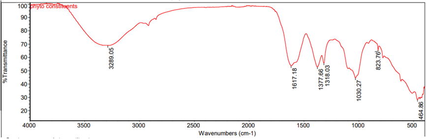
FTIR spectra of AgNPS.
3.4 SEM analysis
The morphology and size of the silver nanoparticles was determined using Scanning Electron Microscopy. Results of SEM analysis inferred, the average size of synthesized AgNPs ranged from 18 to 27 nm and showed the spherical and smooth surface morphology (Fig. 3). It is generally used to provide images about the size, shape, morphology and association of the surface topography of nanoparticles (Egerton, 2008). Similarly, Johnson and Prabu (2015) reported that Commelina benghalensis exhibited the spherical and uniform Ag nanoparticles in SEM analysis. These focused beam of electrons crash with sample surface to create secondary electrons. The evidence on the resulting electrons is exploited to restructure the sample structure morphology (Chandraker et al., 2021).
Scanning Electron Microscopy images of synthesized AgNPs.
3.5 TEM and EDX analysis
To confirm the structure morphology TEM analysis was performed. TEM micrographs of AgNPs revealed that the particle size was observed to be 30–40 nm (Fig 0.4 a). It is the powerful technique for providing exhaustive confirmation about the size and shape of nanoparticles (Dudkiewicz et al, 2011). The TEM characterization result in nm range confirms the synthesis of nanoparticle from the extract. EDX micro analysis is achieved by determining the energy and intensity distribution of X – ray signals produced by electron beam on a specimen. The peaks inferred in EDX analysis is represented in Fig. 4b. The spectra represent the presence of Ag in the synthesized AgNPs, addition to other metal elements like copper (Cu), iron (Fe), and calcium (Ca). The other elements present served as capping organic agents bound to the surface of the silver nanoparticles (Dada et al., 2017). Selected area of electron diffraction (SAED) pattern was recorded from the spherical AgNPs and it evidently shows the ring like electron diffraction patterns (Fig. 4c). Tai and Yang, 2011, reported that the diffraction rings of the AgNPs have been indexed as (1 1 1), (2 0 0) and (2 2 0) consistent with the face centered cubic (fcc) structure of Ag, typical of the polycrystalline AgNPs structure (Mehmood et al., 2014).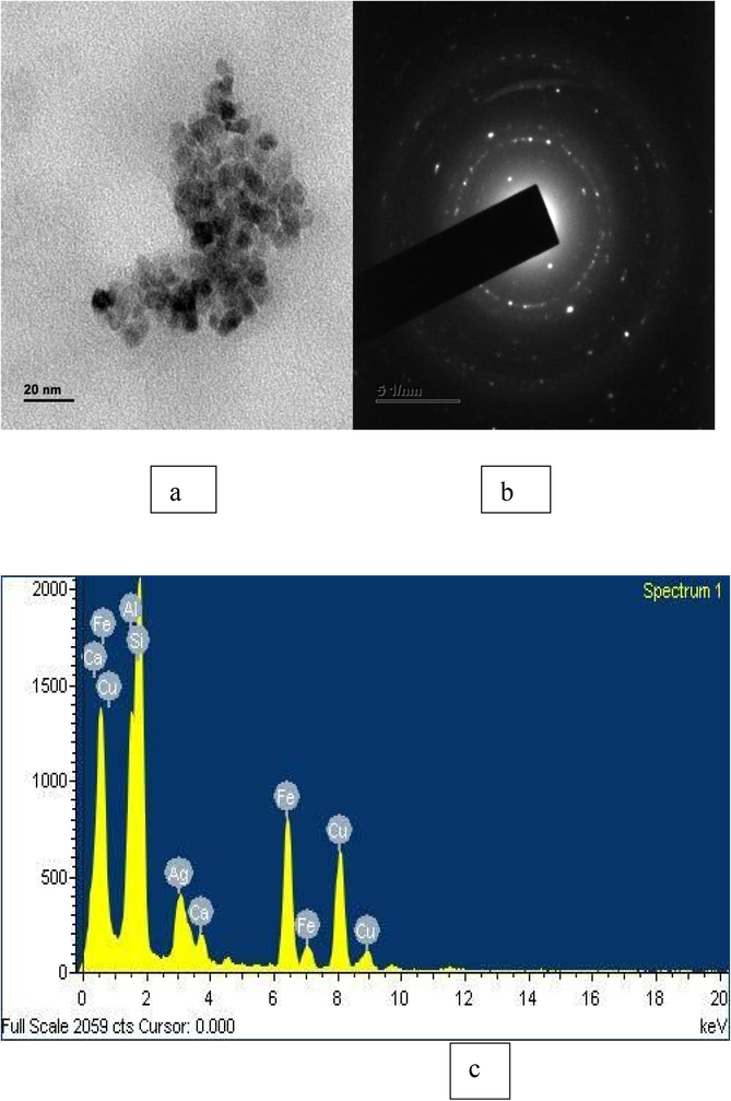
(a) Transmission Electron Microscopy (TEM) image of synthesized AgNPs (b) Selected area of electron diffraction pattern of the synthesized AgNPs showing the rings. (c) EDX analysis EDX analysis displayed the chemical composition of synthesized AgNPs.
3.6 Antimicrobial activity
The antimicrobial activity of AgNPs were tested against two bacteria (Enterococcus fecalis, Pseudomonas aeruginosa) and two fungi such as Candida albicans and Aspergilus niger. Inhibition of Enterococcus fecalis, Pseudomonas aeruginosa in treatment of AgNPs was found to be 62.5 µg/ml and 125 µg/ml respectively. Inhibition of Candida albicans and Aspergilus niger in treatment of AgNPs was found to be 250 µg/ml and 31.2 µg/ml respectively (Table 2). The lipo-solubility property of active compounds present in AgNPs could be responsible for the degradation of the bacterial cell membrane and enter in to the bacterial cell (Elemike et al., 2017). Most AgNPs obtained from medicinal plants have been credited with high antimicrobial properties, especially against drug-resistant human pathogenic and food spoilage microbes (Abdel-Aziz et al., 2014; Reddy et al., 2014). The results from this study correlate with other reports that most plant extract AgNPs exhibit enhanced broad-spectrum antibacterial activities (Mohanta et al., 2017a,b).
S. No
Microorganisms
MIC (µg/ml)
Bacterial species
1
Enterococcus fecalis
62.5
2
Pseudomonas aeruginosa
125
Fungal Species
1
Candida albicans
250
2
Aspergillus niger
31.2
3.7 Cytotoxic activity
The cytotoxic potency of plant extract and AgNPs were evaluated against MCF – 7 (human breast cancer cell lines) by MTT at different concentration. The phase contrast photographs of MCF − 7 (Figs. 5 and 6) at different concentration displayed the abnormal cellular morphologies with detachment of cells and shrinking with a significant reduction in cell viability. Cell growth was inhibited and eventually the cell death occurred and aggregated to form round dead cells at the highest concentration. The plant extract and AgNPs showed the IC50 value of 50.2 µg/ml and 100.5 µg/ml respectively. The present investigations show significant cytotoxic potential of CF-AgNPs against MCF − 7 cell lines. Such potential activity could be due to the effect of biosynthesized nanoparticles and the bioactive components attached to their surface (Valsalam et al., 2019). The result of the present study demonstrates that the cytotoxic activity of AgNPs mediate a dose and time dependent manner.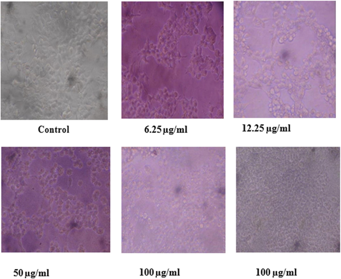
Morphological changes in MCF – 7 cell lines treated with AgNPs at different concentration.
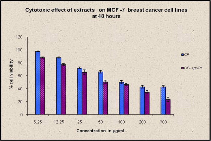
Cytotoxic effect of plant extracts on breast cancer cell lines at 48 h. The AgNPs and plant extract showed the IC 50 value of 50.2 µg/ml and 100.5 µg/ml respectively.
4 Conclusion
The current study presents the environmentally friendly and cost effective procedure for the synthesis of AgNPs by exploiting renewable natural resources. The AgNPs were synthesized from Commelina forskaolii at room temperature and were characterized using UV – vis spectroscopy, FTIR, SEM, TEM and EDX analysis. The UV–Vis spectra confirmed the surface plasmon resonance of green-synthesized silver nanoparticles. The FTIR spectra revealed that the biomolecules were responsible for reducing and capping of AgNPs. Gram negative bacteria are the most common pathogen present in critically ill patients. Bacterial and fungal infections cause crucial diseases in worldwide. AgNPs exhibited good antimicrobial against pathogens and also possess potent cytotoxic activity against MCF − 7 (breast cancer cell lines). Further in vivo studies should be carried out for successful application in pharmaceutical and nanotechnology based industries in emerging of new drugs to fight against bacterial, fungal pathogens and to suppress the proliferation of cancer cells.
Acknowledgements
The authors like to thank the Sree Narayana Guru Educational Trust (SNGET), Coimbatore for providing the financial support. Authors also like to thank the management of Sree Narayana Guru College and Karpagam Academy of Higher Education for providing the laboratory facilities. The authors acknowledge the support from Princess Nourah bint Abdulrahman University Researchers Supporting Project number (PNURSP2022R15), Princess Nourah bint Abdulrahman University, Riyadh, Saudi Arabia.
Declaration of Competing Interest
The authors declare that they have no known competing financial interests or personal relationships that could have appeared to influence the work reported in this paper.
References
- Antioxidant and antibacterial activity of silver nanoparticles biosynthesized using Chenopodium murale leaf extract. J. Saudi Chem. Soc.. 2014;18(4):356-363.
- [Google Scholar]
- Extracellular biosynthesis of silver nanoparticles using the fungus Fusarium oxysporum. Colloids Surf., B. 2003;28(4):313-318.
- [Google Scholar]
- A review on synthesis, optimization, mechanism, characterization, and antibacterial application of silver nanoparticles synthesized from plants. J. Chem.. 2020;2020:1-12.
- [Google Scholar]
- A study of the surface plasmon resonance of silver nanoparticles by the discrete dipole approximation method: effect of shape, size, structure, and assembly. Plasmonics. 2010;5(1):85-97.
- [Google Scholar]
- Antimicrobial and cytotoxic properties of Streptomyces sp. (ERINLG-51) isolated from Southern Western Ghats. South Indian. J. Biol. Sci.. 2015;1(714):5.
- [Google Scholar]
- Green synthesis and characterization of silver nanoparticles using Calotropis procera extract. J. Appl. Chem. Sci. Int.. 2017;8(4):137-143.
- [Google Scholar]
- David, B. L. Ac. Medicine at your feet: healing plants of the Hawaiian Kingdom Commelina diffusa (Honohono). (1998) Publishing web: www.medicineatyourfeet.com.
- Characterization of nanomaterials in food by electron microscopy. Trends Analyt. Chem.. 2011;30(1):28-43.
- [Google Scholar]
- Egerton, 2008, R.F. Physical Principles of Electron Microscopy: An Introduction to TEM.
- Silver nanoparticles mediated by Costus afer leaf extract: synthesis, antibacterial, antioxidant and electrochemical properties. Molecules. 2017;22(5):701.
- [Google Scholar]
- Trease and evans. Pharmacognosy 2002 9th Edition published by Saunders Elsevier, 553
- [Google Scholar]
- Phytochemical and Proximate Studies of Various Parts of Commelina benghalensis L. and Commelina diffusa Burm. f. Int. J. Plant Sci. Ecol.. 2019;5(4):43-46. 2381-6996 (Print); ISSN: 2381-7003
- [Google Scholar]
- Gold and silver nanoparticles from Trianthema decandra: synthesis, characterization, and antimicrobial properties. Int. J. Nanomed.. 2012;7:5375.
- [Google Scholar]
- Comparative assessment of the apoptotic potential of silver nanoparticles synthesized by Bacillus tequilensis and Calocybe indica in MDA-MB-231 human breast cancer cells: targeting p53 for anticancer therapy. Int. J. Nanomed.. 2015;10:4203.
- [Google Scholar]
- Gastroprotective effect of plumbagin and ethanolic extract of plumbaginales in experimentally-induced ulcer. J. HerbMed Pharmacol.. 2016;5(3)
- [Google Scholar]
- Green synthesis and characterization of silver nanoparticles by leaf extracts of Cycas circinalis, Ficus amplissima, Commelina benghalensis and Lippia nodiflora. Int. Nano Lett.. 2015;5(1):43-51.
- [Google Scholar]
- Bacteria as workers in the living factory: metal-accumulating bacteria and their potential for materials science. Trends Biotechnol.. 2001;19(1):15-20.
- [Google Scholar]
- Chandraker et al., 2021, S.K. Chandraker, M.K. Ghosh, M. Lal, et al. A review on plant-mediated synthesis of silver nanoparticles, their characterization and applications Nano Express, 2 (2021), p. 22008.
- Synthesis of silver nanoparticles using plants extract and analysis of their antimicrobial property. J. Saudi Chem. Soc.. 2015;19(3):311-317.
- [Google Scholar]
- Antibacterial efficacy of silver nanoparticles synthesized by a green method using bark extract of Melia azedarach L. J. Pharm. Innov.. 2014;9(3):238-245.
- [Google Scholar]
- Biosynthesis of silver nanoparticles from Protium serratum and investigation of their potential impacts on food safety and control. Front. Microbiol.. 2017;8:626.
- [Google Scholar]
- Antimicrobial, antioxidant and cytotoxic activity of silver nanoparticles synthesized by leaf extract of Erythrina suberosa (Roxb.) Front. Mol. Biosci.. 2017;4:14.
- [Google Scholar]
- Rapid colorimetric assay for cellular growth and survival: application to proliferation and cytotoxicity assays. J. Immunol. Methods. 1983;65:55-63.
- [Google Scholar]
- Nanoporous graphene enriched with Fe/Co-N active sites as a promising oxygen reduction electrocatalyst for anion exchange membrane fuel cells. Adv. Funct. Mater.. 2016;26(13):2150-2162.
- [Google Scholar]
- Particle size determination using TEM: a discussion of image acquisition and analysis for the novice microscopist. Langmuir. 2008;24(20):11350-11360.
- [Google Scholar]
- Size and shape- dependent antibacterial studies of silver nanoparticles synthesized by wet chemical routes. Nanomaterials. 2016;6:74.
- [CrossRef] [Google Scholar]
- Evaluation of antioxidant, antibacterial and cytotoxic effects of green synthesized silver nanoparticles by Piper longum fruit. Mater. Sci. Eng., C. 2014;34:115-122.
- [Google Scholar]
- Saini, R., Saini, S., & Sharma, S. (2010). Nanotechnology: the future medicine. Journal of cutaneous and aesthetic surgery, 3(1), 32. SEM, and AEM, New York: Springer Science, 2008. https://doi.org/10.1007/b136495.
- Green synthesis of silver nanoparticles using Argemone mexicana leaf extract and evaluation of their antimicrobial activities. Dig. J. Nanomater. Bios.. 2010;5(2):483-489.
- [Google Scholar]
- Evaluation of central nervous system (CNS) depressant activity of methanolic extract of Commelina diffusa Burm. in mice. Clin. Phytosci.. 2018;4(1):1-7.
- [Google Scholar]
- Fabrication of paper-based conductive patterns for flexible electronics by direct-writing. J. Mater. Chem.. 2011;21(16):5938-5943.
- [Google Scholar]
- Biosynthesis of silver and gold nanoparticles using Musa acuminate colla flower and its pharmaceutical activity against bacteria and anticancer efficacy. J. Photochem. Photobiol. B. 2019;201:111670
- [CrossRef] [Google Scholar]
- Biosynthesis of antimicrobial silver nanoparticles by the endophytic fungus Aspergillus clavatus. Nanomedicine. 2010;5(1):33-40.
- [Google Scholar]
- Zhang, X.L. and Dai, L.C. A review of chemical constituents and pharmacological effects of Commelina communis Linn. Medicine plant chemistry and traditional Chinese medicine resources sustainable development seminar, (2009) 47–50.
- Nanoparticles in medicine: therapeutic applications and developments. Clin. Pharmacol. Ther.. 2008;83(5):761-769.
- [Google Scholar]
- Simultaneous qualitative and quantitative study of main compounds in Commelina communis linn. by UHPLC–Q-TOF-MS-MS and HPLC–ESI-MS-MS. J. Chromatogr. Sci.. 2018;56(7):582-594.
- [Google Scholar]


