

Translate this page into:
Sero-diagnosis of Corynebacterium pseudotuberculosis affecting camels using recombinant phospholipase D (rPLD) and anti-camels IgY-conjugate
⁎Corresponding author. imoussa1@ksu.edu.sa (Ihab M. Moussa)
-
Received: ,
Accepted: ,
This article was originally published by Elsevier and was migrated to Scientific Scholar after the change of Publisher.
Peer review under responsibility of King Saud University.
Abstract
Caseous lymphadenitis has universally prevalence especially in Saudi Arabia causing pseudotuberculosis or caseous lymphadenitis (CLA) in sheep and camels. The affected camels showed sever emaciation and multiple abscesses. The current study is aimed to evaluate the incidence of Corynebacterium pseudotuberculosis infections in camels in the Kingdome of Saudi Arabia and sero-diagnosis of the disease using recombinant PLD protein as coating antigen and anti-camel conjugated peroxidase as a specific anti species in enzyme linked immuno-sorbant assay. A total of 280 adult one humped camels from different localities; 35 apparently infected camels with CLA and 245 apparently normal camels were examined clinically and subjected to standard bacteriological technique, sero-diagnosis using ELISA and molecular detection and characterization using multiplex PCR. Thirty five out of 280 (12.5%) examined camels had superficial lymph nodes with cheesy whitish to greenish pus while 3 (1.07%) camels had congested internal lymph nodes from apparently normal camels. Using the recombinant PLD as a coating antigens and anti-camel conjugated peroxidase as a specific anti species for camels revealed high sensitivity and specificity and the ELISA technique could detect all the apparently infected animals(100%) and all the apparently normal harbouring the infections. The current study concluded that using of recombinant PLD as a coating antigens and anti-camel IgY conjugated peroxidase as a specific anti-species for camels in ELISA can be utilized for rapid and specific detection of infected and apparently normal camels infected with C. pseudotuberculosis.
Keywords
Sero-diagnosis
Corynebacterium pseudotuberculosis
Caseous lymphadenitis
Recombinant PLD
Anti-camel IgY conjugate

1 Introduction
Corynebacterium pseudotuberculosis biotype ovis strain, is a Gram-positive bacteria most commonly associated with pseudotuberculosis or caseous lymphadenitis (CLA) in sheep and camels (Camelus dromedaries) and in horses and goat (Ulcerative lymphangitis) (Guerrero et al., 2018, Osman et al., 2018, Tejedor et al., 2004).
CLA has universally prevalence especially in Saudi Arabia causing serious economic losses for ovine and caprine husbandries (reduce meat, milk and wool yield, culling of affected animals and contamination of carcasses and skins in slaughter houses) as well as it could be transmitted to human leading to abscess in liver and suppurative inflammation in lymph nodes (Guerrero et al., 2018, Hawari 2008, Tarazi and Al-Ani, 2016). If this disease established in a flock or herd, CLA eradication will be problematic due to the inefficacy of antimicrobial therapy. The best reliable control strategy in case of this disease should be including vaccination of livestock in addition to identification and removing the infected animals.
The affected camels showed sever emaciation and multiple abscesses formation (especially of the superficial Lymph nodes) and the internal organs especially liver and lungs showed granulomatous abscess formation surrounded by fibrous tissues (Borham et al., 2017, Wernery and Kinne, 2016).
C. pseudotuberculosis secretes a phospholipase D (PLD) exotoxin that is responsible for the major virulence component of this bacterium. Rapid detection and characterization of C. pseudotuberculosis affecting camels by enzyme linked immuno-sorbant assay (ELISA) using recombinant PLD (Mara Thais de Oliveira Silva, 2019) and molecular detection of C. pseudotuberculosis using multiplex PCR which could detect three genes, the 16S rRNA gene, rpoB gene; the RNA polymerase β-subunit gene and pld gene, specific for the microorganism, at the same time can facilitate the rapid detection and isolation of the infected animals and the carrier animals harboring the organism (Moussa et al., 2014).
The present study is directed to evaluate the incidence of C. pseudotuberculosis infections in camels in the Kingdome of Saudi Arabia and using recombinant crud PLD as a coating antigens and anti-camel conjugated peroxidase as a specific anti-species for sero-diagnosis of CLA diseased camels using such ELISA kit.
2 Materials and methods
2.1 Standard microbiological techniques for detection and characterization of C. pseudotuberculosis
2.1.1 Collection of lymph nodes and serum samples of infected and apparently normal camels from Abattoir
A total of 280 adults one humped camels from localities; 35 apparently infected camels with Caseous Lymphadenitis (with congested superficial lymph nodes or lymph nodes showing cheesy whitish to greenish pus) and 245 apparently normal camels were examined clinically and subjected to post-mortem examination. Blood samples, superficial lymph nodes as well as deeply affected lymph nodes were collected.
2.1.2 Isolation and identification of Corynebacterium pseudotuberculosis
Under complete aseptic conditions aspirate from the collected lymph nodes showing cheesy whitish to greenish pus were inoculated onto brain heard infusion agar (Oxoid) containing 4 mg/L nalidixic acid and 200 mg/L fosfomycin (Sigma) and incubated aerobically at 37 °C for 48 h. Positive culture was identified according to Tejedor-Junco (2008) using Gram stains and different biochemical test specially urease, catalase, and nitrate reduction test.
2.2 Preparation of Corynebacterium pseudotuberculosis antigens; mainly phospholipase D (PLD) and recombinant PLD for preparation of coating antigens for ELISA
The completely identified strains of C. pseudotuberculosis by bacteriological examination were inoculated onto brain heard infusion broth containing 4 mg/L nalidixic acid and 200 mg/L fosfomycin (Sigma), then incubated aerobically at 37 °C for 72 h and. Culture filtrates were obtained by centrifuged for 30 min. at 10,000×g. The supernatants were filtered through a 0.22 µm membrane and the total protein content were measured using Lowry modified method by using a commercial detergent compatible protein assay kit and then kept at −20 °C for further analysis (Mara Thais de Oliveira Silva, 2019, Moura-Costa et al., 2008). The recombinant DNA was prepared as previously mentioned by Moussa et al. (2014).
2.3 Enzyme linked immunosorbant assay (ELISA) for the detection of specific immunoglobulins against Caseous lymphadenitis in camels
Preliminary work had been done to evaluate the suitable concentration of the crude (PLD) and recombinant PLD (rPLD), the cut off values for the ELISA and the suitable dilution of the serum samples collected from infected and apparently normal camels (Mara Thais de Oliveira Silva, 2019) using a panel of 10 reference positive sera (from camels which had an abscess and culture-positive for C. pseudotuberculosis) and 10 reference negative sera (from camels flocks which had never had a case of CLA). Briefly, ELISA plates were coated with the crude C. pseudotuberculosis antigens diluted 1:100 and filtrate diluted 1:100 in carbonate bicarbonate buffer (pH 9.6) (5 μg/well), and incubated at 4 °C for 12 hr. Then the plates then were washed twice with phosphate buffered saline solution containing 0.05 % Tween 20 (PBST), blocked with 5 % skimmed milk in PBST, and incubated for 2 hr at 37 °C. After 2 additional washes with PBST, 100 ml of serum samples collected from all camels either infected or apparently normal (280 camels) diluted 1:100, in PBST with 1 % skimmed milk were added (each serum sample was tested in duplicate). The same negative and positive controls were used in all plates and the mixture was incubated for 1 hr at 37 °C. The plates were washed 3 times with PBST. Camel IgY antibodies conjugated with horseradish peroxidase, were added at a concentration of 1:5000 (recommended concentration after checker board titration in the preliminary tested plate) and incubated for 1 hr at 37 °C. After that the plates were washed, and the reaction was developed with tetramethylbenzidine chromogen and hydrogen peroxide substrate (100 ml/well) for 15 min, and the reaction was stopped with 1 N sulfuric acid. The Optical Densities (OD) were measured using ELISA reader at 450 nm (Carminati et al., 2003 and Seyffert et al., 2010).
2.4 Molecular detection and characterization of the recovered Corynebacterium pseudotuberculosis from infected camels
2.4.1 Extraction of DNA from the recovered isolates
The genomic DNA from the lymph nodes of infected and apparently normal camels bacteriologicaly positive for isolation of C. pseudotuberculosis as well as the DNA extracted from all the strains recovered by bacteriologically examinations revealed were extracted by DNA extraction Kits.
2.4.2 Multiplex PCR assay
The DNA extracted from the lymph nodes of infected and apparently normal camels bacteriologically positive for isolation of C. pseudotuberculosis as well as the DNA extracted from all the strains recovered by bacteriologically examinations were subjected for direct detection of 16S rRNA gene, rpoB gene and pld gene specific for identification of C. pseudotuberculosis using multiplex PCR according to Moussa et al. (2014). The PCR products were examined by agarose gel electrophoresis after staining of the amplified bands with ethedium bromide.
3 Results
3.1 The post-mortem examination
The post-mortem examination revealed 35out of 280 (12.5 %) examined camels had superficial lymph nodes showed cheesy whitish to greenish pus (inferior cervical, prescapular, mandibular or Pectoral lymph nodes) while 3 (1.07 %) camels had congested internal lymph nodes from apparently normal camels as shown in Table 1 and Figs. 1 and 2.
Test used
Apparently infected camels with Caseous Lymphadenitis (35 camels)
Apparently normal camels
(245 camels)
Postmortem Examination
35 (12.5 %)
3 (1.07 %)
Bacteriological examinations
28/35 (80 %)
0 (0 %)
ELISA
32/35 (94.28 %)
8 (2.86 %)
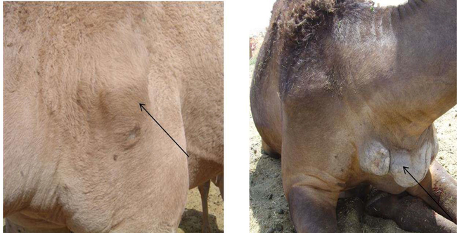
Infected camels showing abscess formation filled with pus.
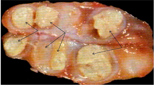
Affected lymph nodes filled with pus and caseated materials.
3.2 Bacteriological examination
Bacteriological examination revealed only 28 (80 %) out of 35 examined lymph nodes from the apparently infected camels with caseous lymphadenitis were positive for isolation of C. pseudotuberculosis although all the three examined congested lymph nodes were negative for isolation of C. pseudotuberculosis as shown in Table1.
3.3 Enzyme linked immunosorbant assay (ELISA)
ELISA could detect 32 (94.28 %) out of 35 apparently infected animals. All 28 serum samples collected from bacteriologically positive animals for C. pseudotuberculosis (100 %). While in case of apparently normal camels, the three serum samples collected from camels with congested internal lymph nodes were positive for ELISA, more over ELISA could detect another 5 positive samples apparently normal without any post mortem finding as shown in Table 1.
3.4 Molecular detection and characterization of the recovered C. pseudotuberculosis from infected camels
All the DNA extracted from the lymph nodes of infected and apparently normal camels bacteriologically positive or negative for isolation of C. pseudotuberculosis 35 apparently infected animals and three apparently normal with congested lymph nodes were positive for multiplex PCR (100 %). The amplification of 816 bp fragments of 16S rRNA, 446 bp fragments of rpoB gene and 203 bp fragments of PLD gene of C. pseudotuberculosis were observed as shown in Fig. 3.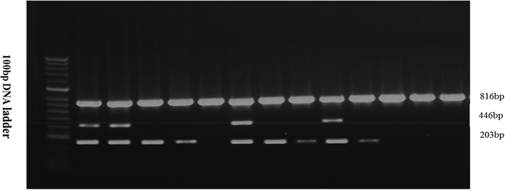
Agarose gel electrophoresis showing multiplex PCR for amplification of 816 bp fragments of 16S Rrna, 446 bp fragments of rpoB gene and 203 bp fragments of PLD gene of C. pseudotuberculosis.
4 Discussion
Caseous lymphadenitis (CLA) in camels has universally prevalence especially in Saudi Arabia causing serious economic losses for camels, therefore, the current study is aimed to evaluate the incidence of (CLA) in camels in the Kingdom of Saudi Arabia and evaluate the currently used test for eradication program of the disease and evaluate the recombinant PLD as a coating antigen and anti-camel conjugated peroxidase for serodiagnosis of (CLA) in camels using ELISA technique.
Diagnosis of (CLA) in camels is based primarily upon clinical observations (external abscesses) and isolation and identification of C. pseudotuberculosis phenotypically and biochemically using standard microbiological technique to differentiate this bacterium from other abscess-inducing pathogenic bacteria (Dercksen et al., 2000a; Dercksen et al., 2000b; Williamson, 2001; Dorella et al., 2009).
Synergistic haemolysis-inhibition test, microagglutination assay, complement fixation test and C. pseudotuberculosis phospholipase D (PLD) antigen-based ELISA have been proposed for diagnosis of suppurative lymphadenitis in camels (Dercksen et al., 2000a; Dercksen et al., 2000b). These tests have a great value for the early detection of subclinically and apparently normal animals, however, most of them have many draw backs, including low sensitivity, and specificity and cannot differentiate between previously exposed animals and those still harboring the pathogen.
In the present study 280 adult camels (35 apparently infected camels with suppurative lymphadenitis and 245 apparently normal camels without any gross lesions) were examined clinically and subjected to post-mortem examination. Blood samples, superficial lymph nodes as well as deeply affected lymph nodes were collected. The post-mortem examination revealed 35out of 280 (12.5 %) examined camels had superficial lymph nodes showed cheesy whitish to greenish pus (inferior cervical, prescapular, mandibular or Pectoral lymph nodes) while 3 (1.07 %) camels had congested internal lymph nodes from apparently normal camels as shown in Table 1 and Figs. 1 and 2. The results of Radwan et al. (1989) support our results in a study in camels of KSA.
Bacteriological examination could detect only 28 (80 %) out of 35 examined lymph nodes from the apparently infected camels with C. pseudotuberculosis although all of which were positive by multiplex PCR and ELISA using recombinant crud PLD (rPLD). Moreover, three examined congested lymph nodes were negative for isolation of C. pseudotuberculosis this information indicated the lower sensitivity of bacteriological methods for detection of the positive cases and it might be due to the many drawback of bacteriological method (Mara Thais de Oliveira Silva, 2019) as shown in Table 1.
Using the recombinant crud PLD as a coating antigens ant anti-camel conjugated peroxidase as a specific anti species for camels revealed high sensitivity and specificity and the test could detect all apparently infected animals (100 %) and all the apparently normal harbouring the infections as shown in Table 1. Our results confirm the conclusions of what been described in previous report (Mara Thais de Oliveira Silva, 2019).
5 Conclusions
Using a recombinant PLD as a coating antigens and anti-camel conjugated peroxidase as a specific anti-species for camels in enzyme linked immuno-sorbant assay can be utilized for rapid and specific detection of infected and apparently normal camels infected with C. pseudotuberculosis.
Acknowledgment
This work was funded by the National Plan for Science, Technology and Innovation (MAARIFAH), King Abdulaziz City for Science and Technology, Kingdom of Saudi Arabia, Award Number (2-17-04-001-0007).
Declaration of Competing Interest
The authors declare that they have no known competing financial interests or personal relationships that could have appeared to influence the work reported in this paper.
References
- Caseous lymphadenitis in Sudanese and Somalian camels imported for meat consumption in Egypt. Alexandria J. Vet Sci.. 2017;55(2):52-59.
- [Google Scholar]
- A comparison of four serological tests for the diagnosis of caseous lymphadenitis in sheep and goats. Vet. Microbiol.. 2000;75(2):167-175.
- [Google Scholar]
- A comparison of four serological tests for the diagnosis of caseous lymphadenitis in sheep and goats. Vet. Microbiol.. 2000;75:167-175.
- [Google Scholar]
- Antigens of Corynebacterium pseudotuberculosis and prospects for vaccine development. Expert Rev. Vaccines.. 2009;8(2):205-213.
- [Google Scholar]
- Isolation and molecular characterization of Corynebacterium pseudotuberculosis from sheep and goats in Mexico. Microb. Pathog.. 2018;117:304-309.
- [Google Scholar]
- Corynebacterium pseudotuberculosis infection (caseous lymphadenitis) in camels (Camelus dromedarius) in Jordan. Am. J. Anim. Vet. Sci.. 2008;3(2):68-72.
- [Google Scholar]
- Mara Thais de Oliveira Silva , Francisco Silvestre Brilhante Bezerra , Rodrigo Barros de Pinho, Caroline de Santana Ferreira , Wanessa Lordêlo Vivas , Ricardo Wagner Dias Portela, Vasco Ariston de Carvalho Azevedo, Sibele Borsuk1, J. Med. Microbiol. 2019;68:1759–1765.
- Evaluation of the humoral and cellular immune response to different antigens of Corynebacterium pseudotuberculosis in Caninde goats and their potential protection against caseous lymphadenitis. Vet. Immunol. Immunopathol.. 2008;126:131-141.
- [Google Scholar]
- Single point mutation as a molecular tool for preparation of recombinant vaccine against Caseous Lymphadenitis. J. Food, Agric. Environ.. 2014;12(2)
- [Google Scholar]
- The epidemiology and pathophysiology of caseous lymphadenitis: A review. J. Vet. Med. Res.. 2018;5(3)
- [Google Scholar]
- Corynebacterium pseudotuberculosis infection in camels (Camelus dromedarius) in Saudi Arabia. Trop. Anim. Health Prod.. 1989;21(4):229-230.
- [Google Scholar]
- An outbreak of dermatophilosis and caseous lymphadenitis mixed infection in camels (Camelus dromedaries) in Jordan. J. Infect. Dev. Ctries.. 2016;10(05):506-511.
- [Google Scholar]
- Pseudotuberculosis in dromedary camels in the Canary Islands. Trop. Anim. Health Prod.. 2004;36(5):459-562.
- [Google Scholar]
- Lymphadenitis (Pseudotuberculosis) in camelids: a review. Austin J. Vet. Sci. Anim. Husb.. 2016;3(1):1-6.
- [Google Scholar]
- Caseous lymphadenitis in small ruminants. Vet. Clin. North. Am. Food Anim. Pract.. 2001;17:359-371.
- [Google Scholar]


