

Translate this page into:
Biofabricated selenium nanoparticles mediated from Goniothalamus wightii gains biomedical applications and photocatalytic degrading ability
⁎Corresponding authors at: Ethnopharmacology and Algal Biotechnology Laboratory, Department of Botany, School of Life Sciences, Periyar University, Salem, Tamil Nadu 636 011, India (A. Maruthupandian); Centre of Excellence for Pharmaceutical Sciences, North-West University, Potchefstroom 2520, South Africa (K.R.R. Rengasamy). Department of Pharmacy, University of Naples Federico II, 80131 Naples, Italy (M. Daglia). rengasamy@iceir.net (Kannan R.R. Rengasamy), maria.daglia@unina.it (Maria Daglia), pandianmdu82@gmail.com (Arumugam Maruthupandian)
-
Received: ,
Accepted: ,
This article was originally published by Elsevier and was migrated to Scientific Scholar after the change of Publisher.
Abstract
Selenium nanoparticles (SeNPs) have attracted recently in different biomedical applications due to its unique role of metabolic function prevents cellular damage and other severe disease of human. In this study, biosynthesis of SeNPs from Goniothalamus wightii (Gw-SeNPs) and characterization were performed on UV–vis spectroscopy showed at 265 nm, it was primarily confirmed the biosynthesis of SeNPs. The XRD pattern revealed that peaks at 29.98 (1 0 1), 33.22 (1 1 1), 47.42 (2 0 0), 65.38 (2 2 0) and 77.40 (3 1 1)) exhibited a highly crystalline structure. The FE-SEM image proved that the formation of Gw-SeNPs which were polygonal shape. Zeta potential and DLS value of SeNPs recorded as −17.6 mV and 20–110 nm. The results described the synthesized SeNPs have good antibacterial potential on both strains of gram-positive as well as gram-negative pathogens. In vitro antioxidant ability of biogenic Gw-SeNPs has been assessed the inhibition of DPPH free radical scavenging ability with IC50 value was reported as 75.62 (µg/mL) and ABTS inhibitory concentration at 50% was 74.12 (µg/mL). The present findings provide important evidence on cytotoxic effects of Gw-SeNPs on MCF-7 cells. Dye degradation ability of Gw-SeNPs against reactive dyes over the 97% in 150 min in methylene blue dye degradation, the experiment was done under the direct sunlight. Finally, the larvicidal activity against fourth instars mosquito larvae was also executed. In this study, the data indicate that biosynthesis of Gw-SeNPs strongly performed a significant and multifunctional role in biomedical and environmental field.
Keywords
Gw-SeNPs
Anticancer activity
Methylene blue
Antioxidant
Larvicidal activity

1 Introduction
Green nanotechnology continuously expelled novel nano fabricated product to improve health and environment against the obstacles. It has been achieved surprising elevations very swiftly and established modern nano science as green nano medicine (Kanwar et al., 2019). The green nano medicine exhibits potential applications may be the combinations of typical bioactive compounds found in biological organisms including plants, bacteria, fungi etc. The selenium has been exploited rapidly in the synthesis of nanoparticles through biological methods very recently. Apart from that, the selenium nanoparticles (SeNPs) are eco-friendly, low cost, biodegradable and possibly accomplish reduction reactions, easy to synthesis and manage but physical and chemical methods are taking a lot of energy to reducing synthesis nanoparticles, overall, this approach was against to green methods (Jahdaly et al., 2021). The SeNPs bear numerous outstanding biomedical features (Korde et al., 2020) and shows enhanced biocompatibility and degradability when compared to other metal nanoparticles (Zhang et al., 2021; Kumar, 2022). The medicinal plant, Goniothalamus leaf extract has chemical derivatives which are promising in targeting cancers. Besides, leaf extract of G. wightii has been analysed for antioxidant, antimicrobial and larvicidal activities (SujanaKA, 2020). It was reported that SeNPs synthesis is a simple, effective and inexpensive method while compared with other methods using microorganisms and enzyme biosynthetic agents (Zhang et al., 2021). The earlier researchers reported that, Goniothalamus genus have verity of bioactive compounds (Palani et al., 2020). Therefore, the present study G. wightii has been selected to synthesis SeNPs by green method. The present study novel synthesized SeNPs from G. wightii leaf extract (Gw-SeNPs) and confirmed by using various characterization methods. Moreover, evaluate their antibacterial, anti-biofilm, antioxidant, larvicidal potential and photocatalytic dye degradation activity along with cell viability, and cytotoxicity potential in human breast cancer cell line (MCF-7).
2 Materials and methods
2.1 Sample collection
The leaves of plant were collected from KMTR (Kalakad Mundanthurai Tiger Reserve Forest) Tirunelveli, India. The plant has been identified using standard flora in the herbarium deposited in Ethnopharmacology and Algal Biotechnology Laboratory, Botany Department, Periyar University, India (Herbarium No: PU/BOT/AVN.177).
2.2 Plant extracts preparation and biosynthesis nanoparticles
The collected plant leaves rinse with fresh water, and deionised water for remove all unwanted impurities. The leaves were air dried at room temperature and then use the kitchen blender to make a powder of G. wightii leaves. After that, the powder of G. wightii leaves (10 g) has been soaked in 100 mL of distilled water for 30 min and then filtered using Whatman No.1 filter paper. The extract was stored at 4°C in the refrigerator until further investigation. Chemicals were purchased (Merck Chemicals ltd., India), and utilized as received with next to no further purging. Double distilled water was utilized all through the analysis. For the biosynthesis of Gw-SeNPs, 10 mL of leaf extract was added drop by drop with 30 mM sodium selenite solution, which is followed by the ascorbic acid (40 mM) added. Let the solution allowed to react at room temperature for 24 h until the light brown changed to orange. This biosynthesised SeNPs was centrifuged for 30 mins at 10,000 rpm. The pellet was allowed to dry at 60°C using hot air oven. After the drying process the Gw-SeNPs powder was utilized for additional examination of characterization and biomedical applications.
2.3 Characterization studies
To observe the optical characters of Gw-SeNPs was examined by UV–vis spectroscopy (ELICO U.V165) of the synthesized SeNPs operated at a resolution of 1 nm and a range between 200 and 800 nm. The XRD example of the dried nanoparticles were gotten utilizing powder x-beam diffractometer (siemens d5000, Germany) as described by standard methodology. The functional and active groups on the synthesised nanoparticles were analyzed by FT-IR (Fourier Transform Infrared Spectroscopy; Spectrum 4000–400 cm−1) and the resolution of 4 cm−1. The DLS (dynamic light scattering) and followed by the zeta potential of Gw-SeNPs particle size distribution was analysed using Nano ZS90 instrument in 25°C water (Malvern, Worcestershire. UK). Furthermore, the biosynthesized of Gw-SeNPs size and structure was determined by SEM and EDX (Cart Zeiss Microscopy GmbH Germany, Modal EVO 18, resolution: 3 nm).
2.4 Antibacterial activity of Gw-SeNPs
2.4.1 Collection of bacterial species
The bacterial stock culture species including Pseudomonas aeruginosa (MTCC-741), Staphylococcus aureus (MTCC-96), Staphylococcus epidermidis (MTCC-435) and Escherichia coli (MTCC-443), for antibacterial and anti-biofilm studies. The isolates were procured from Microbiology Department, Periyar University, India.
2.4.2 Antibacterial efficiency of Gw-SeNPs
The antibacterial efficiency of synthesised nanoparticles were investigated against four selected pathogens (P. aeruginosa, S. aureus, S. epidermidis and E. coli) by agar in disc diffusion method (Palani et al., 2020). The Gw-SeNPs stock solution (1 mg/mL) was mixed and makes dosages of 50 µg were put into the disc along with 50 µL of extracts and tetracycline as a standard drug (positive control). The measurement was done from the center of the disk to the edge of area with zero growth and the measurement was considered in mm and this indicates zone of inhibition.
2.5 Anti-biofilm assay
To observe the influence of Gw-SeNPs on biofilm formation, in this method was practiced b (Palani et al., 2020). The 1 mL of bacterial culture was added in 4 mL of nutrient broth medium using sterile tubes then the make various dosages (10, 20, 30, 40, and 50 mg/mL) of Gw-SeNPs has been added into the tubes additionally and antibiotics (cefotaxime) used as control, and followed in shaking incubator for 24 h at 37°C. Subsequently, the content from the tubes were removed gently and then washed with purified water. All the test tubes were breeze dried, then added crystal violet (5 mL) in each tube and then incubated in 37°C for 45 mins. After the incubation excessive stain has been removed from all tubes. The dye complex cells with adherent nature and stabilized with 5 mL of 95% v/v ethanol. It was measured at 595 nm by spectrophotometer and the percentage of inhibition was estimated with standardized formula. Thought the experimental were analyzed with three times; the data were conveyed as mean ± SE.
Inhibition (%) = (Control OD − Test OD) ∕ (Control OD) × 100.
2.6 In-vitro antioxidant activities of Gw-SeNPs
2.6.1 DPPH assay
The scavenging activity of the nanoparticles were measured using 22 mg DPPH (2,2-diphenyl-picryl-hydrazine). About 18 mL of stock solutions have been taken and the same was diluted to 100 mL by using methanol. The different concentrations of Gw-SeNPs sample have been mixed with 1 mL methanol solution. This was allowed to stand for 30 min after shaken vigorously, the same was measured at 517 nm.
DPPH was calculated as follows:
DPPH radical scavenging ability (%) = [Ac-As/Ac] × 100.
Ac - Control, As - Sample or Standard.
2.6.2 ABTS+ radical scavenging activities
About 2.45 mM potassium persulfate was mixed with 7 mM ABTS+ allowed to stand for 16 h in 37°C. The reaction was started by adding ABTS+ (1.0 mL) of diluted to 10 µL of various concentrations ranging from 5 to 100 µg/mL of SeNPs. The stock solution wasdiluted with ethanol for the preparation working solutionafter incubation. About 10 µL of ethanol and vitamin C were used a control and positive control, respectively. After 6 min the solution was read at 734 nm. The inhibition was estimated by using following formula: Inhibition (%) = (A0-A1)/A0 × 100.
Whereas, A0 - Control, A1- Standard.
2.7 Cell culture
The MCF-7 cell line as preserved in DMEM with 2 mM L glutamine and adjusted to 1.5 g/L Na2CO3, with this 2 mM L glutamine, 1 mM sodium pyruvate, 1.5 g/L balanced salt solution. In addition, adjust and maintain the glucose, 10 mM of (4 (2-hydroxyethyl) 1-piperazine ethane sulfonic acid) and 10 M FBS. Streptomycin and penicillin (100 IU/100 µg) are adjusted to 1 mL/L. This was continued in a humidified CO2 incubator at 37°C with 5 % CO2.
2.7.1 In vitro cytotoxicity of biosynthesized Gw-SeNPs
The MCF-7 cells (1 × 10⁴ cells/well) have been cultured in a 96-well plate for 48 h. The cancer cells were mixed with biosynthesis of Gw-SeNPs in different concentrations, which range from 20 to 100 µg/mL and incubated from 24 to 48 h for MTT assay. The prepared MTT used at standard concentration (5 mg/mL), 2,5 diphenyl tetrazolium bromide, yellow tetrazolium and then add 100 μL MTT to each processed SeNPs and the same was incubated for 4 h. Crystals in the purple area was observed and then the crystals were dissolved with 100 µL of dimethyl sulfoxide and read at 570 nm using multi-well ELISA. Use the following formula to perform vitality sorting percentage for OD value:
Cells are grown-up (1 × 10⁵ cells/cover glass) and the same was incubated with biosynthesized of Gw-SeNPs at their IC50 concentration, besides it was fixed in methanol: acetic acid at 3:1, v/v. The coverslip is carefully placed on the glass slide for the analysis of morphometric. The morphological shifts of MCF-7 cells were examined using confocal fluorescence microscopy (Leica SP5).
2.8 Larvicidal bioassay
Aedes aegypti, Culex quinquefasciatus, and Anopheles stephensi were collected from Entomology Research Laboratory, Periyar University, India. The larvicidal test was carried by the method of WHO, with a slight alteration with literature (Palani et al., 2020). Before the experiment, the larva was fed a mixture of yeast and dog food and whole experiment was carried out at a temperature of 28 ± 2°C, 70–80 % or light and dark conditions. Batches of 25 fourth-instar larvae were placed in paper cups (200 mL) containing various concentrations of Gw-SeNPs (5, 10, 15, 20, and 25 mg/L). The experiment was performed in three times. The mortality of the larvae was recorded after 24 h.
2.9 Photocatalytic capability
The photocatalytic degradation was analysed by method of Cittrarasu et al. (Cittrarasu et al., 2021). Briefly, the dye degradation using Gw-SeNPs was assessed by methylene blue (MB) degradation. About 10 mg/L of MB dye was mixed with 1000 mL of distilled water used as stock solution. About 10 mg of Gw-SeNPs was mixed to MB solution (100 mL). The mixture was kept under stirring for 30 min in dark. Then the mixture was observed under direct sunlight, where the temperature range from 35 to 40°C, and the further reaction was evaluated by using UV–visible spectroscopy. The reaction was kept under sunlight and then degradation percentage were analyzed in every 30 minutes. Absorption values at 660 nm were compared with that of MB dye at various time intervals after filtration. The degradation percentage was calculated using following equation:
Dye degradation (%) = 100*[A-B/B].
Where, A - Initial dye concentration, B - Final concentration.
2.10 Statistical analysis
The data of the present investigation was expressed as mean ± SE. For calculating the significant level ANOVA test followed by Duncan’s multiple range test and the p < 0.05. The average larval mortality data were calculated LC50 and LC90 values and Chi square values were determined by adopting R-software (http://www.r-project.org/) software. The result was rechecked by SPSS software with version 16.
3 Results and discussion
3.1 Synthesis and characterizing of SeNPs
The literature survey clearly demonstrated that the SeNPs found to have UV–vis wavelength between 270 and 400 nm. The Gw-SeNPs synthesis is confirmed by observing the color change from light brown to blurred orange color while incubating with leaf extract of G. wightii (Fig. 1) showed the absorption peak values were recorded at 265 nm for SeNPs and the maximum of absorption in the 200–350 nm range (Cittrarasu et al., 2021; Meenambigai et al., 2022). The SeNPs biosynthesized from the aqueous leaf extract of G. wightii was characterized as shown in Fig. 2. The DLS reveals the size distribution histogram at an average size 80 nm and the size was ranging between 20 and 110 nm (Fig. 2a). The zeta potential of SeNPs demonstrates the electrostatic repulsion with analysis. The biosynthesized SeNPs found to have a strong negative charge at −17.6 Mv, this negative charge of SeNPs can be an effective dye degradation agent (Fig. 2b). The zeta potential value, which are confirms the strong repulsion force among the particles and there by proliferations the stability of the nanoparticles (Filipovic et al., 2021).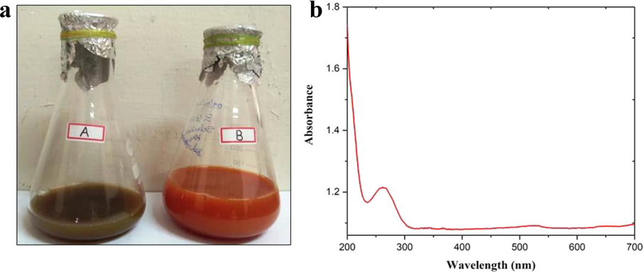
Characterizations of SeNPs synthesized from Goniothalamus wightii leaf extract: a) Color change occurs during SeNPs synthesis, b) Absorption peak noticed for the synthesized SeNPs in UV–vis.
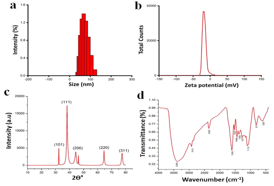
Characterizations of SeNPs synthesized using Goniothalamus wightii leaf extracts a) DLS b) Zeta, c) XRD, d) FT-IR.
The XRD analysis of Gw-SeNPs is demonstrated in Fig. 2c. The XRD spectrum shows diffraction peaks for SeNPs at 29.98°, 33.22°, 47.42°, 65.38° and 77.40° 2θ, similar to the peak values viz., 101, 111, 200, 220 and 311 of cubically oriented crystalline structure. Also mentioned diffraction peaks at 29.98°, 33.22°, 47.42°, 65.38° and 77.40° 2θ, which denote that (1 0 1), (1 1 1), (2 0 0), (2 2 0) and (3 1 1) are corresponding to each other. Furthermore, the collected data suggested that phytochemicals, especially tannins, protect SeNPs from combination and thus maintain the long-term equilibrium of nanoparticles (Meenambigai et al., 2022; Filipovic et al., 2021). Moreover, the FT-IR spectrum of Gw-SeNPs is depicted in Fig. 2d. The FT-IR spectra have several major peaks for many functional groups, such as, 3386 strong broads OH alcohol, 2923 NH amine salt, 2380 OH alcohol, 1606 C⚌C, 1521 N—O stretching nitro compound, 1446 CH alkane, 1380 CH aldehyde, 1110 CO secondary alcohol, 811 C⚌C bending trisubstituted alkene, 599 CI halos of biosynthetic SeNPs in aqueous medium.
The morphological of the SeNPs was also examined by SEM (Fig. 3 a,b,c) which reveals specific points corresponding to the interface layers of Se in diffraction mode, indicating the spherical nature of selenium. The peaks are corresponding to the element in the EDX range was also confirm the presence of elemental as selenium in the reaction mixture (Fig. 3d).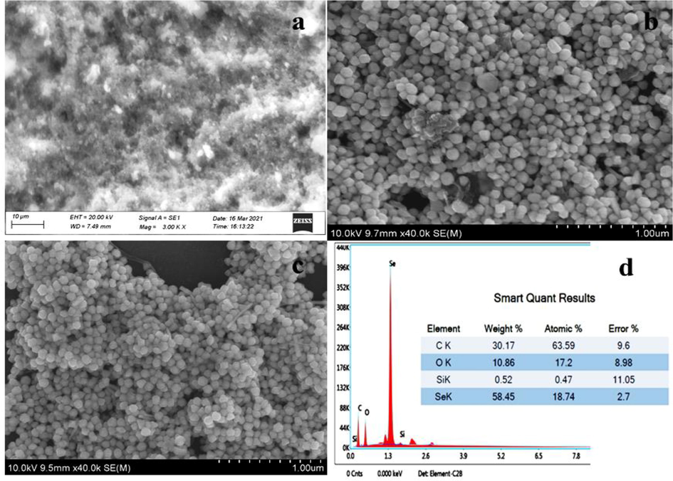
SEM (a, b, c) with EDAX (d) reveals the Gw-SeNPs morphology and elemental percentage.
3.2 Antibacterial activity of Gw-SeNPs
This part of experiment is mainly focusing on selenium toxicity and developing the bio-functionalized selenium nanoparticles by green nanotechnology. The antibacterial activity of G. wightii and Gw-SeNPs were tested against four different bacterial pathogens by following the disc diffusion method. The Gw-SeNPs seems to have good antibacterial effect which is proved by noticing the development of inhibition zone up to 10, 12, 9 and 14 mm for E. coli, P. aeruginosa, S. aureus, and S. epidermidis only 50 µg/mL respectively, when compared with control G. wightii leaf extract and the internal standard Tetracycline. However, the outcomes are displayed in Fig. 4 obviously uncovered the bactericidal property of the integrated SeNPs against gram-positive microbes likewise gram-negative microscopic organisms. Another report detailed that SeNPs shows viable bacterial development restraint against all the test microorganisms (Hernandez-Diaz et al., 2021). These studies agree with our findings which showed the antibacterial property against bacterial pathogens. They noticed that the early phase of the interface SeNPs adhere to the cell wall, subsequently saturate and kill the bacterial cell by damaging the membrane of cells (Cremonini et al., 2016). Therefore, biosynthesized SeNPs is more effective antibacterial agent and could be for different biomedical applications.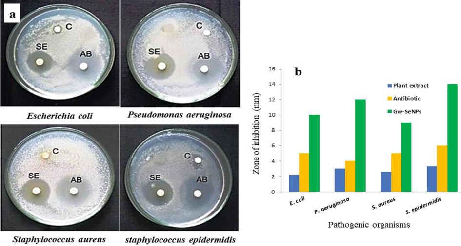
Antibacterial activity of bio synthesized SeNPs from Goniothalamus wightii leaf extract. a) Inhibition zone of different pathogen bacteria and C - Control leaf extract, SE - SeNPs, AB - Antibiotic (Tetracycline). b) Diameter of inhibitory zone for Goniothalamus wightii leaf extract, antibiotic (Tetracycline) and Gw-SeNPs.
3.3 Effect of SeNPs on anti-biofilm assay
The biosynthesized SeNPs (10, 20, 30, 40 and 50 μg/mL) were tried against biofilm creating bacterial strains P. aeruginosa and S. epidermidis. The reduction in biofilm of P. aeruginosa was 61.46% and 62.24% in S. epidermidis at high concentration of 50 μg/mL of SeNPs exposure as shown in Table1. The potential degradation properties of anti-biofilms are confirmed the selected bacterium in P. aeruginosa. Hence, the SeNPs is recommended as a potential anti-biofilm agent in near future as the anti-biofilm effect is more prominent in SeNPs when compared to standard antibiotic cefotaxime. Information (mean ± SE) represents the inhibition zones (mm) for antibacterial ability. Mean values inside the segment followed by various letters in superscript are statistically significant at P < 0.05 level.
Concentration (µg/mL)
Antibiotic (Cefotaxime)
SeNPs
Pseudomonas aeruginosa
10
29.61 ± 0.19d
31.34 ± 0.52e
20
36.39 ± 1.46c
36.35 ± 0.53d
30
38.24 ± 1.09c
42.57 ± 0.14c
40
45.10 ± 2.38b
56.81 ± 0.06b
50
54.90 ± 0.60a
61.46 ± 0.53a
Staphylococcus epidermidis
10
8.96 ± 1.09e
2.56 ± 1.47e
20
15.43 ± 2.13d
11.14 ± 1.52d
30
28.32 ± 1.57c
38.01 ± 1.11c
40
40.58 ± 1.45b
54.01 ± 0.77b
50
52.04 ± 1.72a
62.24 ± 1.11a
3.4 Antioxidant activities of synthesized Gw-SeNPs
3.4.1 DPPH free-radical and ABTS scavenging assay
The preparation of novel natural antioxidants is recent trends among the researchers. The present investigations prove that the biosynthesized Gw-SeNPs might can be a promising antioxidant representative when compared to the standard vitamin C (Fig. 5a). The found results also confirmed that the Gw-SeNPs inhibits in DPPH free radical development in a dose-dependent way at the IC50 value 75.62 µg/mL. The radical scavenges of nanoparticles gradually increased with increasing the concentration. The ABTS scavenging activity of SeNPs shown in Fig. 5b; ABTS radical movement was a great strategy to decide the antioxidant capacity. In this study the IC50 value for SeNPs is determined at 74.12 µg/mL. In the results, SeNPs appeared a noteworthy radical rummaging movement and has been supported (Wen et al., 2021). Observed similar trend for their SeNPs, and also reported that the SeNPs investigates the intense antioxidant activity short of any cytotoxicity to natural cells when contrasted with selenium and selenium dioxide (Cittrarasu et al., 2021; Mellinas et al., 2019; Shahabadi et al., 2021). Thus, the study indicates the biosynthesized nanoparticles are leading antioxidant activity.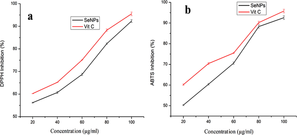
In vitro antioxidant activity of SeNPs synthesized from Goniothalamus wightii leaf extract. a) DPPH radical scavenging activity. b) ABTS+ radical scavenging activity.
3.5 Anticancer activity
The MCF-7 cells have been grown to determine the cytotoxicity of synthesised SeNPs by adopting the MTT protocol 40 µg/mL for 24 h as shown in Fig. 6. The comparative cytotoxicity report was roughly presented (Shahabadi et al., 2021; Alkhudhayri et al., 2020). A greater number of in vitro data have shown that SeNPs remain destructive to cancer cells (Varlamova et al., 2021). In nutshell, the present findings are indication for the cytotoxic effect of biosynthesized Gw-SeNPs opposed to the MCF7 cells. The SeNPs treated MCF-7 cells at IC50 concentration have been observed under confocal microscopy. Which are revealed the destructive property of SeNPs with confirmed through observation of plasma membrane damage (Spyridopoulou et al., 2021; Khan et al., 2018). The Gw-SeNPs were found to have the strongest negative common-sense effects relative to control cells after the 24 and 48 h incubation period. On fluorescence images, the more abnormal cells were found to be dead after being treated with SeNPs. Besides, the control (untreated) cells are energetic as shown in the Fig. 6. This thought shows that nanoparticle-mediated morphological changes might happen inside cells and that morphological changes can confirm the meaning of SeNPs in cancer treatment (Khan et al., 2018).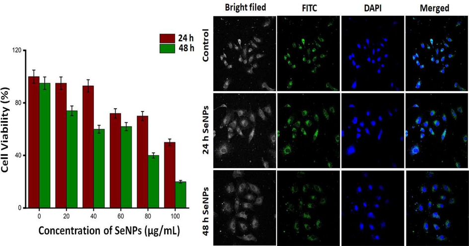
Biosynthesized Gw-SeNPs was assessed by MTT assay and confocal microscopy was observed by staining live and dead apoptotic cells with DAPI and FITC after 24 and 48 h in MCF-7 cell line.
3.6 Larvicidal assay
The various concentrations of Gw-SeNPs were examined on three mosquito fourth instar larvicidal activities against A. aegypti, A. stephensi and C. quinquefaciatus, after 12 and 24 h exposures (Table2). The more potent activity against A. aegypti, A. stephensi through LC50 and LC90values of 4.587, 22.573, 5.218, and 21.173 ppm when compared to C. quinquefaciatus with LC50 and LC90 values of 5.787 and 14.494 ppm, respectively in 24 h exposure. In the current study, the larvicidal activities of SeNPs showed more prominent larvicidal properties against for fourth in star mosquito larvae. Though, the biosynthesized SeNPs serves as insecticidal property because the healing nature of bioactive elements in many herbs can be impacted by a few factors, for instance, the season of assortment, area of species, and climate (Ramya et al., 2020). Similarly, the larvicidal activity of SeNPs utilizing P. corylophilum separate showed that the huge mortality in contrast to the hatchlings of Ae. Aegypti and An. culicifacies at 24 h exposure period (Salem et al., 2021). In addition, SeNPs hampered the functions of mosquito cell walls and membrane, proteins. Hence, this study concludes that the G. wightii leaf extract dependant synthesis of nanoparticles exhibits more potential activity against mosquito larval.
Species
Concentration (mg/L)
LC50 (mg/L)
(LCL-UCL)
LC90 (mg/L)
(LCL-UCL)
χ2
LC50 (mg/L)
(LCL-UCL)
LC90 (mg/L)
(LCL-UCL)
χ2
12 h
24 h
C. quinquefasciatus
25
4.786
(2.931–6.289)21.175
(16.690–31.730)6.621
5.787
(4.245–2.709)14.494
(12.009–19.007)5.787
20
15
10
5
0
A. aegypti
25
10.386
(7.847–12.809)64.979
(40.354–177.530)2.133
4.587
(2.611–6.177)22.573
(17.418–35.982)8.3
20
15
10
5
0
A. stephensi
25
6.499
(4.277–8.273)34.964
(25.107–66.340)8.024
5.218
(3.442–6.661)21.173
(16.873–30.828)11.354
20
15
10
5
0
3.7 Photocatalytic activity
Photocatalytic, refers to the conversion of complex-coloured molecules into simple atoms (or) by-products like carbon dioxide and minerals (Kumar, 2022; Cittrarasu et al., 2021; Menon et al., 2021). The MB is frequently used in many industries for various applications, and this dye was mixed into water sources to cause multi-disease for creatures (Hassanien et al., 2019). In the present investigation MB dye was degraded by biosynthesized of Gw-SeNPs, the experiment was done under the direct sunlight. However, contrasted with the control arrangement. The detailed results are shown in Fig. 7a, which reveals the decolonization of MB and the efficiency, was steadily increased with good degradation rate (97%) in 150 min. The SeNPs was act as acceptors of electrons from the plant extract and as a contributor of electrons for MB. Overall, the SeNPs could bleach the MB within 150 min and to cause intermediary products (Fig. 7b), while expanding the time of SeNPs exposure under sunlight to increasing the ability. An earlier study reported that the biosynthesized SeNPs from plant extract could be effective resources for industrial waste dye degradation (Jahdaly et al., 2021). This result clearly indicates that the effective character of SeNPs in the photo catalysis of the toxic contaminants acquired from textile trades.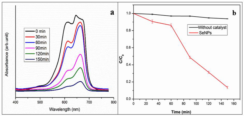
(a) Absorption spectrum of SeNPs biosynthesis from Goniothalamus wightii leaf extract effect on Methylene blue under sun light, (b) Phot catalytic of Methylene blue in the presence Gw-SeNPs and Control (without catalyst).
4 Conclusion
The present study demonstrates that the biosynthesis of SeNPs from the aqueous leaf extract of Goniothalamus wightii through a simple, green, economic and eco-friendly approach. The G. wightii extract was found to have rich amount of alcohol, flavonoids and ketones which favours the biosynthesis of SeNPs. The synthesized Gw-SeNPs have negative charge, a polygonal shape and very stable nanometre size. The Gw-SeNPs have exhibited that strong antibacterial activity against a wide scope of microbes. In addition, Gw-SeNPs have free radical scavenging activity and have been found to an equivalent efficient compare with standard antioxidant vitamin C. The Gw-SeNPs have shown that high toxicity against MCF-7 cells, which are very safe and biocompatible and also very effective on industrial waste dye degradation. Thus biosynthesized Gw-SeNPs have enormous application in the area of pharmaceuticals, food and environmental industries.
Funding information
No external funding was received for this work.
CRediT authorship contribution statement
Chinnaraj Santhosh: Methodology, Data curation, Formal analysis. Balamuralikrishnan Balasubramanian: Conceptualization, Writing – review & editing. Palani Vino: Methodology, Data curation, Formal analysis. Maluventhen Viji: Methodology, Data curation, Formal analysis. Chandrababu Rejeeth: Methodology, Data curation, Formal analysis. Soundarapandian Kannan: Methodology, Data curation, Formal analysis. Hammad Ullah: Writing – review & editing. Kannan R.R. Rengasamy: Writing - review & editing Maria Daglia: Writing – review & editing. Arumugam Maruthupandian: Conceptualization, Writing – review & editing.
Acknowledgement
The authors are significantly appreciative to the Department of Botany, Periyar University, Tamil Nadu, India for giving a University Research Fellowship and specialized help during the research.
Declaration of Competing Interest
The authors declare that they have no known competing financial interests or personal relationships that could have appeared to influence the work reported in this paper.
References
- Selenium nanoparticles induce cytotoxicity and apoptosis in human breast cancer (MCF-7) and Liver (HepG2) cell lines. Nanosci. Nanotechnol. Lett.. 2020;12:330.
- [Google Scholar]
- Green synthesis of selenium nanoparticles mediated from CeropegiabulbosaRoxb extract and its cytotoxicity, antimicrobial, mosquitocidal and photocatalytic activities. Sci. Rep.. 2021;11:15.
- [Google Scholar]
- Biogenic selenium nanoparticles: characterization, antimicrobial activity and effects on human dendritic cells and fibroblasts. Microb. Biotechnol.. 2016;9(6):758-771.
- [Google Scholar]
- Filipovic, N., Usjak, D., Milenkovic, M.T., Zheng, K., Liverani, L., Boccaccini, A.R., Stevanovic, M., 2021. Comparative Study of the Antimicrobial Activity of Selenium Nanoparticles with Different Surface Chemistry and Structure. Front.Bioeng.Biotechnol. 8:624621.
- Eco-friendly approach to synthesize selenium nanoparticles: photocatalytic degradation of sunset yellow azo dye and anticancer activity. Chem Select.. 2019;4(31):9018-9026.
- [Google Scholar]
- Antibacterial activity of biosynthesized selenium nanoparticles using extracts of Calendula officinalis against potentially clinical bacterial strains. Molecules. 2021;26(19):5929.
- [Google Scholar]
- Selenium nanoparticles synthesized using an eco-friendly method: Dye decolorization from aqueous solution, cell viability, antioxidant, and antibacterial effectiveness. J. Mater. Res. Technol.. 2021;11:97.
- [Google Scholar]
- Selenium nanoparticles synthesized using an eco-friendly method: Dye decolourization from aqueous solution, cell viability, antioxidant, and antibacterial effectiveness. J. Mater. Res. Technol.. 2021;11:97.
- [Google Scholar]
- Green nanotechnology-driven drug delivery assemblies. ACS Omega. 2019;4(5):8804-8815.
- [Google Scholar]
- FMSP-nanoparticles induced cell death on human breast adenocarcinoma cell line (MCF-7Cells): morphometric analysis. Biomolecules. 2018;8:32.
- [Google Scholar]
- Plant extract assisted eco-benevolent synthesis of selenium nanoparticles- a review on plant parts involved, characterization and their recent applications. Chem. Rev.. 2020;2:168.
- [Google Scholar]
- Nanomaterial’s synthesis, types and their use in Bioremediation and Agriculture. Nat. Resour. Human Health.. 2022;2(3):349-365.
- [Google Scholar]
- Green synthesis of selenium nanoparticles mediated by nilgirianthus ciliates leaf extracts for antimicrobial activity on foodborne pathogenic microbes and pesticidal activity against Aedes aegypti with molecular docking. Biol. Trace Elem. Res.. 2022;200(6):2948-2962.
- [Google Scholar]
- Microwave-assisted green synthesis and antioxidant activity of selenium nanoparticles using Theobroma cacao L. Bean Shell Extract. Molecules. 2019;24:4048.
- [Google Scholar]
- Catalytical degradation of industrial dyes using biosynthesized selenium nanoparticles and evaluating its antimicrobial activities. Sustain. Environ. Res.. 2021;31:12.
- [Google Scholar]
- Palani, V., Shanmugasundaram, M., Maluventhen, V., Chinnaraj, S., Liu, W., Balasubramanian, B., Arumugam, M., 2020. Phytoconstituents and their potential antimicrobial, antioxidant and mosquito larvicidal activities of Goniothalamuswightii Hook. F. & Thomson. Arab. J. Sci. Eng. 45:4555.
- Actinobacterial enzyme mediated synthesis of selenium nanoparticles for antibacterial, mosquito larvicidal and anthelminthic applications. Part. Sci. Technol.. 2020;38:72.
- [Google Scholar]
- Antibacterial, cytotoxicity and larvicidal activity of green synthesized selenium nanoparticles using Penicillium corylophilum. J Clust Sci. 2021;32(2):351-361.
- [Google Scholar]
- Selenium nanoparticles: Synthesis, in-vitro cytotoxicity, antioxidant activity and interaction studies with ct-DNA and HSA, HHb and Cyt c serum proteins. Biotechnol. Rep.. 2021;30:00615.
- [Google Scholar]
- Anticancer activity of biogenic selenium nanoparticles: apoptotic and immunogenic cell death markers in colon cancer cells. Cancers. 2021;13(21):5335.
- [Google Scholar]
- A new species of Goniothalamus (Annonaceae) from the Western Ghats of Tamil Nadu. India. Taiwania. 2020;65:180.
- [Google Scholar]
- Mechanisms of the cytotoxic effect of selenium nanoparticles in different human cancer cell lines. Int. J. Mol. Sci.. 2021;22:7798.
- [Google Scholar]
- Biosynthesis and antioxidation of nano-selenium using lemon juice as a reducing agent. Green Process. Synth.. 2021;10:188.
- [Google Scholar]
- Antibacterial properties and mechanism of selenium nanoparticles synthesized by Providencia sp. DCX. Env. Res.. 2021;194
- [Google Scholar]


