

Translate this page into:
Hydrothermally synthesized lanthanide-incorporated multifunctional zirconia nanoparticles: Potential candidate for multimodal imaging
⁎Corresponding author at: Department of Physics, GC University 38000, Faisalabad, Pakistan. fakharphy@gmail.com (M. Fakhar-e-Alam)
-
Received: ,
Accepted: ,
This article was originally published by Elsevier and was migrated to Scientific Scholar after the change of Publisher.
Peer review under responsibility of King Saud University.
Abstract
Abstract
Objectives
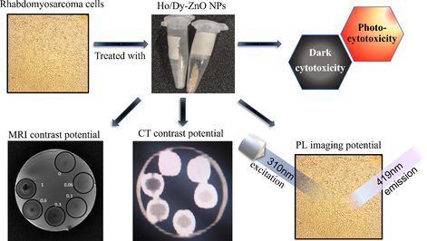
Enabled zirconium oxide Nanoparticles (NPs) are multifunctional nanoparticles that can be employed for multimodal imaging and can show good biocompatibility. In this work, we have synthesized Dysprosium (Dy) and Holmium (Ho) enabled zirconium oxide nanoparticles (DY/Ho-ZrO2 NPs) and have evaluated their potential as candidates for contrast agents in different imaging modalities such as X-ray computed tomography (CT), Photoluminescence (PL) imaging and Magnetic Resonance Imaging (MRI). Rare earth elements have large atomic numbers, and their incorporation can enhance the X-ray attenuation characteristics of their host along with their optical properties. Rare earth elements exhibit paramagnetic character which can be utilized for MRI contrast enhancement. A combination of these imaging modalities can fulfill the limitations faced while using a single imaging technique.
Methods
We have successfully synthesized Dy and Ho incorporated zirconia nanoparticles (Dy/Ho-ZrO2) by a simple one-step hydrothermal method.
Results
Prepared NPs were characterized for their physical properties. X-ray diffraction (XRD) revealed the crystalline phases present in Dy-ZrO2 and Ho-ZrO2 NPs with a crystallite size of 27 nm and 21.54 nm respectively. Scanning Electron Microscopy (SEM) results revealed the morphology of the nanoparticles, while EDS analysis gave the qualitative as well quantitative nature of synthesized nanocrystals. Photoluminescence (PL) data of Dy/Ho-Zirconia NPs have shown emission peaks near 419 nm when excited at 310 nm. Suspensions of prepared NPs with various concentrations were imaged on a CT machine in the clinical setting for contrast study. High CT contrast was observed for these NPs even at very low concentrations. Dysprosium doped sample was further evaluated for its potential as an MRI contrast agent. Dark cytotoxicity and photo-cytotoxicity results performed using Rhabdomyosarcoma (Rd) cancer cell lines revealed good biocompatibility of prepared NPs.
Conclusions
These results strongly signify the potential of these multifunctional Dy/Ho-Zirconia NPs to act as biocompatible multimodal imaging agents for biomedical applications.
Keywords
Multimodal imaging
Zirconia
Nanoparticles
Lanthanides

1 Introduction
The development and synthesis of biocompatible, luminous bioimaging agents for use in diagnostics, imaging, and preventative medicine is critical for biomedicine's future (Mondal et al., 2020). Therefore, the development of nanomaterials suited for use as non-invasive diagnostic tools for biomedical purposes has attracted researchers' curiosity. The use of optically active components in conjunction with other imaging modalities is a particularly appealing technique to create multifunctional probes for biological imaging and identification (Li et al., 2016). The optical imaging method is particularly appealing for its high sensitivity, multicolor imaging, and activatable characteristic. However, its basic limitations are low spatial resolution and shallow tissue penetration (Pu et al., 2014). To overcome this limitation, various imaging modalities such as magnetic resonance imaging and X-ray computed tomography imaging can be combined with optical imaging techniques (Cheng et al., 2014). Because of its excellent spatial resolution and no tissue penetration limit, Computed Tomography (CT) has been widely employed as one of the most reliable imaging techniques (Lee et al., 2013). In terms of availability, efficiency, and affordability, CT has long been a standard technology in clinical imaging, and it can also provide high spatial and temporal resolution 3D structural details of tissues with differential X-ray absorption properties. CT imaging has long been utilized for bone imaging because it can easily discriminate between electron-dense structures (high-Z materials, such as bone) and electron-poor things (e.g., soft tissues). Magnetic resonance imaging (MRI), like CT, has an unlimited detection depth and can create 3D images, but it has a low detection sensitivity and lack multiplexing (James and Gambhir, 2012). However, MRI provides great resolution on soft tissues while CT excels at hard tissues thus MRI and CT are complimentary to each other.
Many currently accessible formulations involve potentially toxic ingredients, and clinical translation of nanotechnology will necessitate nanoparticles with the lowest feasible toxicity (Naahidi et al., 2013). Due to their good biocompatibility, ZrO2 nanoparticles have been used in medical and orthopaedical applications, primarily for the repair and replacement of sick and damaged portions of the human skeleton, including bones, teeth, and joints (Wang et al., 2016). Hence, zirconium dioxide (ZrO2) may be employed as a host matrix for the design and manufacture of novel nanomaterials owing to its low toxicity. Moreover, the incorporation of inorganic nanoparticles with lanthanide ions is emerging as a promising class of new bio probes and has shown great potential in various biomedical applications such as bio labeling, bioimaging, and theragnosis (Huang et al., 2019). Eu3+, La3+, Gd3+, Tb3+, Ce3+, and other rare earth elements have been effectively incorporated within various host nanostructured systems for bioimaging applications (Graeve et al., 2010; Doat et al., 2003). Lanthanide enabled NPs have displayed potential as multimodal imaging contrast agents as a result of their diverse yet tunable optical and magnetic properties (He et al., 2017). With the distinctive 4f manifold of trivalent lanthanide cations (Ln3+), Lanthanide enabled NPs upon excitation, emit light at different wavelengths ranging from the ultraviolet (UV) to near-infrared (NIR) regime (Liu et al., 2013). The incorporation of lanthanides not only enhances the optical properties of inorganic metal oxides but also provides them with better thermal and photostability (Geitenbeek et al., 2019; Chen et al., 2014). The high absorption coefficient for the host and low phonon energy (470 cm−1) of ZrO2 make it an ideal host for lanthanide ions (Liu et al., 2012). The inclusion of lanthanide can also enhance other important features of nanomaterials, such as magnetism and X-ray attenuation coefficient, which raises the potential of nanomaterials to be employed as contrast agents for multimodal imaging. Holmium and Dysprosium are among the elements having the highest magnetic moments, and they can generate significant transverse relaxation of hydrogen protons in free water. For ultra-high field T2 contrast agents, Ho and Dy based nanomaterials are the best options. MRI contrast agents based on high magnetic moment elements are projected to see considerable expansion in the near future, given the ongoing quest of ultra-high field intensity MRI. The combination of photoluminescence, X-ray computed tomography, and magnetic resonance imaging is expected to provide high-resolution 3D details of tissues and cells with high sensitivity (Xing et al., 2012).
Herein, we have followed the hydrothermal route for synthesizing Dy and Ho incorporated zirconia NPs. Low crystallization temperatures enable direct production of nanocrystalline powders with a high degree of precipitation which reduces the content of metal ions in effluents as per environmental regulations, making hydrothermal synthesis a soft chemical route with significant advantages (Robert et al., 2005). Prepared nanoparticles were characterized for their physical properties and were first-ever evaluated for their bi-modal imaging potential by studying their PL, CT and MRI contrast properties. Although bare zirconia NPs have been evaluated previously for their in-vitro cytotoxicity, however, the cytotoxicity of the enabled NPs should also be evaluated to estimate their full potential for biomedical applications. We have evaluated the dark cytotoxicity of prepared nanoparticles on Rhabdomyosarcoma (Rd) cancer cells along with their photo-cytotoxicity using the MTT assay and red laser.
2 Materials and methods
2.1 Chemicals and reagents
Research grade Zirconium chloride, Dysprosium oxide, Holmium oxide, Nitric acid, and Urea were purchased from Sigma Aldrich. Nitrates of Dy and Ho were obtained upon reacting their oxides with nitric acid. Other reagents used for dark cytotoxicity and phototoxicity include Minimum Essential Medium (MEM), Fetal Bovine Serum (FBS), Dimethyl sulfoxide (DMSO), Phosphate Buffered Saline (PBS), and Rd cell lines from the National Institute of Health (NIH).
2.2 Synthesis of Dy/Ho-ZrO2 nanoparticles
Schematics of the synthesis procedure are shown in Fig. 1. Dy/Ho-ZrO2 NPs were prepared by a hydrothermal method as reported previously for Eu doped ZrO2 NPs (Atabaev, 2018). ZrCl4 and Dy(NO3)3·6H2O were taken in mole ratios 0.89:0.11 and dissolved in 70 ml deionized water in a beaker with vigorous magnetic stirring. 1.75 g Urea was then added to the solution. After 20mins of stirring, the solution was transferred into a 100 ml Teflon-lined stainless autoclave reactor which was then placed into a wind drying oven at 140 °C for 2 hrs. Pressurized heating facilitated the production of fine Zirconia nanoparticles. The autoclave was allowed to cool down to room temperature. The white crystalline product was obtained upon opening the Teflon flask which was washed three times with deionized water by centrifuging at 6000 rpm. Obtained 11% Dy-ZrO2 nanoparticles were dried overnight at 65 °C and were further calcinated for 1 h and at 700 °C. The same procedure was followed using Ho(NO3)3·5H2O in place of Dy(NO3)3 to obtain 11% Ho-ZrO2 NPs. The resulting nanoparticles when ultrasonicated for 10 min lead to homogenous suspension of nanoparticles in deionized water.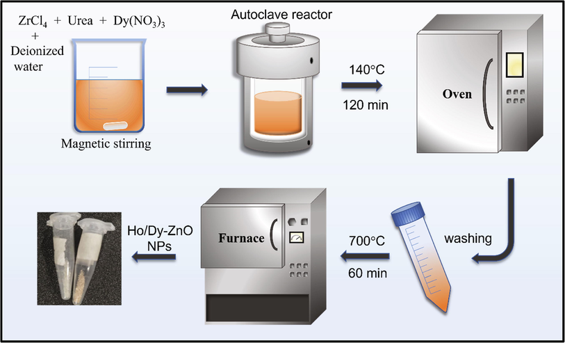
Schematics of Synthesis for Dy/Ho-Zirconia NPs.
2.3 Characterization of Dy/Ho-ZrO2
Physical characterizations for the prepared nanoparticles were performed using powder X-Ray diffraction (Cu-Kα radiations, 0.154 nm), Scanning electron microscopy, and Energy Dispersive X-ray spectroscopy. Tyndall test was performed to examine the colloidal stability of NPs (Stetefeld et al., 2016). The optical properties of the samples were studied using an F-98 fluorescence spectrometer. CT scans of synthesized samples were performed using General Electric’s Discovery SPECT/CT at NORI hospital Islamabad. MRI contrast properties were studied using 1.5 T GE MRI scanner. Cell viability and phototoxicity test were conducted on Rd cancer cell lines. Absorbance data was recorded using a microplate reader. All bio characterizations were performed in a sterile environment.
2.4 Cell culture and cell viability test
Human Rhabdomyosarcoma (Rd) cell lines representing the muscle cancer cell (Stevens et al., 2008) were received from the National Institute of Heal (NIH). Cells were sub-cultured in minimum essential medium (MEM) non-essential amino acids solution containing 10% Fetal Bovine Serum (FBS). Moist environment and 37 °C temperature were provided for facilitating the growth and to ensure adequate adhesion to the substrate. Upon achieving the confluence state, the cells were seeded in a 96 well plate and were placed in an incubator at 37 °C for further growth and adhesion. After 24 h of subculturing, cells were treated with various concentrations (60–240 μg/ml in FBS) of prepared nanoparticles and were further incubated for 24 h. Cell viability was determined by performing an MTT assay in a 96 well plate by a method as described in previous studies (AlSalhi et al., 2020; Atif et al., 2019, 2016, 2012, 2011b,a,c, 2010, 2009; Fakhar-E-Alam et al., 2012; Fakhar-e-Alam et al., 2020; Iqbal et al., 2020; Hayat et al., 2013). All cell culture experiments were performed in a sterile environment inside a laminar flow hood.
3 Results and discussion
Characteristic XRD peaks of Dy-ZrO2 and Ho-ZrO2 can be seen in the spectrum as shown in Fig. 2. Spectrum was analyzed and peaks were found to agree with the reported literature (Atabaev, 2018). Peaks for the Dy-ZrO2 were indexed using ICDD card number 96–900-7449. XRD Peaks of the Ho-ZrO2 sample were indexed using ICDD card number 96–152-5707 and both the samples were found to be in a tetragonal phase with excellent crystallinity. The crystallite size (τ) for both samples was calculated using Scherrer's formula (Fig. 3).
where K is the shape factor, λ is the wavelength of X-rays, β is the full width at half the maximum intensity and θ is the Bragg angle. Dy-ZrO2 possessed a crystallite size of 27 nm while that for Ho-ZrO2 was 21.54 nm.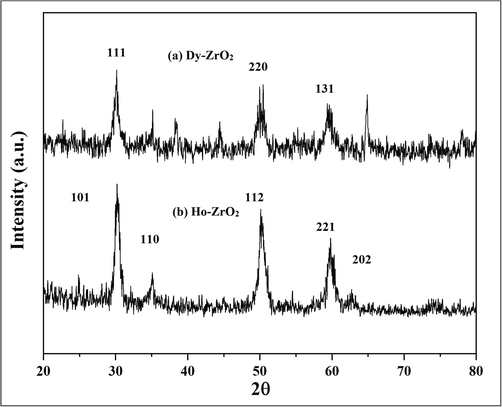
XRD analysis of 11% Dy/Ho-Zirconia Samples.
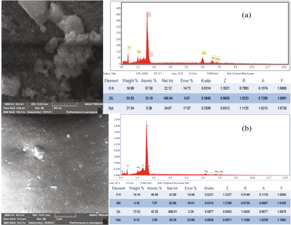
SEM micrograph and EDX results for (a) Dy-ZnO2 and (b) Ho-ZrO2.
SEM for both samples was performed for high-resolution imaging while EDX was performed to study the elemental composition as shown in figure three. EDS results confirmed the successful incorporation of Dy and Ho atoms into vacant sites of tetragonal zirconia crystals was the prerequisite for contrast experiments.
Uniform suspension of synthesized nanoparticles in an appropriate medium is very important for biomedical applications and contrast studies. To determine the colloidal stability Tyndall test was performed using a red laser as shown in Fig. 4(b). The suspension of NPs was exposed to a red laser at varying time intervals up to 30 h and the results indicate good colloidal stability of nanoparticles over an extended period.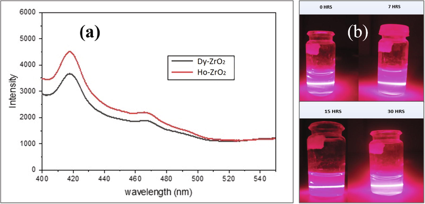
(a) PL Spectra of Dy/Ho-ZrO2 at an excitation wavelength of 310 nm; (b) Tyndall test with a red laser (630 nm) at subsequent time intervals.
3.1 Photoluminescence, CT contrast and MRI contrast study
Optical characteristics were evaluated using an F-98 fluorescence spectrophotometer. Emission wavelength scan shows the photoluminescence spectra of Dy and Ho enabled zirconia nanoparticles recorded in the range of 400–560 nm is shown in Fig. 4(a). Both samples show a prominent and intense emissions spectrum near 419 nm when excited at the wavelength of 310 nm.
CT scans of the suspensions of Dy/Ho-ZrO2 NPs in a water phantom were performed at three different energies 80 KV, 100 KV, and 120 KV as shown in Fig. 5. These energies are normally used for patient scans in clinical settings. Contrast materials present in glass vials were placed in a water-filled phantom. Different concentrations of contrast material (0, 2.1, 2.8, 4.2, 6.3 and 7 mg/ml) were scanned at three energies while the rest of the parameters were similar. Contrast (Hounsfield units) is decreasing with increasing energy as shown in Fig. 5.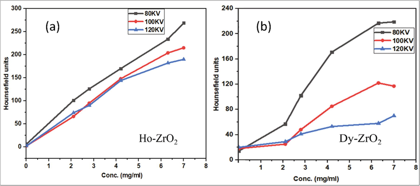
The plot of HU vs Different concentrations of prepared samples at three different energies. (a) for Ho-ZrO2; (b) for Dy-ZrO2.
As energy is increased, more and more photons are passing through the material without making any interaction as a cross-section for photoelectric, and Compton's effect is low at high energies. Also, increasing behavior of the X-ray attenuation coefficient can be observed with the concentration of the NPs. The highest X-ray attenuation i.e. high contrast was observed at 80 KV while the lowest contrast was at 120 KV. These optical and X-ray attenuation results indicate the superior potential of Dy/Ho = ZrO2 NPs to be used as CT contrast agents.
For the study of MRI contrast properties, a 1.5 T GE MRI scanner was used. Inside a cylindrical water-filled phantom, a series of Dy-ZrO2 samples with varying Dysprosium ion concentrations were placed. The water phantom was placed inside the scanner using the knee flex coil. Scans were taken using the Fast Spin Echo (FSE) sequence. T2 weighted scans were conducted since Dysprosium increase the T2 relaxation rate. The recovery time TR was set to 3000 ms, and the echo times TE were 50 ms, 90 ms, 130 ms, 170 ms, 190 ms, 500 ms, and 900 ms, respectively. The slice thickness was 3.5 mm, and the length of the eco train was 35. The results were presented as a series of DICOM images. IMAGEJ software was used to open DICOM images. The appropriate Region of Interest (ROI) was chosen, and the intensity variation with echo time was recorded. T2 time for a specific ROI or vial was calculated using a plot of intensity versus echo time and an exponential curve fit. T2 time for the remaining vials was computed in the same way. Finally, T2 durations were plotted against the concentration of paramagnetic ions, yielding a relaxation rate in mM−1s−1.
The T2 weighted images become darker due to a high concentration of paramagnetic ions. In order to depict clinical conditions, contrast vials were placed in a deionized water medium. Relaxivity rate R2 in units of mM-1S-1 is obtained by plotting 1/T2 times against Dy concentration. The R2 value obtained after the linear fit is 64.4 mM−1s−1 as shown in Fig. 6, which is close to values obtained by Tegafaw et al. (2015).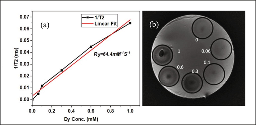
(a) Plot of 1/T2 time vs Dy3+ ion concentration; (b) Contrast vials placed in water medium having concentration 0 to 1 mM Dy concentration.
3.2 Dark cytotoxicity and photo-cytotoxicity studies
MTT Assay and phototoxicity studies were performed after treating the cells with Dy/Ho-ZrO2 NPs. The viability of RD cells, incubated with increasing concentrations of the NPs (60–240 µg/ml) was evaluated using the MTT colorimetric assay. Culture media was replaced with 100 μL PBS solution containing 20 μL MTT stock solution (5.0 mg/mL) and cells were further incubated for 4 h. The MTT working solution was then aspirated and 100 µL of DMSO was added per well to solubilize the formazan crystals formed within the viable cells. The absorbance data was recorded using a microplate reader. The percentage of cell viability was determined from the absorbance data using the following relation.
Cell viability was found to be 88% or more even at higher concentrations up to 240 µg/ml while much better cell viability was observed at low concentrations which is indicative of very less dark cytotoxicity of these NPs as shown in Fig. 7.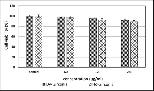
MTT Assay cytotoxicity Results.
For phototoxicity cells were exposed to different doses of red laser after 24 h of treatment with NPs. Cells were further incubated for 24 h before performing MTT assay. The data of phototoxicity at a fixed dose of 60 μg/ml is plotted against varying doses of light as shown in Fig. 8. Cell viability for both samples was found to be more than 80% even at higher light doses which is indicates the low phototoxicity of these samples.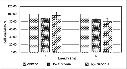
Phototoxicity results, both samples have a concentration of 60 μg/ml.
4 Conclusion
Rare earth metals Dysprosium and Holmium were used to synthesize lanthanide-incorporated zirconia nanoparticles via the hydrothermal method. Conventional characterizations like XRD, SEM revealed the crystalline nature and morphologies of the Dy/Ho-ZrO2 NPs. Tyndall test shows good colloidal stability of these NPs. Dark cytotoxicity and phototoxicity studies of the samples have revealed their nontoxic behavior. Optical properties, X-ray attenuation and MRI relaxivity characteristics signify the potential of these nanoparticles to be used for multimodal imaging i.e., PL/CT/MRI imaging technique. The use of PL, MRI, and CT together can produce medical images with excellent resolution, sensitivity, and information for medical diagnosis with no limit on the target sites detection depth.
Acknowledgements
Researchers Supporting Project number (RSP-2021/397), King Saud University, Riyadh, Saudi Arabia.
Declaration of Competing Interest
The authors declare that they have no known competing financial interests or personal relationships that could have appeared to influence the work reported in this paper.
References
- Synthesis of NiO nanoparticles and their evaluation for photodynamic therapy against HeLa cancer cells. J. King Saud Univ. Sci.. 2020;32:1395-1402.
- [CrossRef] [Google Scholar]
- Fabrication and optical properties of Eu-doped zirconia nanoparticles. Mater. Today Proc.. 2018;6:7-10.
- [CrossRef] [Google Scholar]
- Fluorescence spectra of cultured normal and malignant lung cells. Laser Phys.. 2012;22:1353-1357.
- [CrossRef] [Google Scholar]
- In vitro cytotoxicity of mesoporous SiO2@Eu(OH)3 core-shell nanospheres in MCF-7. J. Nanomater. 2016:1-6.
- [CrossRef] [Google Scholar]
- Role of sensitivity of zinc oxide nanorods (ZnO-NRs) based photosensitizers in hepatocellular site of biological tissue. Laser Phys.. 2011;21(11):1950-1961.
- [CrossRef] [Google Scholar]
- Study of the efficacy of 5-ALA mediated photodynamic therapy on human rhabdomyosarcoma cell line (RD) Laser Phys. Lett.. 2010;7:757-764.
- [CrossRef] [Google Scholar]
- Study of the efficacy of Photofrin®-mediated PDT on human hepatocellular carcinoma (HepG2) cell line. Laser Phys.. 2011;21:1135-1144.
- [CrossRef] [Google Scholar]
- In vitro study of 5-aminolevulinic acid-based photodynamic therapy for apoptosis in human cervical HeLa cell line. Laser Phys. Lett.. 2009;6:886-891.
- [CrossRef] [Google Scholar]
- Cytotoxic and photocytotoxic effect of Photofrin® on human laryngeal carcinoma (Hep2c) cell line. Laser Phys.. 2011;21:1235-1242.
- [CrossRef] [Google Scholar]
- Manganese-doped cerium oxide nanocomposite induced photodynamic therapy in MCF-7 cancer cells and antibacterial activity. Biomed Res. Int. 2019:1-13.
- [CrossRef] [Google Scholar]
- Upconversion nanoparticles: design, nanochemistry, and applications in theranostics. Chem. Rev.. 2014;114:5161-5214.
- [CrossRef] [Google Scholar]
- PEGylated WS2 nanosheets as a multifunctional theranostic agent for in vivo dual-modal CT/photoacoustic imaging guided photothermal therapy. Adv. Mater.. 2014;26:1886-1893.
- [CrossRef] [Google Scholar]
- Europium-doped bioapatite: A new photostable biological probe, internalizable by human cells. Biomaterials. 2003;24:3365-3371.
- [CrossRef] [Google Scholar]
- Sensitivity of A-549 human lung cancer cells to nanoporous zinc oxide conjugated with Photofrin. Lasers Med. Sci.. 2012;27:607-614.
- [CrossRef] [Google Scholar]
- Fakhar-e-Alam, M., Aqrab-ul-Ahmad, Atif, M., Alimgeer, K.S., Suleman Rana, M., Yaqub, N., Aslam Farooq, W., Ahmad, H., 2020. Synergistic effect of TEMPO-coated TiO<inf>2</inf> nanorods for PDT applications in MCF-7 cell line model. Saudi J. Biol. Sci. https://doi.org/10.1016/j.sjbs.2020.09.027.
- Chemically and thermally stable lanthanide-doped Y 2 O 3 nanoparticles for remote temperature sensing in catalytic environments. Chem. Eng. Sci.. 2019;198:235-240.
- [CrossRef] [Google Scholar]
- Luminescence variations in hydroxyapatites doped with Eu2+ and Eu3+ ions. Biomaterials. 2010;31:4259-4267.
- [CrossRef] [Google Scholar]
- The potential of PEGylated BaMnO3 nanoparticles as drug delivery agents. Laser Phys. Lett.. 2013;10(2):025603.
- [CrossRef] [Google Scholar]
- Simultaneous enhancement of photoluminescence, MRI Relaxivity, and CT contrast by tuning the interfacial layer of lanthanide heteroepitaxial nanoparticles. Nano Lett.. 2017;17:4873-4880.
- [CrossRef] [Google Scholar]
- Rare earth ion– and transition metal ion-doped inorganic luminescent nanocrystals: from fundamentals to biodetection. Mater. Today Nano. 2019;5:100031.
- [CrossRef] [Google Scholar]
- Photodynamic therapy, facile synthesis, and effect of sintering temperature on the structure, morphology, optical properties, and anticancer activity of Co3O4 nanocrystalline materials in the HepG2 cell line. J. Photochem. Photobiol. A Chem.. 2020;386:112130.
- [CrossRef] [Google Scholar]
- A molecular imaging primer: modalities, imaging agents, and applications molecular imaging agents applications of molecular imaging guide to performing a molecular. Physiol. Rev. 2012:897-965.
- [CrossRef] [Google Scholar]
- Multimodality imaging in nanomedicine and nanotheranostics. Cancer Biol. Med. 2016
- [CrossRef] [Google Scholar]
- Lanthanide-doped luminescent nanoprobes: Controlled synthesis, optical spectroscopy, and bioapplications. Chem. Soc. Rev.. 2013;42:6924-6958.
- [CrossRef] [Google Scholar]
- Amine-functionalized lanthanide-doped zirconia nanoparticles: Optical spectroscopy, time-resolved fluorescence resonance energy transfer biodetection, and targeted imaging. J. Am. Chem. Soc.. 2012;134(36):15083-15090.
- [CrossRef] [Google Scholar]
- Rare earth element doped hydroxyapatite luminescent bioceramics contrast agent for enhanced biomedical imaging and therapeutic applications. Ceram. Int.. 2020;46:29249-29260.
- [CrossRef] [Google Scholar]
- Biocompatibility of engineered nanoparticles for drug delivery. J. Controlled Release. 2013;166(2):182-194.
- [CrossRef] [Google Scholar]
- Semiconducting polymer nanoparticles as photoacoustic molecular imaging probes in living mice. Nat. Nanotechnol.. 2014;9:233-239.
- [CrossRef] [Google Scholar]
- Dynamic light scattering: a practical guide and applications in biomedical sciences. Biophys. Rev.. 2016;8:409-427.
- [CrossRef] [Google Scholar]
- The British Childhood Cancer Survivor Study: Objectives, methods, population structure, response rates and initial descriptive information. Pediatr. Blood Cancer. 2008;50:1018-1025.
- [CrossRef] [Google Scholar]
- Dual-mode T1 and T2 magnetic resonance imaging contrast agent based on ultrasmall mixed gadolinium-dysprosium oxide nanoparticles: Synthesis, characterization, and in vivo application. Nanotechnology. 2015;26(36)
- [CrossRef] [Google Scholar]
- Good biocompatibility and sintering properties of zirconia nanoparticles synthesized via vapor-phase hydrolysis. Sci. Rep.. 2016;6:1-9.
- [CrossRef] [Google Scholar]
- Multifunctional nanoprobes for upconversion fluorescence, MR and CT trimodal imaging. Biomaterials. 2012;33:1079-1089.
- [CrossRef] [Google Scholar]
Appendix A
Supplementary data
Supplementary data to this article can be found online at https://doi.org/10.1016/j.jksus.2022.102080.
Appendix A
Supplementary data
The following are the Supplementary data to this article:


