

Translate this page into:
Morphological damage and increased ROS production of biosynthesized silver nanoparticle against MCF-7 breast cancer cells through in vitro approaches
⁎Corresponding author at: State Key Laboratory of Biocontrol, Guangdong Provincial Key Laboratory of Plant Resources and Southern Marine Science and Engineering Guangdong Laboratory (Zhuhai), School of Life Sciences, Sun Yat-Sen University, Guangzhou 510275, PR China. liwenjun3@mail.sysu.edu.cn (Wen-Jun Li)
-
Received: ,
Accepted: ,
This article was originally published by Elsevier and was migrated to Scientific Scholar after the change of Publisher.
Peer review under responsibility of King Saud University.
Abstract
In this study of anti-cancer activity, the cytotoxicity effect of the silver nanoparticle was shown excellent anti-cancer activity against MCF-7 breast cancer cells. The IC50 concentration of the synthesized silver nanoparticle against performed MCF-7 breast cancer cells was 1000 µg/ml concentration. Further, the shape modifications in the silver nanoparticle treated MCF-7 cells were viewed by phase contrast microscope and proved after comparison with untreated control. Next, the intracellular proliferation, chromatin condensation, granular membranes cleavage and surface layers damage were effectively seen by fluorescence microscopy using AO/EB fluorescence dyes. The failure of matured nucleus was clearly shown after the treatment of Ag NPs and was effectively proved by fluorescence microscope using Hoechst 33342 stain. Finally, the anti-cancer effect identification through various invitro experiments was clearly favored to performed silver nanoparticle, and suggested that the nanoparticle was very effective against multi drug resistant bacteria, biofilm producing bacteria and cancer cells.
Keywords
Silver nanoparticle
Anticancer activity
MCF-7 breast cancer cells
Cytotoxicity effect
Morphological modification
Fluorescence microscope

1 Introduction
Recent years, nanomaterial and nanoparticles have overwhelmed interest to biomedical researcher due to the impact of invalid drug choice for most of the infections. Also, nanomaterial and their technologies are used in various applications including industrial and biopharmaceuticals especially, cosmetics, biomedical, drug delivery, drug engineering, waste water treatment, food packaging, sensor and other magnetic devices (Saber et al., 2018). Based on their size and shapes, nanoparticles are received increased attention in many applications, especially their unique properties and small sized particles are frequently used (Kumar, et al., 2018). Among the nanoparticles, silver nanoparticles have great interest in all the applications field, because it has smaller sized, larger surface area containing material, also it has high level of unique properties that helped to increase the nature in any applications (Dadashpour et al., 2018).
Based on the advantages, recent researchers are great concerned in silver nanoparticles and used in various applications like waste water treatment, anti-microbial, anti-malarial, anti-viral, anti-cancer and various other applications (Padinjarathil et al., 2018). Before, most of the researchers are concentrated different method to synthesis of silver nanoparticles like chemical, physical and biological through chemical reduction, biological materials reduction and electrochemical reduction. In addition, the photochemical reduction and heat reduced methods of silver nanoparticles was also used in synthesis method (Govindan et al., 2020). Authors are trying to get toxic free materials using these are the capping agents with the free aggregation mode. But, physical and chemical reduction mediated methods are having more toxicity and could not use in any applications properly and their usage is also limit (Rajabnia and Meshkini, 2018). If the toxic level increases, the infections are not decreased, so researchers are searching the options to reduce the toxicity with increased biomedical applications (Ebrahimzadeh et al., 2020).
Synthesis of nanomaterial’s through biological routes including precursors of plant extract, microbe’s means bacteria and fungi, seaweeds, actinomycetes, some yeasts and various plant and tree parts (Govindan et al., 2021). Usually, all the biological mediated nanomaterial synthesis is very efficient and their biomedical application capacity is also very high. Among these, plant mediated nanomaterial synthesis is the excellent selection, because of the ease of scale up, cost effective and importantly no toxic level (Kim et al., 2018). For these advantages, presently all the researchers are concentrated green synthesized nanomaterials with excellent biomedical applications in various field (Lakhan et al., 2020). In addition, most recent reports are reported evidently with low toxic nanomaterials, and their inhibition efficiency against various biomedical applications are also very excellent (Kahsay et al., 2018; Kup et al., 2020; Govindan et al., 2019).
Further, the most advantages in plant mediated synthesis are, phytochemical derivatives, protein, hormones, amino acids, bioactive compounds, alkaloids, organic compounds, polysaccharide materials, amino acids and other plant growth promoting hormones with their nutrients (Sahoo et al., 2020; Lakshmanan et al., 2018). All these materials are acted as a reducing and capping agents which helped to make unique properties, larger surface area and smaller sized particles. Among the nanoparticles, silver nanoparticle is an excellent nanoparticle that has excellent properties in the application field of biomedical, biopharmaceutical, industrial, food packaging, waste water treatment and agricultural field (Danjie et al., 2020). Apart from this, recent reports of Shah et al. (2020) concentrated in anti-microbial and Mohanta et al. (2020) described the silver nanoparticle efficiency against anti-biofilm properties. In addition, the anti-oxidant and anti-cancer abilities of the green synthesized silver nanoparticle were reported by Govindan et al. (2020); Shan et al. (2020); Singh et al. (2018). All those results are also indicated that the green synthesized silver nanoparticle is very efficient, low cost synthesis and decreased toxicity level.
Altogether, the present study is aimed to confirm the anti-cancer activity of previously reported silver nanoparticles performed against MCF-7 human breast cancer cells through cytotoxicity and morphological evidences.
2 Materials and methods
2.1 Needed media and solutions
All the chemicals and glass wares of initial experimental set up was purchased from Merck Scientific & Co with the help of local supplier Ponmani @ Co, Tiruchirappalli, Tamil Nadu, India. The cytotoxicity stain of MTT solution, DMEM medium, FBS, and respective antibiotics for deform the bacterial contamination were purchased from Merck Company of local supplier Suresh Scientific @ Co, Tiruchirappalli, Tamil Nadu, India. For morphological observation using fluorescence dye of AO/EB was used and procured from Invitrogen linked with local supplier of Priya Scientific @ Co, Tiruchirappalli, Tamil Nadu, India. The MCF-breast cancer cells were supplied by NCCS, Maharashtra, India.
2.2 Synthesis of nanoparticles
In this study, the silver nanoparticle was effectively synthesized using plant act as a precursor molecule. The Ag NPs was synthesized based on the method proposed by previously reported author of Moteriya and Chanda (2018). For methodology, 0.1 mL of plant extract plus 0.9 mL of 1 mM silver nitrate were taken together in fresh sterile test tube and hold in room atmosphere for 1 h time duration. Then color formation was monitored, which converted to pale yellow to yellow, clear green to mixed green color through the naked eye observation. Next, the color changed solution as dried in air and washed by distilled water using hot air oven at 45 °C for 1 day. The collected powder samples were maintained in deep freezer for further experiment.
2.3 Characterization of silver nanoparticles
Usually, the confirmation of silver nanoparticle was indicated by UV-spectrometer and then supported by XRD. The, the SEM and TEM of morphological evidences were used to confirm the size and shape of the silver nanoparticles. All those physiochemical parameters of spectroscopic results were confirmed the converted silver nanoparticles were derived from plant extract (Kumar et al., 2019).
2.4 Biomedical applications of synthesized silver nanoparticles against cancer cells
2.4.1 Cytotoxicity assay of silver nanoparticles against MCF-7 breast cancer cells
Initially, MCF-7 breast cancer cells were used to detect the anti-cancer effect of silver nanoparticles by 96-well of microtitre plate assay. This cytotoxicity assay was used from previously reported evidences of Krishnaraj et al. (2014). In this experiment, initially, 2 × 10⁴ of MCF-7 breast cancer cells were gradually added equal amounts in all the 96-wells of the plate and allowed to attach the cells using 35 °C with input of CO2 incubator. Then, different concentration 100–1000 µg/mL of synthesized silver nanoparticles were added into all the MCF-7 breast cancer cells containing plate and treated was hold 1 day in CO2 incubator and the humidity was maintained at 95% humidity. In this process, the control well was containing absence of silver nanoparticles. The both the control and treated wells were diluted by MTT solution for bind with attached cells and form formazan crystals in one day time duration. The temperature was allowed only 37 °C. Next, intracellular formazan mediated color production formation was monitored by 540 nm O.D value using microtitre plate reader. Then, the inhibition percentage of IC50 concentration was detected based on the control minus test values, which multiple with 100. The mean values of the result were noted for further work.
2.4.2 Effect of silver nanoparticles against morphology of MCF-7 breast cancer cells
After successful treatment with MCF-7 breast cancer cell with synthesized silver nanoparticle, the morphology and shape of the MCF-7 breast cancer cells were viewed by phase contrast microscope (Govindan et al., 2019). The treated or untreated cancer cells morphology was effectively viewed by phase contrast microscope initially with the use of IC50 concentration. The clean, fresh cover slip was fully filled by MCF-7 breast cancer cells and treated by synthesized silver nanoparticle using IC50 concentration. This process was performed in 6-well and time of 24 h. Then, the glass cover slip was taken form well and washed by PBS and dried in room atmosphere. After clean dry nature, the cover slip culture was directly observed in phase contrast microscope with 40x magnification lens. Finally, the image viewed in the phase contrast was confirmed and supported the IC50 concentration of silver nanoparticles and differentiate the damage or non-damage.
2.4.3 AO/EB model morphological damage
Among the various morphological observation study, the acridine orange and ethidium bromide combination of damaged or undamaged cancer cells differentiation is an important study (Algebaly et al., 2020). Because, it is used to shown the intracellular organelle observation of morphology based on the florescence dye. In our study, the acridine orange and ethidium bromide combination of silver nanoparticle inhibiting MCF-breast cancer cells were viewed by fluorescence microscope. One day later of silver nanoparticle treated MCF-7 breast cancer cells of cover slip was washed by PBS. The treatment was taken IC50 concentration of silver nanoparticle. Then each 10 µg/mL of acridine orange and ethidium bromide were added on the cover slip containing MCF-7 breast cancer cells in dark room and held 5–10 min for observation, and then directly seen the damaged or undamaged MCF-7 pictures in 40x magnified fluorescence microscope.
2.4.4 Hoechst staining
After confirmation with morphological modification effect by phase contrast microscopic images, the intracellular nucleus damages were seen under florescence microscope using Hoechst 33342 staining assay (Erdogan et al., 2019). In 96-well plate containing cover slip was filled by DMEM medium and MCF-7 breast cancer cells with IC50 concentration of silver nanoparticle and hold in CO2 incubator at 37 °C for 1 day. Then 1 × 10⁴ cells were taken in centrifuge tubes and made 10, 000 rpm for 10 min. Then the cells of the pellet were treated by PBS solution and dry the sample in air atmosphere. Consecutively add the 1 mg/ml mixed Hoechst stain and also maintained at 37 °C for 15 min. Then, directly visualized in fluorescence microscopic observation to detect the nucleus differentiation. Usually, 40x magnification is the best magnification for detect the exact nucleus damage.
3 Result
3.1 Synthesis and characterization of silver nanoparticles
In this study of silver nanoparticle synthesis was very important, because it was used to perform against MCF-7 human breast cancer cells. In this study, the Gracilaria corticata mediated silver nanoparticles was obtained from School of life Sciences, Sun Yat-Sen University, China, In addition, all spectroscopical characterization, and their anti-biofilm efficacy was already reported in our previous article (Rajivagndhi et al., 2020). Shortly, the 424 nm exhibited Ag NPs peak was analyzed by UV-spectrometer. Based on the color variation and UV-spectrometer reading, the silver nanoparticle was confirmed. In addition, the FT-IR and XRD results of previous reports were clearly confirmed that the synthesized has no any impurities and also crystalline nature. Further, the confirmation of Ag NPs morphology was exhibited with extremely agglomerated structure and supported by available elemental composition of EDX. Finally, the TEM images were more supported to the previous published result and suggested that the material was Ag NPs. All these UV-spectroscopy, FT-IR, XRD, SEM with EDX and TEM results were already published in our previous article Rajivagndhi et al. (2020). In addition, the synthesized silver nanoparticle has very effective biomedical application and anti-bacterial activity against gram negative K. pneumonia. Also, it has the anti-biofilm ability that tested by various invitro experiments. Also, the anti-oxidant activity of the result was highly useful compared to positive control. Based on the excellent biomedical properties, previously published synthesized silver nanoparticle has inhibition effect against MCF-7 breast cancer cells or not was confirmed by various invitro and morphological experiments. For confirmation of this study, the morphological evidence of synthesized silver nanoparticle was again viewed by scanning and transmission electron microscope. After careful view of microscopic observation, the synthesized silver nanoparticle was shown more agglomerated artifact structure (Fig. 1a, b). The image was shown with drying nature and surface plasmon resonance also very close to silver nanoparticle structure, and confirmed by Ahila et al., 2016; Ali Khan et al., 2021). Also it shown with clears shape of the silver nanoparticle and reduced size of the nanoparticle. Transmission electron microscope images were also more support to previous result and confirmed that the obtained sample was silver nanoparticles (Fig. 2a, b). The spherical morphology of silver nanoparticle was clearly observed by TEM and it suggested that the shape was arranged around 34–35 nm diameter size. In this size, the Ag NPs has highest surface area and excellent intensity (Ravichandran et al., 2018; Khorrami et al., 2020).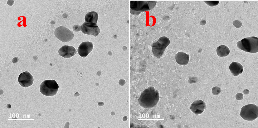
Morphological confirmation of silver nanoparticle by scanning electron microscope (a, b).
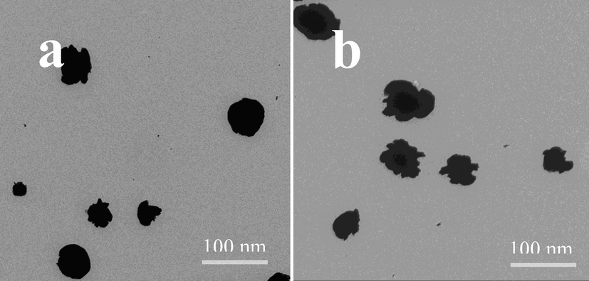
Detection of size and shape of silver nanoparticle by transmission electron microscope (a, b).
3.2 Anti-cancer studies
3.2.1 Cytotoxicity effect of silver nanoparticle
After seen the 96-well plate of treated and untreated culture, we have suggested that the synthesized silver nanoparticle has anti-cancer property against performed MCF-7 breast cancer cells due to the more formazan production. The color intensity was very higher after added the MTT stain and it shown increased death cells. In addition, when the silver nanoparticle concentration was increased, the culture was shown with more confluence or turbidity formation. Almost, the complete death cells were shown in increasing concentration and it was confirmed the silver nanoparticle has anti-cancer properties against MCF-7 breast cancer cells. In the result of O.D value was clearly indicated that the primary growth inhibition process was started at 250 µg/mL concentrations only. Hopefully, the IC50 concentration was fixed in the concentration of 1000 µg/mL and the inhibition rate was 54% against MCF-7 breast cancer cells (Fig. 3). In this concentration, the proliferation efficiency was very high compared with any other concentration. The larger surface area of the silver nanoparticle was very efficient against cancer cells and it similar to the statement of (Ebrahimzadeh et al., 2020). Biosynthesized silver nanoparticle has excellent anti-cancer property against MCF-7 breast cancer cells due to the over production of MMP and ROS Algebaly et al. (2020).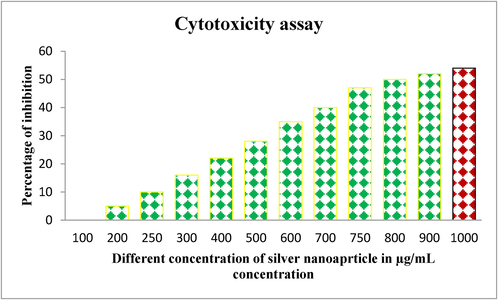
MTT assay based cytotoxicity effect using Ag NPs against MCF-7 breast cancer cells by microtiter plate.
3.3 Morphological evidences by phase contrast microscopy
In phase contrast microscopic, the contrary images were shown between the treated and control MCF-7 breast cancer cells. In control, the well matured, smooth, punch of growth like colonies was observed (Fig. 4a). Whereas, the treated was shown with more unclear structure and rough surface, which affected by silver nanoparticles. Compared with control, the treated result was conveyed that the apoptosis was done partially in some places and completely in some places due to the ROS production in the process and it caused the internal membranes damages (Fig. 4b). The surface changed, rough morphology of the same MCF-7 breast was suggested, the synthesized silver nanoparticle not only inhibit the bacterial cells, and it has the ability to inhibit the cancer cells also (li et al., 2020; Krishnaraj et al., 2014).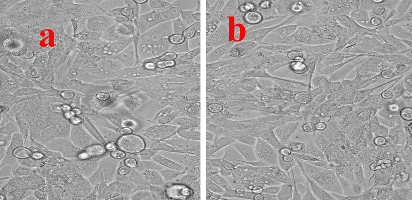
Morphological modification of Ag NPs against MCF-7 breast cancer cells by phase contrast microscope. The clear smooth image of untreated (a) and irregular, modified surface of Ag NPs treated (b) cells of MCF-7.
3.4 Fluorescence microscopy for detection of damaged cells
The IC50 concentration of synthesized silver nanoparticle was very effective against performed MCF-7 breast cancer cells due to the intracellular membrane damages and it shown in fluorescence microscope. After entry of silver nanoparticle, it stimulates the apoptosis for cancer cell death and also it was altered by ROS production. When compared with control, the fluorescence dye ethidium bromide was entered into the internal organelles and leads to bind in nucleus. The nucleus of the cells was also damaged and cell death was increased. The control cells were shown well grown, complete structure and multiple natures of each other was taken short time only. In addition, the proliferation capacity was very high and leads to condensed chromosome, tedious nucleus morphology, condensed chromatin and disassembled nucleus with cytoplasm were observed (Singh et al., 2018; Ahmadian et al., 2018). After this process, the ethidium bromide was undergone to nucleus and binds all the damaged parts of the cells and shown red color. Among the color variation, the greenish yellow of some places and clear yellow or orange color of some places were clearly conveyed information that the MCF-7 cells were damaged through early and late apoptosis respectively (Das et al., 2020; Kumar et al., 2018). The Fig. 5a, b was clearly indicated the differentiation of damaged or undamaged MCF-7 breast cancer cells with nucleus damage and condensed cells with color variation.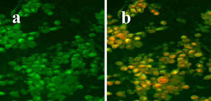
The untreated MCF-7 breast cell line shown with normal, clearly morphology (a) and Ag NPs treated MCF-7 breast cancer cells shown with more damages in intracellular membrane (b) by fluorescence microscope using acridine orange and ethidium bromide stains.
3.5 Nuclear damage in MCF-breast cancer cells using Hoechst 33342 staining
Total nucleus deformation using the effect of silver nanoparticle was viewed by fluorescence microscopic assay using Hoechst 33342 staining. After careful viewed, the nucleus was totally damaged and the regeneration was absent in the cells. The complete fragmentation of MCF-7 cells after treatment of IC50 concentration of synthesized silver nanoparticle was seen in Fig. 6b. The control nucleus in the MCF-7 was shown clear nucleus in the fluorescence image was observed after staining with Hoechst 33342 stains (Fig. 6a). In this assay, the early apoptosis and late apoptosis were clearly showed the membrane organelles damages. Hoechst 33342 staining assay was concluded that the synthesized silver nanoparticle was very efficient against MCF-7 breast cancer cells through phase contrast and fluorescence microscopic observations. Previously, Jeyaraj et al. (2015) reported that the Ag NPs was very effective against MCF-7 breast cancer cells and shown the nuclear belebbing, condensed nucleus and shorted chromatin. Also, it grown with confluent colonies and more patches of rarely sloughing cells.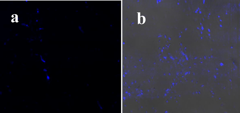
Nucleus image of untreated (a) and Ag NPs treated (b) MCF-7 breast cancer cells by fluorescence microscope using Hoechst 33342 staining.
4 Conclusion
The cytotoxicity result was suggested that the obtained silver nanoparticle is simulated the apoptosis process through ROS production, mitochondrial membrane damage, belbing formation, nucleus cleavages and chromatin condensation in MCF-7 breast cancer cells. The IC50 concentration of silver nanoparticle treated MCF-7 breast cancer cells images were clearly shown, the morphology was damaged. Further, the nucleus deformation effect due to silver nanoparticle influence are also evidently proved by florescence images after stained Hoechst 33342 stain. Hence, the present result was suggested that the obtained silver nanoparticle not only inhibit the multi-drug resistant bacteria and biofilm producing bacteria. It also very effective against MCF-7 breast cancer cell through the morphological confirmation. Finally, the seaweed synthesized silver nanoparticle was very effective and has more biological properties due to the seaweed nutrients enhancement.
Acknowledgment
All the authors gratefully acknowledge the National Natural Science Foundation of China (Project Approval Numbers: 41950410573, 91951205 and 31670009) and Postdoctoral Science Foundation of China (Project Approval Number: 2019M663213) for financial support for this work. Wen-Jun Li was also supported by Introduction project of high-level talents in Xinjiang Uygur Autonomous Region. The authors extend their appreciation to the Researchers Supporting Project number (RSP-2021/70), King Saud University, Riyadh, Saudi Arabia.
Declaration of Competing Interest
The authors declare that they have no known competing financial interests or personal relationships that could have appeared to influence the work reported in this paper.
References
- Synthesis of stable nanosilver particles (AgNPs) by the proteins of seagrass Syringodium isoetifolium and its biomedicinal properties. Biomed. Pharmacot.. 2016;84:60-70.
- [Google Scholar]
- Effect of silver nanoparticles in the induction of apoptosis on human hepatocellular carcinoma (HepG2) cell line. Mater. Sci. Eng.: C. 2018;93:465-471.
- [Google Scholar]
- Biogenic synthesis of silver nanoparticles: Antibacterial and cytotoxic potential. Saudi J. Biol. Sci.. 2020;27:1340-1351.
- [Google Scholar]
- Pomegranate peel induced biogenic synthesis of silver nanoparticles and their multifaceted potential against intracellular pathogen and cancer. Saudi J. Biolog. Sci.. 2021;28(8):4191-4200.
- [Google Scholar]
- Biomimetic synthesis of silver nanoparticles using Matricaria chamomilla extract and their potential anticancer activity against human lung cancer cells. Mat. Sci. Eng. C. 2018;92:902-912.
- [Google Scholar]
- Marine algae Caulerpa Taxifolia mediated silver nanoparticles enhances the anti-cancer activity against A549 lung cancer cell line through cytotoxicity effect/morphological damage. Saudi J. Biol. Sci.. 2020;27:3421-3427.
- [Google Scholar]
- Biosynthesis, and potential effect of fern mediated biocompatible silver nanoparticles by cytotoxicity, antidiabetic, antioxidant and antibacterial, studies. Mat. Sci. Eng. C. 2020;114:111011.
- [Google Scholar]
- Green and facile synthesis of Ag nanoparticles using Crataegus pentagyna fruit extract (CP-AgNPs) for organic pollution dyes degradation and antibacterial application. Bioorg. Chem.. 2020;94:103425.
- [Google Scholar]
- Green synthesis of silver nanoparticles via Cynara scolymus leaf extracts: the characterization, anticancer potential with photodynamic therapy in MCF7 cells. PLoS ONE. 2019;14:e0216496
- [Google Scholar]
- Biosynthesized silver nanoparticles for inhibition of antibacterial resistance and biofilm formation of methicillin-resistant coagulase negative Staphylococci. Bioorg. Chem.. 2019;89:103008.
- [Google Scholar]
- Graphene/nickel oxide nanocomposites against isolated ESBL producing bacteria and A549 cancer cells. Mater. Sci. Eng. C. 2019;102:829-843.
- [Google Scholar]
- Photocatalytic reduction and antibacterial activity of biosynthesized silver nanoparticles against multi drug resistant Staphylococcus saprophyticus (MN310601) Mater. Sci. Eng. C. 2020;114:11024.
- [Google Scholar]
- Enhanced anti-biofilm activity of facile synthesized silver oxide nanoparticles against K. pneumonia. J. Inorg. Organometal. Polymers Mater.. 2021;31:3921-3933.
- [Google Scholar]
- Biogenic metal nanoformulations induce Bax/Bcl2 and caspase mediated mitochondrial dysfunction in human breast cancer cells (MCF 7) RSC Adv.. 2015;5:2159.
- [Google Scholar]
- Synthesis of silver nanoparticles using aqueous extract of Dolichos lablab for reduction of 4-Nitrophenol, antimicrobial and anticancer activities. OpenNano. 2018;3:28-37.
- [Google Scholar]
- Bacteriostatic activity of aquatic extract of black peel pomegranate and silver nanoparticles biosynthesized by using the extract. Biocat. Agricult. Biotech.. 2020;25:101620.
- [Google Scholar]
- Caspase-3/MAPK pathways as main regulators of the apoptotic effect of the phyto-mediated synthesized silver nanoparticle from dried stem of Eleutherococcus senticosus in human cancer cells. Biomed. Pharmacother.. 2018;99:128-133.
- [Google Scholar]
- Acalypha indica Linn: Biogenic synthesis of silver and gold nanoparticles and their cytotoxic effects against MDA-MB-231, human breast cancer cells. Biotechnol. Rep.. 2014;4:42-49.
- [Google Scholar]
- Green synthesis of silver nanoparticles using leaf extract of Holoptelea integrifolia and preliminary investigation of its antioxidant, anti-inflammatory, antidiabetic and antibacterial activities. J. Environm. Chem. Eng.. 2019;7:103094
- [Google Scholar]
- Cytotoxicity of phloroglucinol engineered silver (Ag) nanoparticles against MCF-7 breast cancer cell lines. Mat. Chemist. Phy.. 2018;220:402-408.
- [Google Scholar]
- Biosynthesis of silver nanoparticles using leaf extract of Aesculus hippocastanum (horse chestnut): Evaluation of their antibacterial, antioxidant and drug release system activities. Mat. Sci. Engin. C. 2020;107:110207
- [Google Scholar]
- Eco-friendly green synthesis of clove buds extract functionalized silver nanoparticles and evaluation of antibacterial and antidiatom activity. J. Microbiolog. Methods. 2020;173:105934.
- [Google Scholar]
- Plant-mediated synthesis of silver nanoparticles using fruit extract of Cleome viscosa L.: Assessment of their antibacterial and anticancer activity. Karbala Int. J. Modern Sci.. 2018;4:61-68.
- [Google Scholar]
- Antibacterial and cytotoxic activities of a green synthesized silver nanoparticles using corn silk aqueous extract. Colloids Surf. A: Physicochem. Eng. Asp.. 2020;598:124827.
- [Google Scholar]
- Anti-biofilm and antibacterial activities of silver nanoparticles synthesized by the reducing activity of phytoconstituents present in the Indian medicinal plants. Front. Microbiol.. 2020;11:1143.
- [Google Scholar]
- Biosynthesis of silver nanoparticles formation from Caesalpinia pulcherrima stem metabolites and their broad spectrum biological activities. J. Genet. Eng. Biotech.. 2018;16:105-113.
- [Google Scholar]
- Galactomannan endowed biogenic silver nanoparticles exposed enhanced cancer cytotoxicity with excellent biocompatibility. Int. J. Biolog. Macromol.. 2018;118:1174-1182.
- [Google Scholar]
- Fabrication of adenosine 5′-triphosphate-capped silver nanoparticles: enhanced cytotoxicity efficacy and targeting effect against tumor cells. Process Biochem.. 2018;65:186-196.
- [Google Scholar]
- Anti-oxidant, anti-bacterial and anti-biofilm activity of biosynthesized silver nanoparticles using Gracilaria corticata against biofilm producing K. pneumonia. Colloids and Surfaces A. 2020;600:124830
- [Google Scholar]
- Phyto-mediated synthesis of silver nanoparticles using fucoidan isolated from Spatoglossum asperum and assessment of antibacterial activities. J. Photochem. Photob. B: Biol.. 2018;185:117-125.
- [Google Scholar]
- Green synthesis of silver nanoparticles using Trapa natans extract and their anticancer activity against A431 human skin cancer cells. J. Drug Deliv. Sci. Tech.. 2018;47:375-379.
- [Google Scholar]
- Biogenic silver nanoparticle synthesis with cyanobacterium Chroococcus minutus isolated from Baliharachandi sea-mouth, Odisha, and in vitro antibacterial activity. S. J. Biolog. Sci.. 2020;27(6):1580-1586.
- [Google Scholar]
- Synthesis of AgNPs coated with secondary metabolites of Acacia nilotica: an efficient antimicrobial and detoxification agent for environmental toxic organic pollutants. Mat. Sci. Eng. C. 2020;111:110829.
- [Google Scholar]
- Anti-cancer activity of biosynthesized silver nanoparticles using Avicennia marina against A549 lung cancer cells through ROS/mitochondrial damages. Saudi J. Biol. Sci.. 2020;27:3018-3024.
- [Google Scholar]
- Spectroscopic, microscopic characterization of Cannabis sativa leaf extract mediated silver nanoparticles and their synergistic effect with antibiotics against human pathogen. Alexandria Eng. J.. 2018;57:3043-3051.
- [Google Scholar]
- Attenuation of diethylnitrosamine (DEN) – Induced hepatic cancer in experimental model of Wistar rats by Carissa carandas embedded silver nanoparticles. Biomed. Pharmacoth.. 2018;108:757-765.
- [Google Scholar]
- Phytofabrication of Silver nanoparticles: Novel Drug to overcome hepatocellular ailments. Toxicol. Rep.. 2018;5:333-342.
- [Google Scholar]


