Translate this page into:
Ultrastructure of sensory receptors on the antennae of Oryctes elegans Prell (Coloeptera: Scarabidae)
-
Received: ,
Accepted: ,
This article was originally published by Elsevier and was migrated to Scientific Scholar after the change of Publisher.
Abstract
Oryctes elegans is one of the economically important pest of palm trees in Saudi Arabia. Chemoreceptors are very important organs for mating, feeding or oviposition in insects. We described the morphology ultra structure and description of antennae among males and females using scanning electron microscopy. Observation have been described determined that the antennae of O. elegans were lamellate type and composed of nine segments. There were three types of trichoid sensilla found on the antennae. These were elongated, curved and articulate, respectively. Fourth type was placoid sensillae concentrated on the lateral segments of antennae. These sensillae were abundant distributed on pedicle and the 1st flagellomeres. Sensillae found on females and males antennae were similar but the antennal segments of males were thinner than females.
Keywords
Oryctes elegans
Scanning electron microscope
Antennae
Sensillae
Morphology
Ultrastructure
Sensory receptors
1 Introduction
Oryctes elegans (the fruit stalk borer) is one of the most economically significant pests on palm trees in the Kingdom of Saudi Arabia (Gharib, 1970). Larvae attack roots and trunk of the date palm and the adults attack palm branches and fronds, it also, infested on coconut trees and oil palm. Adults are active at night during April to December in Al-Hasa, and are attracted to light. Females lay eggs in tunnels which are made by adults on the stalk, and between trunks and palm offset.
Females lay between 30 and 50 eggs which are hatching after 6–8 days. These insects yearly have one generation, Martin (1968) stated that these insects may have two generation yearly, in Iraq. The adults attacked middle (heart) of the steam and infestations caused deformation of leaves. Larvae damages more than the adults, because there live the palm stalk and tunnels of the steam which the adults made at leaving it. In this study, scanning electron microscope was used to study sense organs on antennae males and females of O. elegans because these sensillae are very important for oviposition, feeding and mating.
2 Materials and methods
2.1 Insects
O. elegans was reared on the palm tree. We obtained its males and females from Ministry of Water and Agriculture of Al-Kharj in Saudi Arabia.
2.2 Scanning electron microscope (SEM)
Preparatory techniques for SEM by Salama and Abdel Aziz (2001). Freshly emerged adults of O. elegans which were killed by glutaraldehyde 5%. Antennae of O. elegans were carefully excised from the antennal sockets with fine forceps. The antennae were first kept in 70% ethanol for 24 h then dehydrated in a graded acetone series of 70%, 80%, 90% and 100% in each case for 1 h. The antennae were mounted with ventral or dorsal sides on the sticky tape. The dehydration processes was followed by air drying. (Fine Serial No. PGO – 329 – Supply KPFREQ), then examination was made under scanning electron microscope (SEM) (JEOL – JSM 636 OLV). The estimation of sensillum numbers was based on observations from four males and four females.
3 Results
3.1 General description of antennae of O. elegans
The antennae of O. elegans are lamillate in shape and consist of the basic segments: scape, pedicle and flagellum. This antennae form is the characteristic of Family: Scarabidae. Flagellum consists of nine segments, six segments are of equal size but the lateral segments of flagellomeres are slender like oval loops (Figs. 1a,b and 2A–C). In this study, we observed antennae of males and females and found the almost in form and structure but segments of males are slender than females.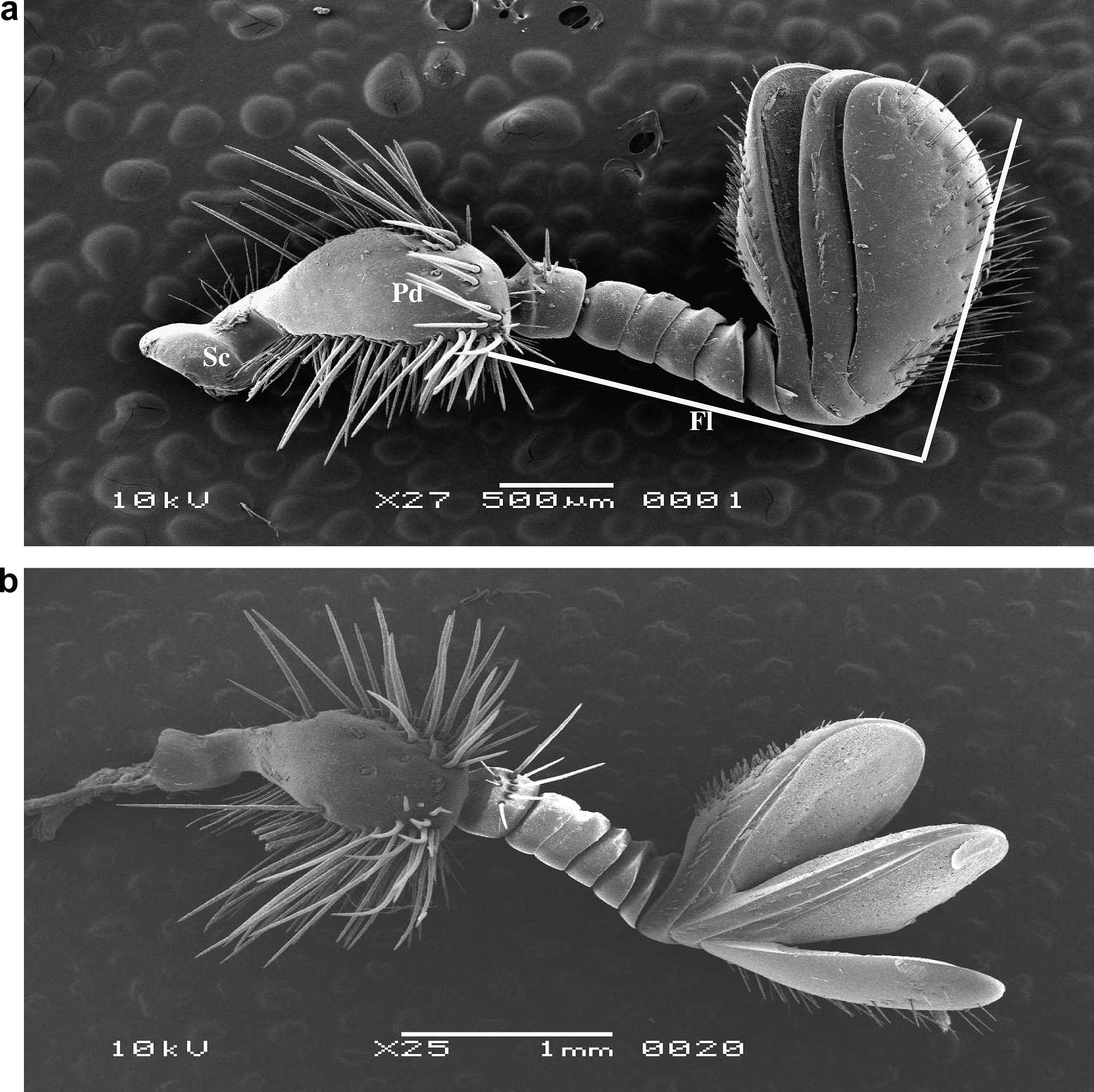
(a) Scanning electron micrograph (SEM) of an excised antenna of female O. elegans showing the scape, pedicle and flagellum. (b) Scanning electron micrograph (SEM) of an antenna of male O. elegans showing the scape, pedicle and flagellum.
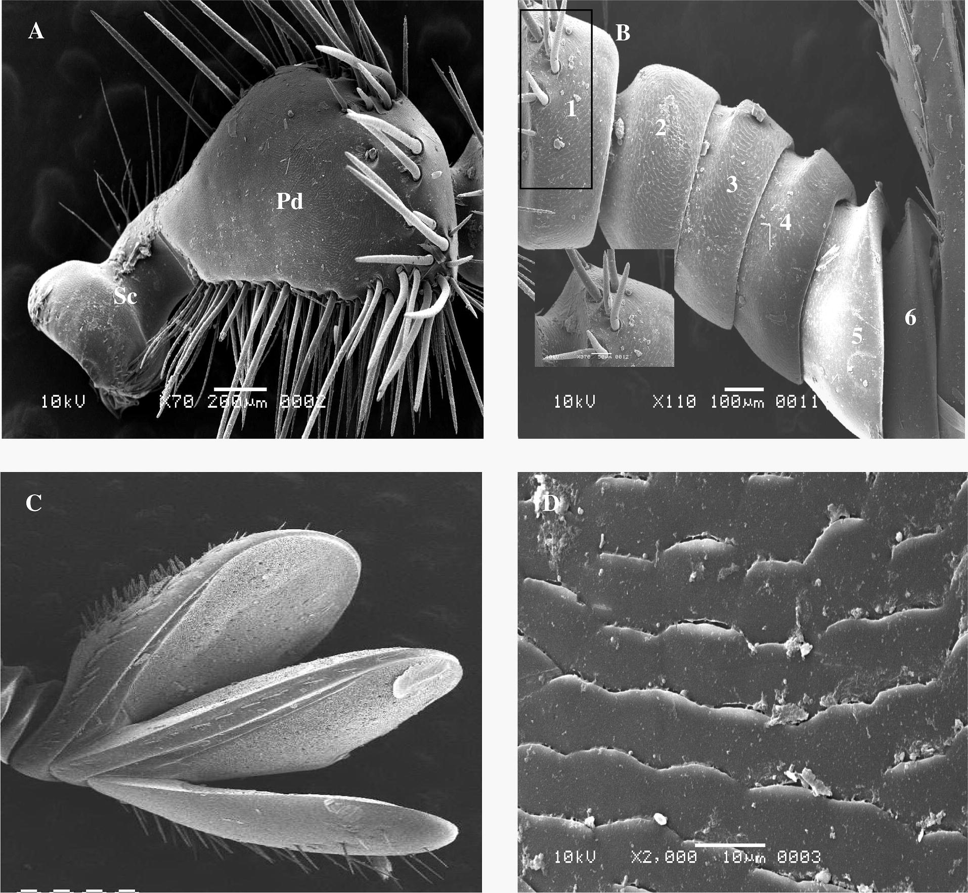
Scanning electron micrograph (SEM) shows: (A) Scape (Sc) and Pedicle (Pd). (B) Funicle show 1st flagellomeres with hairs but another segments without hairs. (C) Club (segmental opening). (D) Cuticle of antenna.
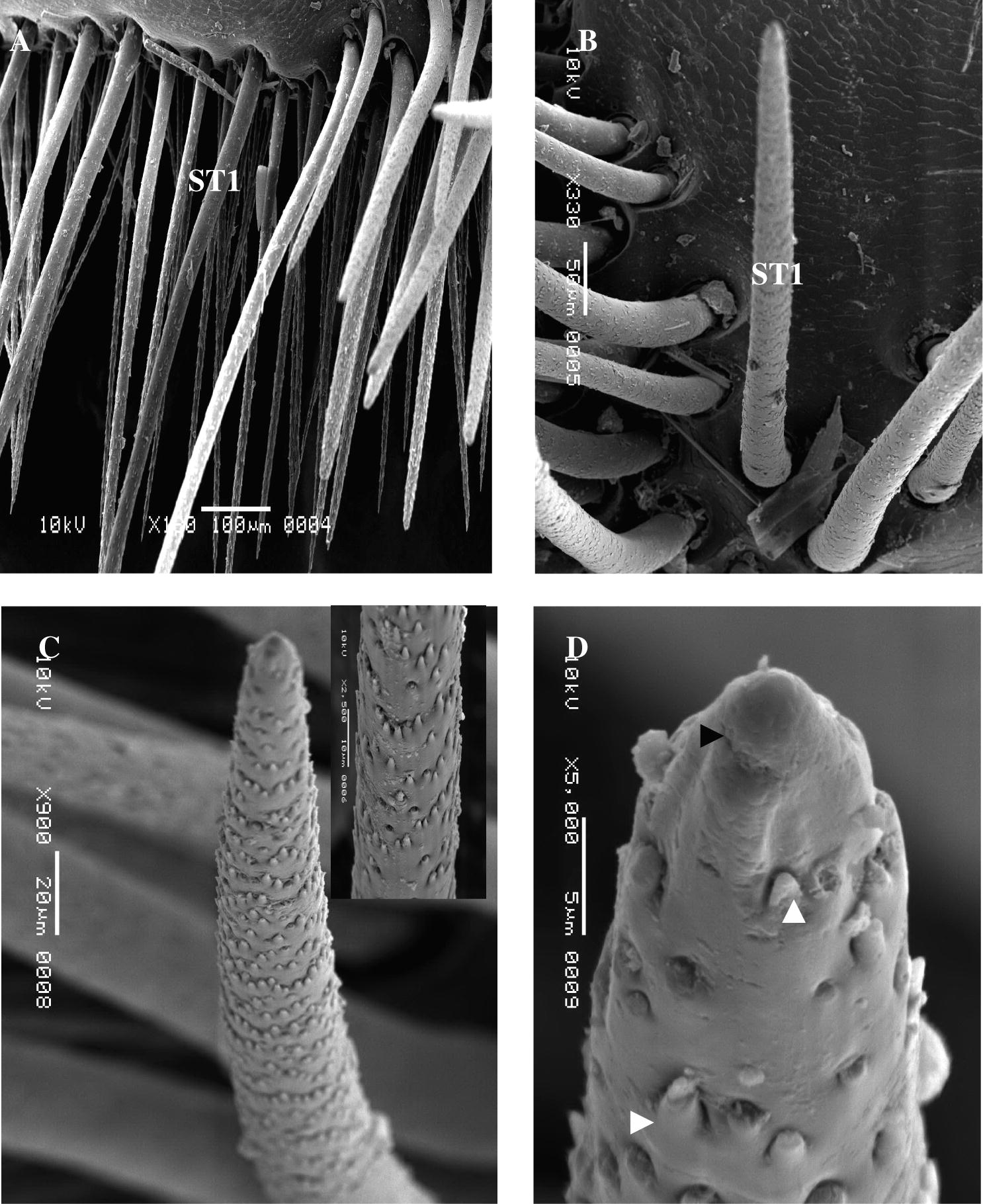
Scanning electron micrograph (SEM) shows of O. elegans ST1: (A) ST1 (100×). (B) ST1 (330×). (C) ST1 (900×). (D) ST1 (500×).
The cuticle of the antennae was black in color and characterized by irregular spiny lines which gave it a feature of the arid land (Fig. 2D), just as Fig. 2A demonstrated scape is lacking sensillae, whereas the pedicle consist of many sensillae concentrated on the segment board, and these hairs were found on the 1st and lateral segment of flagellomeres only.
3.2 Sensillae types
Four morphologically different types of sensilla were recorded on the antennae of male and female O. elegans. These include three types of trichoid sensillae (types elongated, articulate and curved). These are multi porous, while placoid sensillae are like plates and with or without pores. Sensilla trichoidea are long, articulate and curved certain of basis. The distribution of the different sensillae types on each antennal segment is shown in Table 1.
3.2.1 Sensilla trichoidea (ST1) (Fig. 3A–D)
Sensilla trichoidea type I occur on the pedicle and lateral segments, antennomeres in males and antennal flagellum of females they are elongated, growing out from socket of cuticle, tapered edge and these are multi porous. The pores density increases gradually, and the pores are arranged in a regular pattern. The surface of cuticle consists of many projections. The ST1 range from 333.33 to 573.80 μm in length, depending upon their location on the antennae (Table 1). Number and location of the various sensillae observed on the antennae of male and female O. elegans. Sc: scape, Pd: pedicle, F: flagellum. Cu: curved, El: elongated, Ar: articulate. T.F.: total number of sensillae of flagellomeres. T.S.: total number of sensillae of antenna.
Gender
Sensilla
Antennal segments
Sc
Pd
Flagellum
F1
F2
F3
F4
F5
F6
F7
F8
F9
T.Fl.
T.S.
Female
ST1 (Cu)
–
6
–
–
–
–
–
–
–
–
–
–
6
ST2 (El)
–
66
5
–
–
–
–
–
–
39
65
109
175
ST3 (Ar)
–
1
–
–
–
–
–
–
–
–
–
–
1
SP (Pla)
–
–
–
–
–
–
–
–
Many sensilae
–
–
Male
ST1 (Cu)
–
3
–
–
–
–
–
–
–
–
–
–
3
ST2 (El)
–
69
4
–
–
–
–
–
–
37
60
101
170
ST3 (Ar)
–
3
–
–
–
–
–
–
–
–
–
–
3
SP (Pla)
–
–
–
–
–
–
–
–
Many sensilae
3.2.2 Sensilla trichoidea (ST2)
These sensillae are found few in number on the pedicle between another hairs of trichoidea. Sensilla trichoidea 2 characterize articulated, surface of cuticle was similar to ST1. The length of ST2 ranges from 112 to 250 μm (Fig. 4).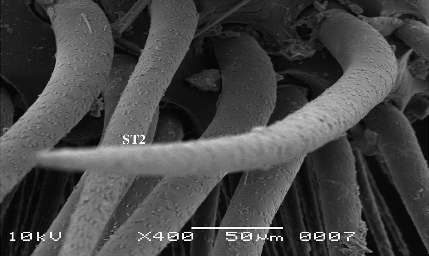
Scanning electron micrograph showing sensilla trichodea articulated ST2.
3.2.3 Sensilla trichodea (ST3)
This type of sensillae occurs on corner of pedicle having different lengths, multi porous, with high density of spines, with curved base in and it socket of cuticle (Fig. 5). Lateral segments of flagellomere consist many hairs of Tr elongate and concentrated on side of 1st and 3rd flagellomere, the length range from 155.55 to 333.33 μm (Fig. 1).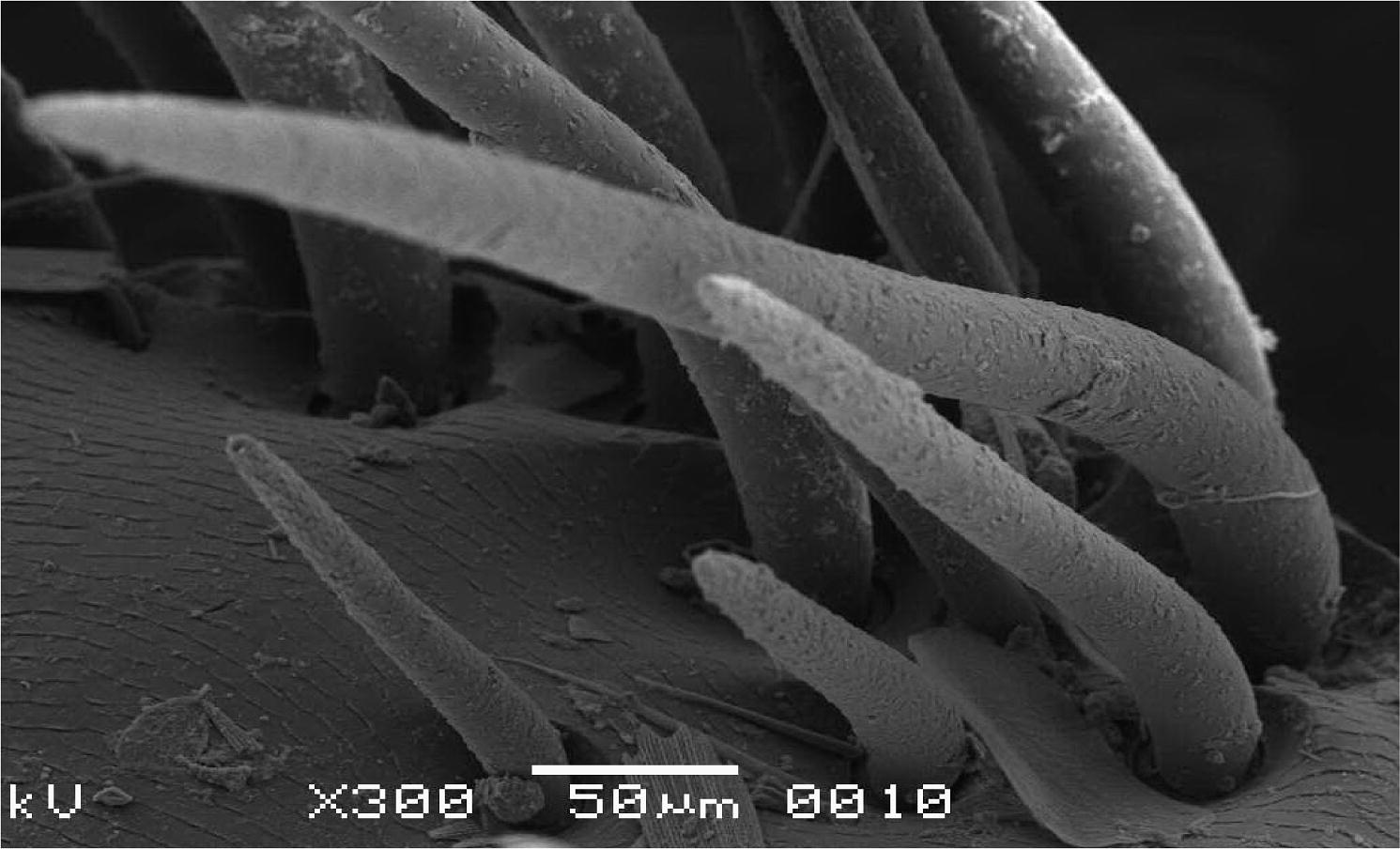
Scanning electron micrograph showing sensilla trichoidea curved ST3.
3.2.4 Placoid sensillae
Placoid sensillae are found on internal surface of lateral segments of flagellomere. These organs are conical pegs and lack hairs, with or without pores (Fig. 6A and B). These sensillae are densely distributed on the ventral surface of both sexes.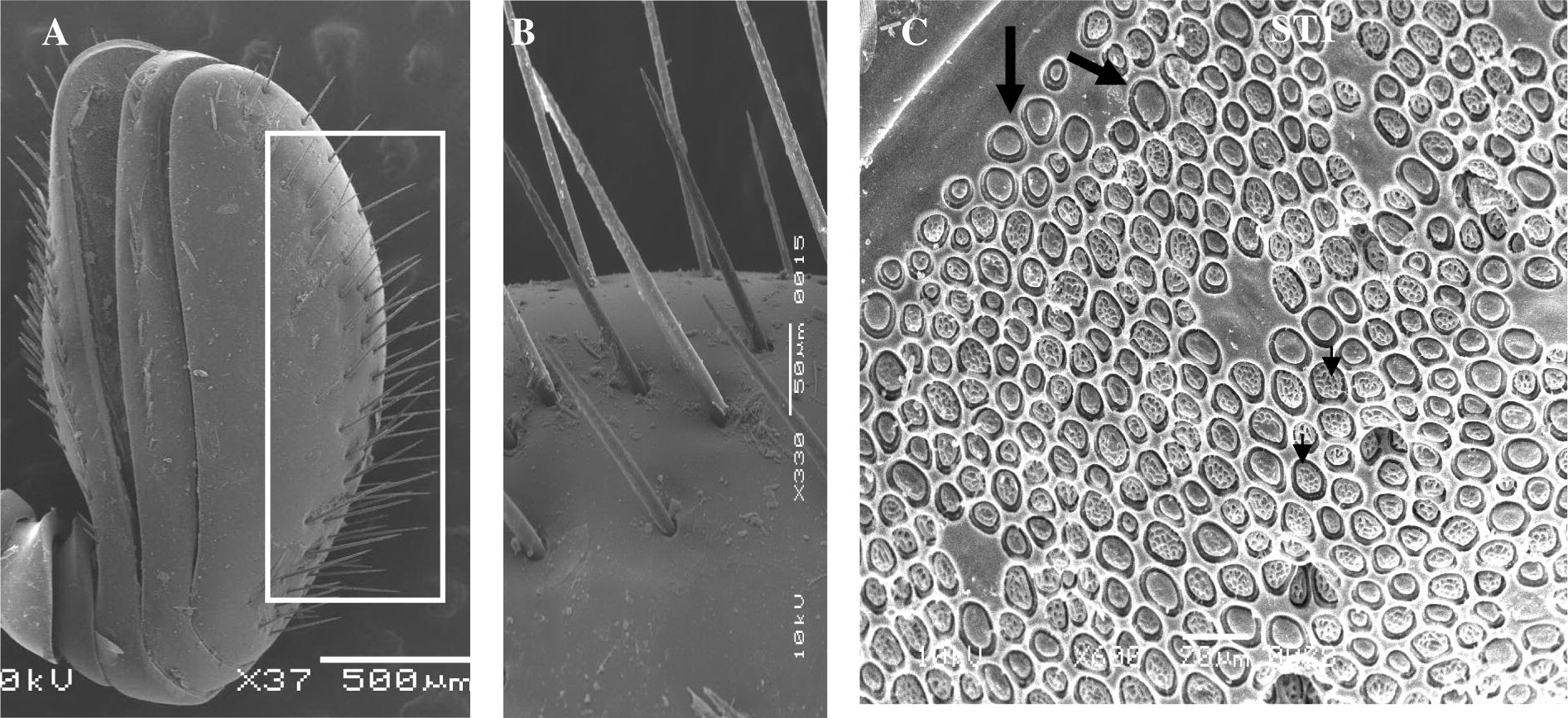
Scanning electron micrograph of antenna: (A) Club. (B) Edge of club show ST1. (C) General view of surface of club show numerous placoid sensillae (small arrow pores and big arrow unpores).
4 Discussion
The external morphology, types and distribution of sensillae on the antennae of male and female O. elegans recorded in this study are largely in conformity with those reported for other coleopterous insects (Alm and Hall, 1986; Merivee et al., 1999; Sharaby and Al-Dosary, 2007).
The antennae of insects have been typically described as consisting of basic three segments, scape, pedicle and flagellum. Our study revealed four morphologically different types of sensillae on the antennae of male and female O. elegans. In general, a great number of ST1 occur on the males and females. The multiporous sensilla trichoidea have previously been described with different names including multiporous sensilla trichoidea with wall pores (Wible et al., 1984; Petterson et al., 2001; Ryan 2002; Onagbola and Fadamiro, 2007; Bleeker et al., 2004; Onagbola and Fadamiro, 2007). In general antennal sensilla trichoidea are presumed to function as olfactory receptors in many insects (Steinbreacht, 1987, 1997; Bleeker et al., 2004), and electrophysiological studies have confirmed sex pheromone receptor function for the trichoid sensilla of Neodiprion sertifer Geoffory (Hymenoptera: Diprionidae) (Hansson et al., 1991). Petterson et al. (2001) proposed a pheromone receptor function for the sensilla trichoidea of Rhopalicos tutela. The greater occurrence of the ST on the antennae of male O. elegans relative to the females may indicate a probable role in mate location, possibly for the detection of females sex pheromones (Chapman, 1982; Bleeker et al., 2004; Onagbola and Fadamiro, 2007; Onagbola et al., 2008).
Placoid sensilla have been found in various sizes and shapes on the antennae. Sensilla placodea of O. elegans are oval disk and like inflow density and commonly occurred in both sexes. The function of sensilla placoidea (SP) is assumed to be olfactory because they posses a multiple cuticular pore system (Steinbrecht, 1984), or possibly these disks have a secretory role, similar to that of glandular openings (Martin, 1977; Suttcliffer and Mitchell, 1980). Single sensillum research shows that sensilla placoidea in Micropitis croceipes are indeed olfactory receptors which responded in a dose-dependent manner to plant volatiles (Ochieng et al., 2000). Their specific function in O. elegans however, has yet to be confirmed electrophysiologically.
References
- Antennal sensory structures of Conotrachelus nenuphar (Coleoptera: Curculionidae) Ann. Entomol. Soc. Am.. 1986;79:324-333.
- [Google Scholar]
- Antennal sensilla of two parasitoid wasps: a comparative scanning electron microscopy study. Micros. Res. Tech.. 2004;63:266-273.
- [Google Scholar]
- Chemoreception: the significance of receptor number. In: Berridge M.J., Treherne J.E., eds. Advances in Insect Physiology. Vol vol. 16. London: Academic Press Inc.; 1982. p. :247-357.
- [Google Scholar]
- Oryctes elegans Prell. (Coloeptera, Dynastidae) Entomol. Phytopath. Appl. (Tehran). 1970;29:10-12. ((in French), 10–19 (in Iranian))
- [Google Scholar]
- Sex some parasitoid hymenoptera with hypothesis on their role sex and host recognition. J. Hymen. Res. 1991;5:206-239.
- [Google Scholar]
- Martin, H., 1968. Report to the Government of Iraq. On Cereal and Date Palm Tree Pests. FAO No. TA 2339.
- Glandes dermiques chez deux coleopteres cavernicoles (Catopidae) Int. J. Insect. Morphol. Embryol.. 1977;6:179-189.
- [Google Scholar]
- Antennal sensilla of the click beetle Melanotus villosus (Geoffroy) (Coleoptera: Elateridae) Int. J. Insect Morphol. Embryol.. 1999;28:41-51.
- [Google Scholar]
- Functional morphology of antennal chemoreceptors of the parasitoid Microplitis croceipes (Hymenoptera: Braconidae) Arthropod Struct. Dev.. 2000;29:231-240.
- [Google Scholar]
- Scanning electron microscopy studies of antennal sensilla of Pteromalus cerealellae (Hymenoptera: Pteromalidae) Micron. 2007;39(5):526-535.
- [Google Scholar]
- Morphological characterization of the antennal sensilla of the Asian citrus psyllid, Diaphorina citri kuwayama (Hemiptera: Psyllidae), with reference to their probable functions. Micron. 2008;39(8):1184-1191.
- [Google Scholar]
- Evidence for the importance of odour-perception in the parasitoid Rhopalicus tutela (Walker) (Hym., Pteromalidae) J. Appl. Entomol.. 2001;125:293-301.
- [Google Scholar]
- Insect Chemoreception: Fundamental and Applied. The Netherlands: Kluwer Academic Publishers; 2002. p. 303
- Distribution of the Sensillae of the red palm weevil, Rhynchophorus ferrugineus (Oliv) (Coleoptera: Curculionidae) Insect Sci. Appl.. 2001;21(2):179-188.
- [Google Scholar]
- Ultra morphological structure of sensory sensillae on the legs and external genitalia of the red palm weevil Rhynchophorus ferrugineus (Oliv.) Saudi J. Biol. Sci.. 2007;14(1):29-36.
- [Google Scholar]
- Arthropods: chemo-, thermo, and hygro-receptors. In: Bereiter-Hahn J., Matolsty A.G., Richards K.S., eds. Biology of the Integument. Vol vol. 1. Berlin: Springer-Verlag; 1984. p. :523-553.
- [Google Scholar]
- Functional morphology of pheromone-sensitive sensilla. In: Prestwich G.D., Blomquist G.J., eds. Pheromone Biochemistry. Orlando: Academic Press; 1987. p. :353-383.
- [Google Scholar]
- Pore structures in insect olfactory sensilla: a review of data and concepts. Int. J. Insect Morphol. Embryol.. 1997;26:229-245.
- [Google Scholar]
- Structure of galleal sensory complex in adults of the red turnip beetle Entomocelis Americana Brown (Coleoptera: Chrysomelidae) Zoomorphology. 1980;96:63-76.
- [Google Scholar]
- Scanning electron microscopy of antennal sense organs of Nasonia vitripennis (Hymenoptera: Pteromalidae) Trans. Am. Micros. Soc.. 1984;103(4):329-340.
- [Google Scholar]


